
Sputum Sample: Collection, Transport, Staining, and Culture
How to collect a good sputum sample, why the first morning specimen matters, the ideal-to-compromise order when a patient cannot expectorate, the cell criteria that decide accept or reject, and correct transport timing.

On this page
The hardest part of a sputum culture is getting sputum. Most rejected specimens are saliva, spit from the mouth rather than coughed from the lungs, and a culture run on saliva reports mouth flora that can send treatment in the wrong direction. Everything below, the timing, the coaching, the cell criteria, exists to answer one question: did this really come from the lower airway?
Sputum, also known as phlegm, is a thick type of mucus in a person’s lungs suffering from an infection or chronic illness affecting the lungs or airways. The first morning expectorated sputum sample is always best for diagnosing lower respiratory tract infections. It is essential that the specimen for culture be sputum and not saliva or nasopharyngeal secretions.
Sputum has been the primary means of determining the causes of lower respiratory tract infections such as bronchitis, bronchiolitis, and pneumonia. The symptoms suggestive of pneumonia include fever, chills, chest pain, and cough. In addition to respiratory symptoms, 10% to 30% of patients with pneumonia complain of headache, nausea, vomiting, abdominal pain, diarrhea, and myalgias.
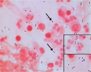 Figure: Streptococcus pneumoniae, Gram positive diplococci
Induced sputum, endotracheal or tracheostomy suction specimen, transtracheal aspirates are other alternatives for expectorated sputum when the patient is uncooperative, pediatric patients or has special conditions/requirements. Sputum samples can be obtained using bronchoscopy, e.g., bronchial washings, aspirates, and bronchoalveolar lavage (BAL).
Sample Collection
Expectorated Sputum
- Do not allow the patient to eat food for 1 to 2 hours before expectoration
- Ask the patient to brush teeth and then rinse mouth with saline or water just before expectoration
- Give the patient a sterile, wide-mouthed, screw-top container to collect a sputum sample
- Instruct the patient to provide a deep-coughed specimen by collecting the coughed material (sputum) into a sterile container by avoiding contamination with saliva.
- Transport the sputum sample immediately to the laboratory. Delay in transport may result in loss of viable infectious agents and recovery of pathogens.
Induced sputum
Induced sputum is used to diagnose P. jirovecii pneumonia, and LRTI caused by mycobacteria or fungal agents. If a patient cannot produce sputum, respiratory therapists may use postural drainage and thoracic percussion to stimulate the production of acceptable sputum.
- Have the patient inhale nebulized hypertonic saline (3% to 7% NaCl) through an ultrasonic nebulizer until a strong cough reflex is triggered, usually within 10 to 20 minutes. Normal saline (0.9%) is not concentrated enough to induce sputum.
- Collect the specimen in a sterile container.
Sputum sampled collected this way may appear watery, resembling saliva but are usually adequate for culture. The induced sputum sample should be labeled accordingly so that the laboratory accepts it without prescreening.
Lower respiratory tract samples collected via tracheal aspirate, bronchoalveolar lavage, or lung biopsy bypass the possibility of contamination with normal upper respiratory tract flora and are more likely to provide an accurate microbiological diagnosis.
Endotracheal or tracheostomy suction specimens
Patients with tracheostomies cannot produce sputum in the normal fashion, but lower respiratory tract secretions can easily be collected in a Lukens trap. The laboratory should treat tracheostomy aspirates or tracheostomy suction specimens as sputum. The clinical significance of the culture isolates should be evaluated based on clinical signs and symptoms as these patients get colonized by gram-negative bacilli or nosocomial pathogens rapidly.
Bronchoscopy specimens
Respiratory physicians can obtain bronchial washings or aspirates, bronchoalveolar lavage (BAL) samples during bronchoscopy by inserting a small amount of sterile physiologic saline into the bronchial tree and withdrawing the fluid. These specimens may get contaminated with normal flora of the upper respiratory tract. Sample collected through bronchoscopy is suitable for detecting most major respiratory tract pathogens, including Pneumocystis jirovecii cysts and fungal elements.
Which sample, and what to do when the patient cannot cough one up
Expectorated sputum is the first choice because it is non-invasive and cheap. But many patients cannot produce it: young children swallow their sputum, intubated patients cannot cough on command, and immunocompromised patients may need a sample clean enough to trust. The rule is to step up the invasiveness only as far as the clinical question demands.
| Rank | Sample | When to use it | How it is collected | What you trade off |
|---|---|---|---|---|
| 1 (ideal) | Expectorated sputum | Cooperative adult who can cough productively | First morning, deep cough into sterile wide-mouth container after rinsing mouth | Easily contaminated by saliva; useless if the patient cannot cough deeply |
| 2 | Induced sputum | Patient cannot expectorate, or Pneumocystis or TB suspected with a dry cough | Nebulized 3% to 7% hypertonic saline until cough triggers | Looks watery like saliva; must be labeled "induced" so the lab does not reject it on cell criteria |
| 3 | Endotracheal or tracheostomy aspirate | Intubated or tracheostomized patient | Suction into a Lukens trap | These patients are colonized by gram-negative and nosocomial flora within days, so a positive culture may be colonization, not infection; interpret with clinical signs |
| 4 (most invasive) | Bronchoscopy: BAL, bronchial wash, protected brush | Immunocompromised host, non-resolving pneumonia, or when a reliable lower-airway sample is essential | Bronchoscope instills and withdraws sterile saline from the target segment | Invasive, needs a specialist; BAL still passes the upper airway so some contamination remains, though far less than expectorated sputum |
Children are the clear exception to the ranking. Infants and young children swallow sputum rather than expectorate it, so the usual step 1 is often unavailable. For suspected TB, a gastric aspirate (collected early morning before the swallowed overnight sputum leaves the stomach) is the classic pediatric substitute; for other infections, bronchoscopy with washings is often the optimal pediatric specimen. Do not force repeated expectoration attempts on a child who cannot produce a deep-cough sample.
One labeling trap worth repeating: an induced sputum is watery and cell-poor and will look like saliva under the microscope. If it is not labeled as induced, the lab may reject it using the standard squamous-cell screen. Always mark induced samples so they bypass that screen.
Note: Its always a good idea to contact the laboratory before collecting for fastidious pathogens.
Sputum Transport
Sputum has no preservative and no transport medium. It goes to the lab in the same sterile wide-mouth container it was collected in, and the clock starts at collection.
- Transport within 1 to 2 hours of collection at room temperature. Prompt processing protects fastidious organisms and stops mouth commensals from overgrowing the true pathogen.
- If delay is unavoidable, refrigerate at 2 to 8 degrees C for up to 24 hours for routine bacterial and mycobacterial culture. Refrigeration slows commensal overgrowth.
- Do not refrigerate if a fastidious organism is suspected, for example when Streptococcus pneumoniae or Haemophilus influenzae is the target, since cold reduces their recovery. When in doubt, transport fast and warm rather than cold.
- For tuberculosis, refrigeration is acceptable and standard when samples must be batched or shipped; keep them cool and out of light.
The single rule that covers most situations: process fresh whenever possible, refrigerate if you cannot, and warn the lab if a fragile organism is in the differential so the sample is not held cold.
Sputum Processing
Staining and Direct Examination
The screen that decides accept or reject. Before culture, the lab checks a Gram-stained smear under low power to confirm the sample came from the lower airway. A good expectorated sputum has more than 25 neutrophils (pus cells) and fewer than 10 squamous epithelial cells per low power field. Many squamous epithelial cells mean the sample is mostly saliva and will be rejected, because culturing it reports mouth flora, not the true pathogen. This screen is applied to expectorated samples only, not to induced sputum, tracheal aspirates, or BAL, which are judged differently.
Direct visual examinations of Lower respiratory specimens: KOH preparation or periodic acid-Schiff-stained smears for fungal element detection, Gram staining (for bacteria and yeasts), Ziehl Neelsen staining for Mycobacterium tuberculosis (or Kinyoun carbolfuchsin stain, auramine or auramine-rhodamine) is commonly used direct examination methods for the detections of pathogen causing lower respiratory tract infections. The selection of a particular method depends on the clinical presentation as per the request of a physician.
The quality of the sputum specimen can be determined by examining Gram-stained smears. A reliable expectorated sputum sample has more than 25 neutrophils (pus cells) and fewer than 10 squamous epithelial cells per low power field. The culture and interpretation of an unreliable sputum sample can be misleading and should be rejected by the laboratory. If large numbers of typical organisms are seen in a good sputum sample, a preliminary assessment of the cause of pneumonia can be made.
Culture of Respiratory tract sample:
Sputum culture is performed to determine the infectious etiologies of pneumonia or pulmonary tuberculosis. The most frequent bacterial cause of community-acquired pneumonia is Streptococcus pneumoniae, whereas S. aureus, and gram-negative rods, such as K. pneumoniae and P. aeruginosa, are the common causes of hospital-acquired pneumonia.
If a few colonies of Gram-negative rods are isolated from the sputum or throat swab of a hospitalized patient, further identification and anti-microbial susceptibility testing are of no clinical relevance, since neither procedure will have any effect on the treatment of the patient.
Routinely used culture media to isolate common etiologic agents of lower respiratory tract infections are; 5% sheep blood agar, MacConkey agar (for isolation and differentiation of gram-negative bacilli), and chocolate agar (for Haemophilus and Neisseria spp). For suspected cases of legionnaires’ disease, buffered charcoal-yeast extract agar (BCYE), and Lowenstein Jensen medium is used for pulmonary tuberculosis.
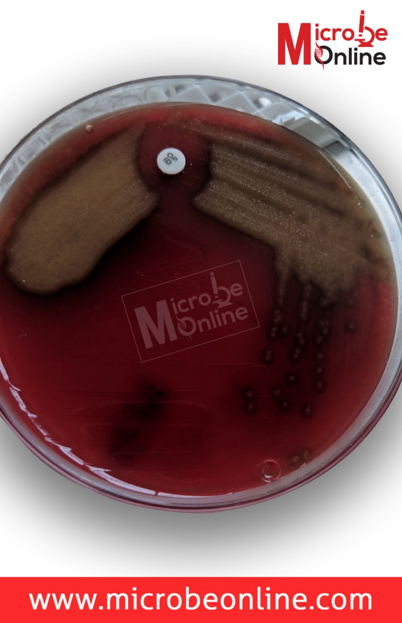 Figure: Optochin sensitive colonies of S. pneumoniae
- Blood cultures accompanying sputum specimens may occasionally be helpful, particularly in high-risk community-acquired pneumonia patients.
- The laboratory should be contacted for specific instructions before collecting specimens for fastidious pathogens such as Bordetella pertussis.
- The range of pathogens causing exacerbations of lung disease in cystic fibrosis patients has expanded, and specimens for mycobacterial and fungal cultures should be collected in some patients.
- A broad diagnostic approach based on invasively obtained specimens is suggested in the immunocompromised host.
- Most negative rapid antigen test results should be confirmed by another method. NAATs have largely replaced rapid antigen tests and cultures for respiratory virus detection.
- Calcium alginate swabs are not acceptable for nucleic acid amplification testing.
- Bronchoscopy with washings is the optimal diagnostic specimen in pediatrics.
List of Bacteria causing Respiratory Tract Infection
A: Definitive pathogens
- Bordetella pertussis
- Chlamydia trachomatis (neonatal pneumonia)
- Chlamydophila pneumoniae
- Mycoplasma pneumoniae
- Mycobacterium tuberculosis
- Corynebacterium diphtheriae (toxin-producing)
- Legionella spp.
B: Possible pathogens
- Streptococcus pneumoniae
- Staphylococcus aureus
- Neisseria meningitidis
- Haemophilus influenzae
- Haemophilus parainfluenzae
- Pseudomonas spp
- Enterobacteriaceae
- Moraxella catarrhalis
List of fungal pathogens causing lower respiratory tract infections
- Pneumocystis jirovecii (formerly Pneumocystis carinii)
- Histoplasma capsulatum
- Coccidioides immitis
- Cryptococcus neoformans (may also be recovered from patients without disease)
- Blastomyces dermatitidis
Note: Nocardia species are sometimes sought on the same fungal-type specimens because they are partially acid-fast and grow on fungal media, but they are aerobic actinomycete bacteria, not fungi.
How to Remember
The whole page is one question: airway or mouth? Every rule (first morning, rinse first, deep cough, the 25/10 cell count) is a filter to keep saliva out. If you remember the goal, you can reconstruct the rules.
25 in, 10 out. More than 25 pus cells means the immune system is active in the lower airway (good, keep it). Fewer than 10 squamous cells means little mouth contamination (good, keep it). Flip either number and the sample is saliva. "Lots of pus, little skin."
Step up, don't skip up. Expectorated to induced to aspirate to bronchoscopy. Each step is more invasive and more reliable. You climb the ladder only as far as the clinical question forces you, not straight to the top.
Induced sputum is the liar that tells the truth. It looks like saliva (watery, cell-poor) but is a real lower-airway sample. Label it, or the lab throws out a good specimen.
Key exam facts in one table
| Point | Fact |
|---|---|
| Best routine sample | First morning expectorated sputum, deep cough |
| Container | Sterile, wide-mouth, screw-cap, no preservative |
| Prep before collection | Rinse mouth with water or saline; do not eat 1 to 2 hours before |
| Acceptance screen (expectorated) | More than 25 neutrophils and fewer than 10 squamous epithelial cells per LPF |
| Screen does NOT apply to | Induced sputum, tracheal aspirate, BAL |
| Induction fluid | Nebulized hypertonic saline 3% to 7% (not normal saline) |
| Transport time | 1 to 2 hours at room temperature |
| If delayed | Refrigerate 2 to 8 C up to 24 h, except when fastidious organisms (pneumococcus, Haemophilus) are suspected |
| Pediatric TB substitute | Early-morning gastric aspirate |
| Commonest CAP pathogen | Streptococcus pneumoniae |
| Commonest HAP pathogens | S. aureus, K. pneumoniae, P. aeruginosa |
| TB stains | Ziehl-Neelsen, or fluorochrome (auramine/auramine-rhodamine) |
| Nocardia | Bacterium (actinomycete), not a fungus; partially acid-fast |
| NAAT caution | Calcium alginate swabs are not acceptable for NAAT |
Where Students Get Confused
"Sputum vs saliva, how does the lab even tell?" Under the microscope, saliva is full of squamous epithelial cells (flat mouth-lining cells) and few pus cells. Real sputum is the reverse. That is what the 25/10 screen measures. It is not about how the sample looks to the eye; a watery induced sputum can be a perfectly good lower-airway sample.
"Why first morning?" Secretions pool in the airways overnight, so the first cough of the day yields the most concentrated lower-airway material. For TB especially, overnight pooling raises the bacillary yield.
"If tracheal aspirates and BAL are cleaner, why not use them for everyone?" Because they are invasive. Tracheal aspiration needs an artificial airway; bronchoscopy needs a specialist and sedation. You accept the small contamination of expectorated sputum in routine cases and reserve the invasive routes for patients where the answer must be reliable (immunocompromised, non-resolving pneumonia).
"A hospitalized patient grew a few gram-negative rods, do we identify and test them?" Often not. Hospitalized and intubated patients get colonized by gram-negative and nosocomial flora quickly. A few colonies without matching clinical signs usually reflect colonization, and full identification and susceptibility testing would not change management. The culture is read against the clinical picture, not in isolation.
"Is refrigeration always safe for delayed samples?" No. Cold protects most samples by slowing commensal overgrowth, but it reduces recovery of fragile organisms like pneumococcus and Haemophilus. If one of those is the target, transport fast and warm instead.
References
- Tille PM. Bailey & Scott's Diagnostic Microbiology. 15th ed. St. Louis: Elsevier; 2022. (Sputum quality screening criteria, respiratory specimen collection and transport.)
- Leber AL, editor. Clinical Microbiology Procedures Handbook. 4th ed. Washington, DC: ASM Press; 2016. DOI: 10.1128/9781683670438.CMPH (Respiratory specimen processing.)
- Shen F, Sergi C. Sputum Analysis. In: StatPearls [Internet]. Treasure Island (FL): StatPearls Publishing; 2023. Available from: https://www.ncbi.nlm.nih.gov/books/NBK563195/
- Datta S, Shah L, Gilman RH, Evans CA. Comparison of sputum collection methods for tuberculosis diagnosis: a systematic review and pairwise and network meta-analysis. The Lancet Global Health. 2017;5(8):e760 to e771. https://doi.org/10.1016/S2214-109X(17)30201-2
- Joyce SM. Sputum analysis and culture. Annals of Emergency Medicine. 1986;15(3):325 to 328. https://doi.org/10.1016/s0196-0644(86)80576-5
- Nascimento CPD, Hadad DJ, Castellani LGS, et al. Sputum sample collected over a period of 5 h: a reliable procedure for early bactericidal activity studies. Diagnostic Microbiology and Infectious Disease. 2018;92(1):25 to 30. https://doi.org/10.1016/j.diagmicrobio.2018.04.013
Frequently Asked Questions
Why is the first morning sputum sample preferred?
Why is the first morning sputum sample preferred?
Secretions pool in the lower airways overnight, so the first deep cough of the day gives the most concentrated lower-airway material. For tuberculosis this raises the bacillary yield.
How does the laboratory decide a sputum sample is really sputum and not saliva?
How does the laboratory decide a sputum sample is really sputum and not saliva?
It screens a Gram-stained smear under low power. A good expectorated sample has more than 25 neutrophils and fewer than 10 squamous epithelial cells per low power field. Many squamous cells indicate saliva, and the sample is rejected. This screen applies to expectorated sputum only.
Why is induced sputum sometimes rejected even though it is a valid sample?
Why is induced sputum sometimes rejected even though it is a valid sample?
Induced sputum is watery and cell-poor, so it fails the squamous-cell screen that is designed for expectorated samples. If it is labeled as induced, the lab bypasses that screen and processes it. Unlabeled, it can be wrongly rejected.
What sample is used when a patient cannot cough up sputum?
What sample is used when a patient cannot cough up sputum?
Step up the invasiveness only as far as needed: induced sputum first, then endotracheal or tracheostomy aspirate in intubated patients, then bronchoscopy with BAL for immunocompromised or non-resolving cases. In young children, gastric aspirate (for TB) or bronchoscopy is often used because children swallow their sputum.
How quickly must sputum reach the laboratory, and can it be refrigerated?
How quickly must sputum reach the laboratory, and can it be refrigerated?
Process within 1 to 2 hours at room temperature. If delayed, refrigerate at 2 to 8 degrees C for up to 24 hours, except when a fastidious organism such as pneumococcus or Haemophilus influenzae is suspected, since cold lowers their recovery.
Why might a hospitalized patient's gram-negative growth be ignored?
Why might a hospitalized patient's gram-negative growth be ignored?
Intubated and hospitalized patients are colonized by gram-negative and nosocomial flora within days. A few colonies without matching clinical signs usually mean colonization, not infection, so identification and susceptibility testing would not change treatment.
What concentration of saline is used for sputum induction?
What concentration of saline is used for sputum induction?
Nebulized hypertonic saline, 3% to 7% NaCl. Normal saline (0.9%) is too dilute to irritate the airway and trigger a productive cough.

Tankeshwar Acharya, MSc (Medical Microbiology)
Tankeshwar Acharya is an Assistant Professor in the Department of Microbiology at Patan Academy of Health Sciences (PAHS), Nepal, where he has been teaching and practicing clinical microbiology for over 14 years. He is the founder of Microbe Online, one of the leading free microbiology education resources on the web, covering bacteriology, mycology, parasitology, immunology, and clinical laboratory diagnostics written from direct experience in both the classroom and the diagnostic laboratory.
Comments
No comments yet. Be the first to share your thoughts.
Leave a comment
All comments are reviewed before they appear.