
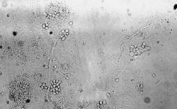
Fungal Staining Methods and Uses
Fungal staining methods: complete comparison of 18 techniques including KOH, LPCB, calcofluor white, GMS, PAS, and India ink. Master reference table with organism-specific stain se...
Fungal staining methods: complete comparison of 18 techniques including KOH, LPCB, calcofluor white, GMS, PAS, and India ink. Master reference table with organism-specific stain se...
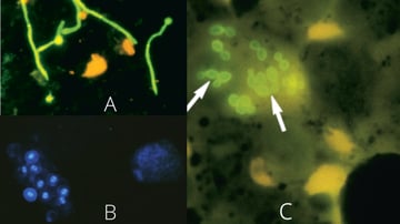
Calcofluor white binds chitin in fungal cell walls, producing bright fluorescence under UV light. Learn the principle, KOH-CFW combined method, specimen-specific applications, and...
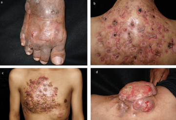
Mycetoma (Madura foot) is a chronic subcutaneous infection caused by fungi (eumycetoma) or bacteria (actinomycetoma). Learn the types, grain colors, causative agents, clinical feat...
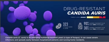
Candida auris is an emerging, often multidrug-resistant yeast that spreads in hospitals and is easily misidentified. Learn how it spreads, who is at risk, how it differs from Candi...
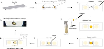
A step-by-step guide to the fungal slide culture technique: how to set it up, why it preserves conidial arrangement, the dimorphic-pathogen safety rule, and how to read the mount.
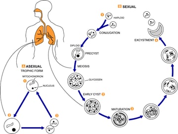
Pneumocystis jirovecii is an atypical fungus (once thought a protozoan) that causes pneumonia in immunocompromised patients. Learn why it was reclassified, why antifungals fail, it...
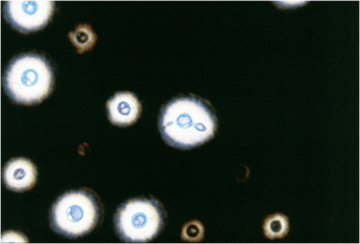
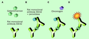
Laboratory diagnosis of fungal infections: direct microscopy, culture, antigen detection (galactomannan, cryptococcal antigen, histoplasma antigen), serology, and molecular methods...
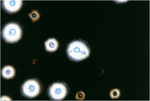
Cryptococcus neoformans and C. gattii: capsule and melanin virulence, narrow-based budding, why it causes fungal meningitis in HIV/AIDS, and lab diagnosis by India ink, birdseed ag...
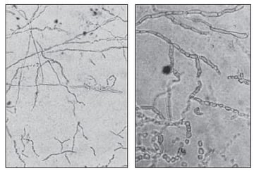
KOH preparation: principle, procedure, 10% concentration rationale, organism-specific findings (dermatophytes, Candida, Mucorales, Coccidioides), troubleshooting artifacts vs true...
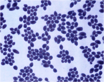
Candida albicans stains Gram-positive and appears as budding yeast cells with pseudohyphae. Learn its microscopy, why it stains as it does, virulence and pathogenesis, the diseases...
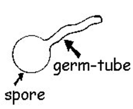
The germ tube test (Reynolds-Braude phenomenon) presumptively identifies Candida albicans by a tube-like outgrowth with no constriction at its base. Learn the principle, procedure,...
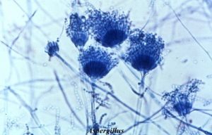
LPCB is the standard mounting medium for microscopic fungal identification: staining chitin in cell walls to reveal hyphae, conidia, and spore arrangements. Learn the tease mount p...
A practical morphology key for identifying fungi under the microscope: reading hyphae (septate vs aseptate, branching angle) and yeast cells (budding base, capsule, size), with a s...