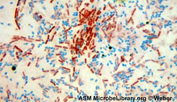
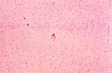
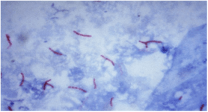
A smear from a sputum specimen is fixed to a glass slide, flooded with carbol fuchsin, heated, decolorized with acid-alcohol, and counterstained with methylene blue. If acid-fast bacilli are present, they retain the red stain against a blue background and a patient with suspected tuberculosis is now one step closer to a confirmed diagnosis.
Staining techniques transform invisible microorganisms into visible, interpretable findings. They are among the oldest tools in diagnostic microbiology and remain essential in every clinical laboratory, including in resource-limited settings where molecular testing is unavailable.
This section covers all major staining methods in clinical and research microbiology:
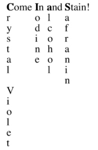
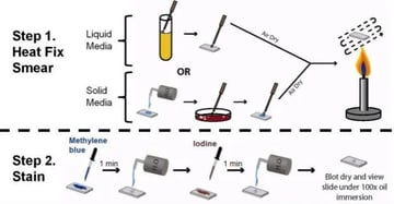
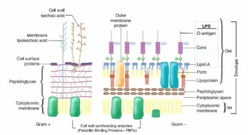
- Gram stain: principle of differential staining based on cell wall composition, step-by-step procedure, results interpretation, common errors and their causes
- Ziehl-Neelsen (acid-fast) stain: for Mycobacterium and Nocardia; hot and cold methods; modified protocols for Cryptosporidium
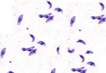
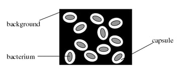
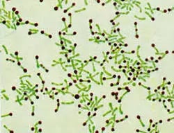
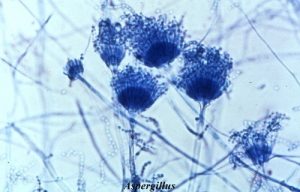
- Special stains: Albert's stain for diphtheria, India ink for Cryptococcus, lactophenol cotton blue for fungi, Giemsa for blood parasites and Chlamydia, Wayson's stain, and others
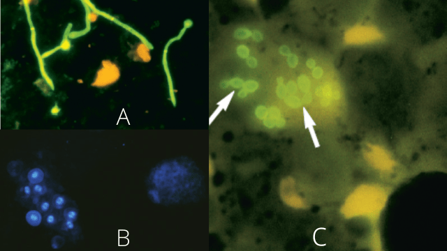
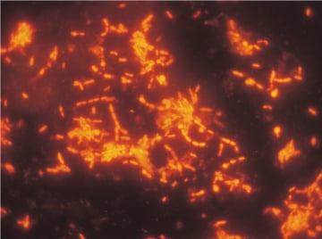
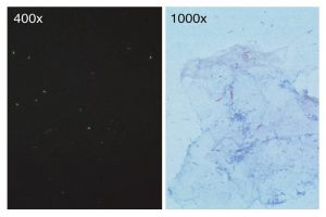
- Fluorescent staining: auramine-rhodamine as a screening stain for acid-fast bacilli; acridine orange; and calcofluor white for fungi
Each article covers the chemical principle behind the stain, the step-by-step procedure, how to interpret the result, what a false-positive or false-negative looks like, and how this stain fits into the diagnostic algorithm for the relevant organisms.