
Capsule Stain: Principle, Procedure, and Results
Capsule staining detects bacterial capsules. Learn the India ink and Anthony's methods, clinically important capsulated organisms, and the Quellung reaction for pneumococcal identification.

On this page
A 4-year-old child presents with fever, bulging fontanelle, and a petechial rash. CSF is cloudy. A Gram stain shows gram-positive lancet-shaped diplococci. A drop of CSF mixed with anti-pneumococcal antiserum under the microscope reveals swollen, refractile capsules around the diplococci, a positive Quellung (capsular swelling) reaction. Streptococcus pneumoniae serotype confirmed. High-dose ceftriaxone is started immediately.
The bacterial capsule is not just a laboratory curiosity. It is one of the most important virulence factors in clinical microbiology: the reason S. pneumoniae, Neisseria meningitidis, Klebsiella pneumoniae, and Haemophilus influenzae can evade the immune system and cause invasive disease. Capsule detection, whether by India ink, Anthony's stain, or the Quellung reaction, is a direct window into bacterial pathogenicity.
Capsule stain is a type of differential stain which uses acidic and basic dyes to stain the background and bacterial cells, respectively, so that presence of the capsule is easily visualized. The capsule is synthesized in the cytoplasm and secreted outside the cell, where it surrounds the bacterium.
Most capsulated bacteria have a capsule made up of a polysaccharide layer, but some bacteria have a capsule made up of polypeptide or glycoprotein. Capsules are associated with virulence in several microorganisms, including Streptococcus pneumoniae and Neisseria meningitides, because capsules resist phagocytosis.
In capsule staining procedure “we do not heat fix and rinse the smear with water” as heat and water may dislodge capsules from bacteria.
Principle of Capsule Stain
Bacterial capsules are non-ionic, so neither acidic nor basic stains will adhere to their surfaces. Therefore, the best way to visualize them is to stain the background using an acidic stain*(e.g., Nigrosine, congo red) and to stain the cell itself using a basic stain (e.g., crystal violet, safranin, basic fuchsin, and methylene blue).
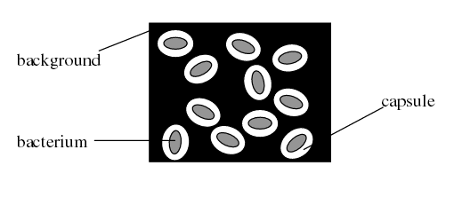 Figure: Capsule Staining (source-microbugz)
Various types of methods are available for the demonstration of bacterial capsules. The results (stain of the cells, background, and capsule) depend on the method used. Two commonly used methods are discussed here:
A. India ink method
In this method, two dyes, crystal violet, and India ink are used. The capsule is seen as a clear halo around the microorganism against the black background. This method is used for demonstrating Cryptococcus.
- The background will be dark (color of India ink).
- The bacterial cells will be stained purple (bacterial cells take crystal violet-basic dyes as they are negatively charged).
- The capsule (if present) will appear clear against the dark background (capsule does not take any stain).
Why capsules resist staining: Bacterial capsules are composed primarily of polysaccharides (occasionally polypeptides, as in Bacillus anthracis). Polysaccharides are non-ionic (they carry no net electrical charge) so neither cationic (basic) nor anionic (acidic) dyes bind to them. This is why capsule staining is always an indirect technique: we stain everything except the capsule and reveal it by contrast. The capsule appears as an unstained halo or clear zone.
B. Anthony’s stain method
In this type of capsule staining procedure, the primary stain is crystal violet, and all parts of the cell take up the purple crystal violet stain. There is no mordant in the capsule staining procedure. A 20% copper sulfate solution serves a dual role as both the decolorizing agent and counterstain. It decolorizes the capsule by washing out the crystal violet, but will not decolorize the cell. As the copper sulfate decolorizes the capsule, it also counterstains the capsule. Thus, the capsule appears as a faint blue halo around a purple cell.
Quellung Reaction (Neufeld Reaction)
The Quellung reaction is the most specific method for detecting and serotyping encapsulated bacteria, particularly Streptococcus pneumoniae. When a capsulated organism is mixed with its type-specific anti-capsular antiserum and methylene blue, the capsule appears swollen and refractile under the microscope. It is the reference standard for pneumococcal serotyping and can be performed directly on CSF for rapid meningitis diagnosis.
For the full procedure, principle, and clinical applications: Quellung Reaction: Principle, Procedure, Results
Materials and Reagents
- Test bacteria: 36-48 hour culture of capsulated bacteria e.g. Klebsiella pneumoniae growing on a slant of EMB agar or culture of other capsulated bacteria and non-capsulated bacteria [Note: Growing Klebsiella pneumoniae in milk-based media (e.g. Skim milk) increase its capsule size, making it easier to visualize.]
- Stain solutions: Depending on the method used (crystal violet, India ink, Nigrosin, copper sulfate, Basic carbol fuschin solution, methylene blue solution, etc).
- Microscopic slides
- Inoculating loop
- Light Microscope with 100x objective lens (oil immersion)
- Immersion oil
- Gas burner
- Tissue paper
Capsule Stain procedure
A. India Ink Method
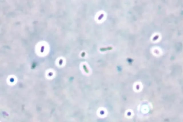 Figure: Capsule staining by India ink method (at 1000x magnification)
- Place a single drop of India ink on a clean microscope slide, adjacent to the frosted edge.
- Using a flamed loop and sterile technique, remove some Klebsiella pneumoniae from culture tube or plate and mix it into the drop of India ink. Be sure there are no large clumps of organism, but try to avoid spreading the drop.
- Place the end of another clean microscope slide at an angle to the end of the slide containing the organism. Spread out the drop out into a film. This is done by contacting the drop of India ink with the clean microscope slide and using the capillary action of the dye/ slide to spread the India ink across the smear.
- Allow the film to air dry (will take 5-7 minutes). DO NOT heat or blot dry! Heat will melt the capsule!
- Saturate the slide with crystal violet for 1 minute and rinse slightly & very gently with water. Be cautious water may remove the capsule from the cell.
- Let the slide air dry for a few minutes. DO NOT blot the slide! Blotting will remove the bacteria from the slide and/or distort the capsule.
- Observe the slide under oil immersion.
Results: Look for purple cells surrounded by a clear halo on a dark background. The halo is the capsule. You may need to decrease the amount of light in order to make the capsule easier to see.
B. Anthony’s stain method
- Place a single drop of crystal violet on a clean microscope slide, adjacent to the frosted edge.
- Using a flamed loop and sterile technique, add three loopful of test bacterium (any capsulated bacteria such as Klebsiella pneumoniae, Streptococcus pneumoniae) from broth culture. If you are adding bacteria from a culture plate make sure that there are no large clumps of the organism, but try to avoid spreading the drop.
- Place the end of another clean microscope slide at an angle to the end of the slide containing the organism. Spread out the drop out into a film. This is done by contacting the drop of crystal violet with the clean microscope slide and using the capillary action of the dye/ slide to spread the crystal violet across the smear.
- Allow the film to air dry (will take 5-7 minutes). DO NOT heat or blot dry! Heat will melt the capsule!
- Tilt the slide and rinse with 20% copper sulfate solution. DO NOT RINSE WITH WATER! Water will remove the capsule from the cell.
- Let the slide air dry for a few minutes. DO NOT blot the slide! Blotting will remove the bacteria from the slide and/or distort the capsule.
- Observe the slide under oil immersion.
Results: Look for purple cells surrounded by a clear or faint blue halo on transparent background. The halo is the capsule. You may need to decrease the amount of light in order to make the capsule easier to see.
Points to remember
- Clean your microscope with lens cleaner, removing all oil from lenses.
- Dispose of staining waste and slides in designated waste containers.
- Be cautious while handling the slide, since the organisms have not been killed.
Troubleshooting Capsule Staining
| Problem | Likely Cause | Action |
|---|---|---|
| No capsule visible (known capsulated organism) | Heat fixation used, capsule melted | Never heat fix for capsule staining; air dry only |
| Capsule visible but poorly defined | Rinsing with water | Use copper sulfate (Anthony's) or no rinse (India ink); water dislodges capsule |
| India ink too dense (cannot see organisms) | Too much India ink | Use a single small drop; organisms should be visible |
| India ink preparation drying too quickly | Smear too thin; warm environment | Work quickly; use a slightly larger drop |
| No Quellung reaction with positive control organism | Antiserum expired or wrong serotype pool | Check antiserum expiry; use omniserum pool first before specific pools |
| False Quellung appearance | Capsule visible even with non-specific antiserum | Compare with non-specific antiserum or saline control; true Quellung is more pronounced with specific antiserum |
How to Remember
"Capsule staining is always indirect stain everything except the capsule": The capsule is non-ionic and takes no stain. You stain the cell (crystal violet, basic fuchsin) and the background (India ink, nigrosin). The capsule appears as the unstained zone between them. If you remember why the capsule can't be stained directly, you understand the principle without memorizing it.
"No heat, no water, no hurry" the three rules of capsule staining:
- No heat (melts the capsule)
- No water rinse (dislodges the capsule)
- No hurry (allow to air dry, do not blot)
Quellung = Quellung-swelling. The capsule appears to swell when the specific antibody binds. Quellung is the only capsule test that also identifies the serotype.
Crypto = India ink: Cryptococcus neoformans in CSF requires India ink. Clear halo on black background = capsule = Cryptococcus until proven otherwise in an immunocompromised patient.
Key Exam Facts
| Feature | Detail |
|---|---|
| Why capsules don't stain | Non-ionic (polysaccharide or polypeptide), no charge to bind acidic or basic dyes |
| Appearance of capsule in India ink method | Clear halo on dark background; cell stains purple (crystal violet) |
| Appearance of capsule in Anthony's stain | Faint blue halo around purple cell (copper sulphate decolorizes capsule) |
| Critical rule | Never heat fix; never rinse with water as both destroy capsule |
| Most important clinical application | India ink for Cryptococcus in CSF; Quellung for pneumococcal serotyping |
| Quellung reaction principle | Type-specific anti-capsular antibody + methylene blue → capsule appears swollen and refractile |
| Quellung clinical use | Pneumococcal serotyping; rapid CSF diagnosis; H. influenzae typing |
| Cryptococcus capsule detection | India ink gold standard; latex agglutination antigen test for CSF |
| B. anthracis capsule composition | It is made up of Poly-D-glutamic acid which is a polypeptide, not polysaccharide |
| Capsule and virulence | Resists phagocytosis, inhibits complement, prevents opsonisation without specific antibody |
| Vaccine connection | Pneumococcal (PCV13/PCV15/PPSV23) and Hib vaccines target capsular polysaccharides |
References and further reading
- Murray PR, Rosenthal KS, Pfaller MA. Medical Microbiology. 9th ed. Elsevier; 2020.
- Forbes BA, Sahm DF, Weissfeld AS. Bailey & Scott's Diagnostic Microbiology. 14th ed. Elsevier; 2023.
- Koneman EW, Allen SD, Janda WM, Schreckenberger PC, Winn WC. Koneman's Color Atlas and Textbook of Diagnostic Microbiology. 6th ed. Lippincott Williams & Wilkins; 2006.
- Henrichsen J. Six newly recognized types of Streptococcus pneumoniae. J Clin Microbiol. 1995;33(10):2759–2762.
- Kapoor G, Saigal S, Elongavan A. Action and resistance mechanisms of antibiotics: A guide for clinicians. J Anaesthesiol Clin Pharmacol. 2017;33(3):300–305.
Frequently Asked Questions
Why can bacterial capsules not be stained directly?
Bacterial capsules are composed primarily of polysaccharides (occasionally polypeptides), which are non-ionic. Since conventional dyes are either cationic (basic dyes) or anionic (acidic dyes), they have no charged surface to bind to on the capsule.
Capsule staining is therefore always indirect: the bacterial cell is stained with a basic dye and the background is stained with an acidic dye, revealing the capsule as an unstained clear halo between them.
How is India ink used to diagnose Cryptococcal meningitis?
How is India ink used to diagnose Cryptococcal meningitis?
India ink (or nigrosin) is mixed with a drop of CSF on a microscope slide and examined under oil immersion. Cryptococcus neoformans appears as a yeast cell (round to oval, 4-20 μm) surrounded by a clear capsule halo against the dark ink background. The halo can be dramatically large relative to the cell body.
India ink is a rapid, inexpensive bedside diagnostic test with approximately 50-80% sensitivity in cryptococcal meningitis. A negative India ink does not exclude cryptococcal meningitis; the cryptococcal antigen latex agglutination test is more sensitive and should be performed when clinical suspicion is high.

Tankeshwar Acharya, MSc (Medical Microbiology)
Tankeshwar Acharya is an Assistant Professor in the Department of Microbiology at Patan Academy of Health Sciences (PAHS), Nepal, where he has been teaching and practicing clinical microbiology for over 14 years. He is the founder of Microbe Online, one of the leading free microbiology education resources on the web, covering bacteriology, mycology, parasitology, immunology, and clinical laboratory diagnostics written from direct experience in both the classroom and the diagnostic laboratory.
Comments
No comments yet. Be the first to share your thoughts.
Leave a comment
All comments are reviewed before they appear.