
Streak Plate Method: Principle, Types, Procedure, and Common Errors
The streak plate method isolates bacteria into pure cultures by progressive dilution across an agar surface. Learn quadrant, T-streak, radiant, and continuous methods, common errors that prevent isolated colonies, and when each method is used clinically.
A wound swab from a diabetic foot ulcer arrives in the microbiology laboratory. The Gram stain shows Gram-positive cocci in clusters, Gram-negative rods, and Gram-positive rods; a mixed infection with at least three different organisms. Before any biochemical identification test, any antibiotic susceptibility test, or any clinical report can be issued, the laboratory must do one thing: isolate each organism into a pure culture.
A biochemical test performed on a mixed culture gives an uninterpretable result. A susceptibility test performed on a mixed culture gives a misleading result. The streak plate is the fundamental technique that transforms a clinically useless mixed specimen into individual, identifiable organisms and every other step in diagnostic microbiology depends on it being done correctly.
For organisms that grow well on agar plates, a streak plate is the method of choice for obtaining pure culture. The streak plate technique is used to isolate the organisms (mostly bacteria) from a mixed population into a pure culture. The inoculum is streaked over the agar surface to “thin out” the bacteria. Some individual bacterial cells are separated and well-spaced from each other.
By streaking, a dilution gradient is established across the surface of the agar plate. Because of this, confluent growth occurs on the part of the plate where the bacterial cells are not sufficiently separated; in other regions where few bacteria are deposited, separate macroscopic colonies develop. Each well-isolated colony is assumed to arise from a single bacterium and represent a clone of a pure culture.
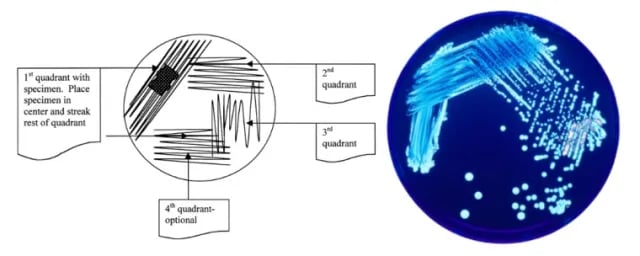 Figure: Appropriate method to streak plate for isolation of bacteria.
Why Pure Culture Matters
A pure culture contains only one species (or strain) of microorganism. Clinical diagnostic microbiology requires pure cultures for three reasons:
1. Biochemical identification tests require a pure culture. API panels, VITEK systems, and MALDI-TOF mass spectrometry are calibrated and validated for single organisms. If two organisms are present in the test inoculum, results are composite and uninterpretable — the system may report a false identification or no identification.
2. Antibiotic susceptibility testing (AST) requires a pure culture. Kirby-Bauer disc diffusion and MIC methods are performed on a suspension of a single organism at a standardized inoculum density (0.5 McFarland). Mixed inocula produce mixed inhibition zones and misleading MIC values that cannot be reported.
3. Colony morphology assessment requires pure culture. Identifying an organism by colony appearance (color, size, hemolysis, odor) is only meaningful when all colonies on the plate are from the same organism. Mixed plates produce ambiguous results.
The clinical consequence of skipping this step: A laboratory that reports antibiotic susceptibility results from a mixed culture may inadvertently recommend an antibiotic that is active against only one of the organisms present in the infection — contributing to treatment failure.
Principle of Streaking
The inoculum is diluted by streaking it across the surface of the agar plate. While streaking in successive areas of the plate, the inoculum is diluted to the point where only one bacterial cell is deposited every few millimeters on the surface of the agar plate. An isolated colony is formed when these lone bacterial cells divide and give rise to thousands and thousands of new bacterial cells. Pure cultures can be obtained by picking well-isolated colonies and re-streaking these on fresh agar plates.
A common assumption is an isolated colony of bacteria is the progeny of a single bacterial cell (i.e. colony is the clone). However, this is not necessarily true. With species in which the cells form a characteristic grouping during cell divisions, the colony-forming unit may develop from a group of cells rather than form a single cell. For example, clusters of staphylococci, chains of streptococci, etc.
Materials required
- A source of bacteria (stock culture, previously streaked agar plate, or any other inoculum)
- Inoculation loop
- A striker/lighter
- Bunsen burner
- Lysol (10%v/v)
- Agar plate (nutrient agar or any other agar medium)
- Paper towels
Tips for the best results
- Use only a small amount of inoculum.
- Streak lightly so that you do not gouge the agar.
- Flame the loop after you streak each quadrant.
- Make sure the surface of the plate is free of droplets of condensed moisture.
Purpose of streaking
The purpose of the streak plate is to obtain isolated colonies from an inoculum. Isolated colonies represent a clone of cells derived from a single precursor.
- To produce isolated colonies of an organism (primarily bacteria) on an agar plate. This is useful when we separate organisms in a mixed culture (to purify/isolate a particular strain from contaminants) or to study an organism’s colony morphology.
- To identify the organism: biochemical tests to identify bacteria are only valid when performed on pure cultures.
Types of Streaking Methods
Many different streaking patterns can be used to separate individual bacterial cells on the agar surface. There are four basic types of streaking methods;
- Quadrant streaking
- T-streak
- Continuous streak
- Radiant streak
Quadrant streaking
As the original sample is diluted by streaking it over successive quadrants, the number of organisms decreases. Usually, by the third or fourth quadrant, only a few organisms are transferred, giving discrete colony-forming units (CFUs).
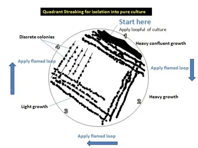 Figure: Quadrant Streaking for isolation into pure culture
Procedure
- Sterilize the inoculating loop in the bunsen burner by putting the loop into the flame until it is red hot or by incinerating it in a micro incinerator. Allow it to cool.
- Pick a small portion of an isolated colony (you do not need a large amount) and streak it gently over the first quadrant, about a quarter of the plate, using a back-and-forth motion (see area 1 in the figure above).
- Flame the loop again and allow it to cool. Returning to the edge of area 1 that you just streaked, extend the streaks into the second quarter of the plate (area 2).
- Flame the loop again and allow it to cool. Returning to the area you just streaked (area 2), extend the streaks into the third quarter of the plate (area 3).
- Flame the loop again and allow it to cool. Returning to the area you just streaked (area 3), extend the streaks into the center fourth of the plate (area 4).
- Flame your loop once more.
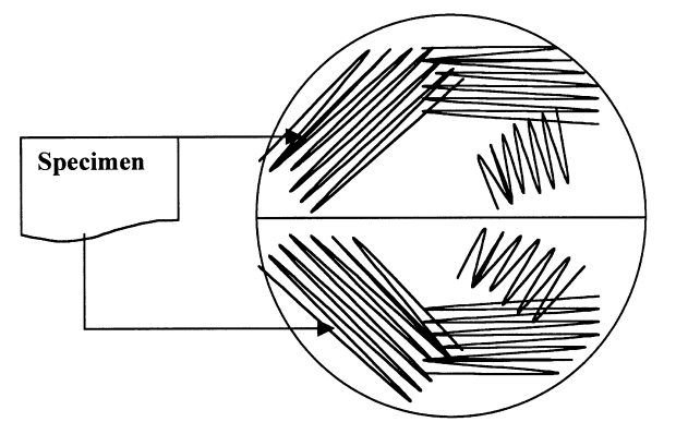 Figure: Bi-plate streaking
Note: Bi-plate inoculation of samples from sterile sites is often done in diagnostic laboratories to save time and space.
Results
The streaked plate is incubated at 35–37°C for 18–24 hours. Examine the colonies grown on the plate carefully. All colonies should have the same general appearance. If there is more than one colony type, each type should be streaked again on a separate plate to obtain a pure culture.
T Streak **Procedure**
- Draw a T shape on the bottom of the plate with a marker, dividing it into three sections (one top section and two bottom sections).
- Sterilize the loop, let it cool, and pick up the inoculum.
- Lift the lid just enough to insert the loop, and streak the inoculum in a zig-zag pattern across the top section, staying within it. Close the lid, then flame the loop and let it cool.
- Rotate the plate 90°. Drag the loop from the edge of the first (top) section into the second section, then zig-zag to fill it. Close the lid, flame the loop, and let it cool.
- Rotate the plate again. Drag the loop from the edge of the second section into the third section and zig-zag to fill it.
- Flame the loop until red hot before setting it down.
Radiant Streak

Procedure
- An agar plate is taken and appropriately labeled.
- Using a sterile (flamed) loop, spread a loopful of sample carefully along the edge of the agar (take care not to gouge the agar).
- Flame the loop, and after it cools, streak 7 to 8 straight lines from area 1 across to the opposite side of the plate.
- The loop flamed again, and cross streaking is done over the previous streaks when cool sufficiently. (start from area 1)
- When setting down the loop, it should be flamed till red hot.
Continuous Streak

Procedure:
- Using a sterile loop with the loopful sample, the organism is spread from edge (A) to the middle of the labeled plate. (gouging should be avoided)
- The plate is then rotated 180°, keeping the already-inoculated portion away from your hand.
- The same inoculum loop is used, and the process of spreading is repeated from the edge (B) to the middle.
- The loop is then flamed and placed aside.
NOTE: Another method of streaking commonly practiced in hospital settings is the “semi-quantitative method of urine culture”:
A commonly used method of streaking with calibrated loop (4mm in diameter) to semi-quantify the bacteria isolated from the urine specimen. The streaking is similar to continuous streaking. The difference is that the primary inoculum is made by drawing a vertical line from the top to the bottom of the plate with a calibrated loop.
Common Errors and How to Avoid Them
Streak plate failure is almost always due to one of five errors. Understanding the mechanism of each error makes them preventable rather than random.
| Error | What happens | Why it happens | Prevention |
|---|---|---|---|
| Too much inoculum in Area 1 | Confluent growth across entire plate; no isolated colonies anywhere | More bacteria than the dilution can disperse | Use the tip of the loop, not a loaded loop; pick a small portion of a single colony |
| Loop not cooled before picking up bacteria from previous area | Area 2 onwards grows same heavy density as Area 1; no progressive dilution | Hot loop kills bacteria in the previous area rather than picking them up | Wait 10–15 seconds after flaming; touch the edge of the agar (away from colonies) to test — if it sizzles, wait longer |
| Gouging the agar surface | Torn agar fragments mixed into streak lines; multiple colony types appear | Too much pressure on the loop | Streak with the side of the loop at a shallow angle (~30°); barely graze the surface |
| Streaking back into a previously streaked area | No dilution gradient established; Area 4 as heavy as Area 1 | Incorrect loop entry point into next area | Enter the next area only from the very edge of the previous area's last few streaks |
| Condensation on agar surface | Spreading growth; colonies merge; satellite colonies around primary growth | Plate not pre-warmed or has moisture droplets | Remove plate from refrigerator 30 minutes before use; dry plates (lid slightly ajar, upside down) in incubator for 10 minutes before inoculation |
Choosing a Streaking Method
| Method | Pattern | Best for | When used |
|---|---|---|---|
| Quadrant streak | Four areas, 90° rotations, loop flamed between each | Most clinical specimens; standard isolation | Universal first choice for most diagnostic specimens |
| T-streak | T-shape dividing plate into three sections | Teaching; moderate inoculum specimens | Common in teaching labs; alternative to quadrant for smaller plates |
| Radiant streak | Central area, then radial lines outward | Dense specimens; heavy mixed flora | Food and water samples; faecal specimens with very heavy growth |
| Continuous streak | Single continuous back-and-forth | Semi-quantitative urine culture (with calibrated loop) | Urine culture in clinical labs; also used for preliminary isolation when colony count is needed |
Semi-quantitative urine culture note: The continuous streak with a calibrated 4 mm loop (delivering approximately 10 µL) is the standard method in clinical urine culture. The number of colonies in the primary streak is counted and reported as approximate CFU/mL (e.g., >10⁵ CFU/mL if confluent growth in primary streak, 10⁴–10⁵ if isolated colonies in primary streak only).
Application of Streak Plate
- It is used for determining the causative agent of the disease using clinical specimens.
- It can be applied to isolate a pure culture of bacteria from the mixture of the bacterial suspension.
- Furthermore, identification using biochemical tests could be done of the isolated colonies.
Limitation of Streak Plate Technique
- It works only for organisms that grow on solid agar. Anaerobes can be isolated by streak plate too, but they must then be incubated in an anaerobic environment; the plate itself is not the limitation, the incubation atmosphere is.
- Only viable (living) organisms form colonies, so the inoculum must contain live bacteria.
- It is qualitative, not quantitative. The streak plate isolates and purifies, but does not by itself give a colony count (the calibrated-loop urine method is the one semi-quantitative exception).
How to Remember
The streak plate does one thing: progressive dilution across a plate surface. Every feature of the technique — flaming between areas, entering from the edge of the previous area, using a small inoculum — exists to establish and maintain this dilution gradient. If any step breaks the gradient, isolated colonies will not form.
The three critical rules:
- Small inoculum — less is more; a loaded loop gives confluent growth
- Cool loop — a hot loop kills; bacteria can only be picked up by a cool loop
- Edge entry — start each new area from the very last few streaks of the previous area, not from the middle
The clinical chain this technique enables:
Mixed specimen → streak plate → isolated colonies → pure culture → biochemical ID → susceptibility testing → targeted antibiotic therapy
Every step after "streak plate" is only valid if this step is done correctly. The streak plate is the foundation on which all clinical microbiology identification rests.
Where Students Get Confused
- "More inoculum means more colonies to isolate." Backwards. A heavy inoculum floods the whole plate with confluent growth and no isolated colonies form anywhere. The technique works by dilution, so less is more: a small pickup gives clean isolation.
- Not flaming (or not cooling) the loop between areas. The loop must be flamed between quadrants to reduce the carryover, then cooled before touching the agar. A hot loop kills the very cells you are trying to move, and skipping the flame carries too many cells forward, both break the dilution gradient.
- Entering the next area from the middle. Each new area must be started from the last few streaks of the previous area, not from its dense center. Entering from the middle carries a heavy load forward and the fourth area ends up as crowded as the first.
- "An isolated colony is always one original cell." Not always. For organisms that grow in groups (staphylococcal clusters, streptococcal chains), a colony-forming unit may start from a clump of cells, not a single cell. The colony is still a pure clone, but "one colony = one original cell" is an approximation.
- Confusing streak plate with spread plate. The streak plate is qualitative: it isolates and purifies. The spread plate is quantitative: it gives a count. They look similar but answer different questions.
- Gouging the agar. Pressing too hard tears the surface, and torn agar disrupts the streak lines and traps organisms, producing false extra colony types. Streak with the flat side of the loop at a shallow angle so it glides.
Key exam facts in one table
| Concept | Key exam fact and why it holds |
|---|---|
| What it is | A qualitative technique that isolates a pure culture by progressively diluting the inoculum across an agar surface until single cells are far enough apart to form separate colonies. |
| Principle | Mechanical dilution. Each successive area carries fewer cells, so by the last area individual cells are spaced out and grow into isolated colonies. |
| Why pure culture matters | Biochemical ID, antibiotic susceptibility testing, and colony-morphology reading are all valid only on a single organism; a mixed culture gives uninterpretable or misleading results. |
| Four methods | Quadrant (universal first choice), T-streak (teaching, smaller plates), radiant (dense/heavy flora), continuous (semi-quantitative urine with a calibrated loop). |
| Three critical rules | Small inoculum; cool loop before picking up cells; enter each new area from the edge of the previous one. Breaking any of these destroys the dilution gradient. |
| Calibrated-loop urine | A 4 mm calibrated loop delivers about 10 µL; colony count × dilution factor (100 for a 10 µL loop, 1000 for a 1 µL loop) estimates CFU/mL for significant bacteriuria (≥10⁵ CFU/mL). |
| Colony = clone | Each well-isolated colony is assumed to be a pure clone. Caveat: grouped organisms (staph clusters, strep chains) may start a colony from several cells, not one. |
| Anaerobes | Can be isolated by streak plate, then incubated anaerobically. The technique is not limited to aerobes; the incubation atmosphere is what changes. |
| Incubation | Typically 35–37°C for 18–24 hours for clinical isolates. |
| Most common failure | Too much inoculum (confluent growth) and a hot or uncooled loop; both prevent isolated colonies. |
References
- Clinical Microbiology Procedures Handbook (4th ed.). (2016). American Society of Microbiology. https://doi.org/10.1128/9781555818814 (keep)
- Mahon, C. R., Lehman, D. C., & Manuselis, G. (2018). Textbook of Diagnostic Microbiology (6th ed.). Elsevier.
- Sanders, E. R. (2012). Aseptic laboratory techniques: plating methods. Journal of Visualized Experiments, (63), e3064. https://doi.org/10.3791/3064
- Image source: CDC Public Health Image Library. Image credit: CDC/James Gathany (PHIL #: 7925)
Frequently Asked Questions
Why is it essential to flame and cool the inoculating loop between each streaking area?
Why can a biochemical identification test or antibiotic susceptibility test not be performed on a mixed culture?
What is a semi-quantitative urine culture and how does the streak plate technique enable it?
Why does too much inoculum prevent isolated colonies?
What is the difference between the streak plate and the spread plate?
Can the streak plate be used to isolate anaerobic bacteria?

Tankeshwar Acharya, MSc (Medical Microbiology)
Tankeshwar Acharya is an Assistant Professor in the Department of Microbiology at Patan Academy of Health Sciences (PAHS), Nepal, where he has been teaching and practicing clinical microbiology for over 14 years. He is the founder of Microbe Online, one of the leading free microbiology education resources on the web, covering bacteriology, mycology, parasitology, immunology, and clinical laboratory diagnostics written from direct experience in both the classroom and the diagnostic laboratory.