

A patient has a painless genital ulcer. The lab preps a wet mount of fluid from it and looks under a standard bright-field microscope, the kind used for every Gram stain that day. Nothing shows up. Not because nothing is there, but because Treponema pallidum is roughly 0.1 to 0.18 μm wide, thinner than the ~0.2 μm resolving limit of light microscopy itself. No amount of focusing or magnifying fixes that; it's a physical floor, not a technique problem.
Switch to a dark-field microscope, and the same slide suddenly shows a bright, corkscrew-shaped thread rotating and flexing against total darkness. The organism hasn't gotten any wider. What changed is the question being asked of it. Bright-field microscopy tries to resolve the organism's structure, and fails, because it's below the resolution limit. Dark-field microscopy instead detects the light the organism scatters, the same way a single dust mote glinting in a sunbeam is visible without ever being resolved. You can't see its shape in fine detail, but you can absolutely see that it's there, and that it's moving.
That distinction, detecting something below the resolution limit versus resolving its structure, is the whole reason dark-field microscopy exists as a separate technique, and it's the reason it remains, a century after it was developed, still the fastest way to catch syphilis before antibody tests would even turn positive.
Dark-field microscopy is a technique that can be used for the observation of living, unstained cells and microorganisms. In this microscopy, the specimen is brightly illuminated while the background is dark. It is one type of light microscope, others being bright-field, phase-contrast, differential interface contrast, and fluorescence.
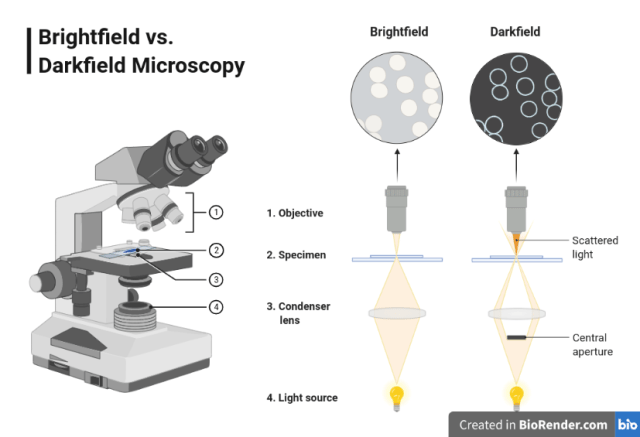 Figure: Components of Bright field microscope and difference in illumination with darkfield microscope.(Image source: Created with BioRender.com)
Principle
Dark-field microscopy uses a light microscope with an extra opaque disc underneath the condenser lens, or a special condenser having a central blacked-out area, due to which the light coming from the source cannot directly enter into the objective.
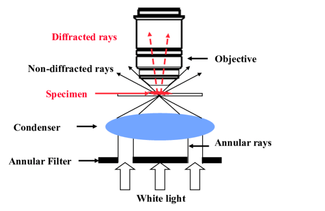 Figure: Principle of Dark-Field Microscopy(Image courtesy: Olivier Haeberlé)
The path of the light is directed in such a way that it can pass through the outer edge of the condenser at a wide-angle and strike the sample at an oblique angle. Only the light scattered by the sample reaches the objective lens for visualization. All other light that passes through the specimen will miss the objective, thus the specimen is brightly illuminated on a dark background.
Uses of Dark-Field Microscopy
Dark-field microscopes are used in the microbiology laboratory for the following purposes;
- Visualization of spirochetes such as Treponema pallidum (syphilis), Borrelia burgdorferi (Lyme borreliosis), and Leptospira interrogans (leptospirosis) in clinical samples.
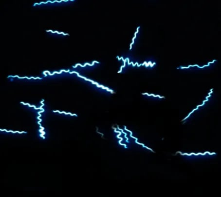 Figure: Borrelia in dark field microscopy(Image courtesy S Bhimji)
Spirochetes can not be seen by light microscopy because of their thin dimensions. This is a detection problem, not a resolution problem. A structure narrower than the ~0.2 μm resolving limit of light microscopy can still scatter light strongly enough to be seen, even though its fine internal structure can't be resolved. For the physics behind this resolving limit, see Working Mechanism of the Light Microscope.
Observation of microbial motility; tufts of bacterial flagella can often be seen in unstained cells by dark-field or phase-contrast microscopy
Observation of internal structure in larger eukaryotic microorganisms such as algae, yeasts, etc.
Advantages of Dark-Field Microscopy
- Dark-field microscopy does not improve the microscope's fundamental resolving power, but it dramatically improves contrast and the detectability of structures at or below the resolving limit, which is why extremely thin organisms like spirochetes become visible at all.
- It improves image contrast without the use of stain, and thus does not kill the cells.
- Allows direct detection of fastidious or difficult-to-culture organisms (such as Treponema pallidum) directly in patient samples.
- No sample preparation is required.
- It requires no special setup, even a light microscope can be converted to dark field.
Limitations of Dark-Field Microscopy
- Necessity to examine wet, moist specimens containing living organisms very quickly, because visualization of the moving bacteria is essential to detection.
- The sample must be very strongly illuminated, which can cause damage to the sample.
- Besides the sample, dust particles also scatter the light and appear bright.
- Sample material needs to be spread thinly, dense preparations can grossly affect the contrast and accuracy of the dark field’s image.
How to Remember
- Detection vs. resolution, the concept this whole technique depends on: resolving something means seeing its fine structure; detecting something means seeing that it's there at all. Dark-field trades the first for the second, on purpose, for exactly the organisms too thin to resolve any other way.
- Why the background flips: bright-field lets direct light straight through, so the specimen appears dark against brightness. Dark-field blocks that direct light entirely and shows you only what the specimen scatters, so the specimen glows against total darkness. Same specimen, opposite lighting logic.
- A dust mote in a sunbeam: you can't resolve the shape of a single dust particle floating in a shaft of sunlight, but you can absolutely see it glinting. That's the same physical principle letting a 0.1 μm-wide spirochete become visible under dark-field despite being well below the classical resolving limit.
Key exam facts
| Concept | Detail | Why it's tested |
|---|---|---|
| Principle | Opaque disc/special condenser blocks direct light; only scattered light from the specimen reaches the objective | The mechanism question most commonly asked about this technique |
| What dark-field actually improves | Detectability and contrast for structures at or below the light microscope's ~0.2 μm resolving limit, not the resolving limit itself | Common misconception worth correcting directly |
| Classic organisms | Treponema pallidum, Borrelia burgdorferi, Leptospira interrogans | All spirochetes too thin to resolve by bright-field |
| Sample requirement | Living, unstained, examined quickly | Non-motile or fixed specimens defeat the purpose, since motility is often the identifying feature |
| Key limitation | Dust and debris also scatter light and can be mistaken for organisms | A frequently tested source of false readings |
Where Students Get Confused
- Assuming dark-field improves resolution. It doesn't change the light microscope's fundamental resolving power; it improves the detectability of structures already below that limit through scattered light, a distinct optical concept from resolution itself.
- Assuming any thin organism will simply "show up" under dark-field. The organism still needs to be alive and motile in most diagnostic uses, since motility, not just visibility, is often what confirms the identification.
- Treating "no staining required" as "no preparation required at all." The sample still needs to be fresh, thinly spread, and free of excess debris; dust and thick preparations both degrade the image just as much as they would in any other technique.
- Assuming dark-field is a routine, always-available lab test. It requires a special condenser setup, immediate examination of a living specimen, and a trained microscopist, which is exactly why it's reserved for specific point-of-care situations rather than routine diagnostic panels.
References
- Procop, G. W., & Koneman, E. W. (2017). Koneman's Color Atlas and Textbook of Diagnostic Microbiology (7th ed.). Wolters Kluwer.
- Tille, P. M. (2022). Bailey & Scott's Diagnostic Microbiology (15th ed.). Elsevier.
- Madigan, M. T., Bender, K. S., Buckley, D. H., Sattley, W. M., & Stahl, D. A. (2018). Brock Biology of Microorganisms (15th ed.). Pearson.
- Willey, J. M., Sherwood, L. M., & Woolverton, C. J. (2016). Prescott's Microbiology (10th ed.). McGraw-Hill Education.

Comments
No comments yet. Be the first to share your thoughts.
Leave a comment
All comments are reviewed before they appear.