
Burkholderia pseudomallei: Melioidosis, Pathogenesis, Diagnosis, Treatment
How Burkholderia pseudomallei causes melioidosis, why diabetes is the main risk factor, why it can reactivate years later, how it is diagnosed, and why treatment has two phases.

On this page
A rice farmer in Thailand with poorly controlled diabetes comes to hospital during the monsoon season with a high fever and cough that have worsened over a week. A chest x-ray shows pneumonia, and blood cultures grow a gram-negative organism. He recalls no injury, but he wades barefoot through flooded fields every day. The clues line up: heavy rain, wet soil, bare feet, and diabetes, the strongest risk factor of all. An organism living in that soil has entered his body and taken advantage of his weakened defenses. This is melioidosis, caused by Burkholderia pseudomallei, a disease that can also lie silent for years before it appears.
Introduction
Burkholderia pseudomallei causes melioidosis (also called Whitmore's disease), a serious and often life-threatening infection. A closely related organism, B. mallei, causes glanders, a similar disease chiefly of horses and, rarely, humans.
One idea organizes the whole topic: B. pseudomallei lives freely in soil and water in the tropics, so people are infected from the environment, and it does its most serious damage in people whose defenses are weakened, above all by diabetes. It is also an intracellular organism that can survive quietly in the body and reactivate years later, which is why melioidosis has been nicknamed the "Vietnamese time-bomb." Because of its potential for aerosol spread, the CDC classifies B. pseudomallei (and B. mallei) as a Category B bioterrorism agent.
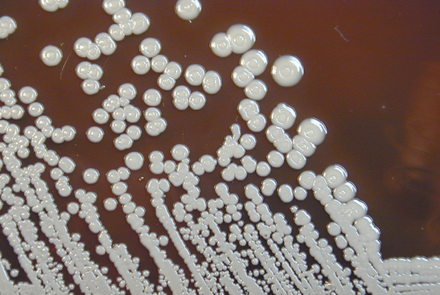 Figure: B.pseudomallei on Blood Agar
Properties
- Aerobic, non-fermentative gram-negative bacillus (it does not ferment carbohydrates).
- Gram-negative rod with a characteristic bipolar or "safety pin" appearance (the ends stain more darkly than the middle), best seen with methylene blue.
- Motile (an important difference from Pseudomonas-like non-fermenters it can be confused with, and from B. mallei, which is non-motile)
- It is a member of the family Burkholderiaceae, which includes other genera (Cupriavidus, Lautropia, Pandoraea, and Ralstonia). More than 87 species of Burkholderia have been identified. Other medically important species are Burkholderia cepacia, Burkholderia gladioli, etc.
- Burkholderia pseudomallei is a saprophyte of soil and water and is spread to humans and animals through direct contact with contaminated sources.
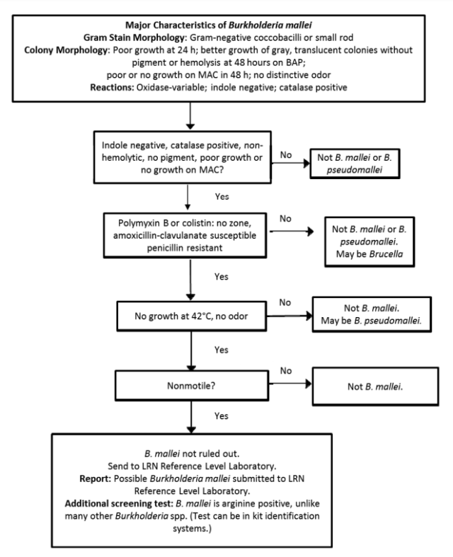 Figure: Flow diagram to rule out or refer B. mallei. Image source: Laboratory Response Network (LRN)
Pathogenesis
B. pseudomallei is a saprophyte of soil and water in endemic regions. People are infected from the environment by three routes:
- Inoculation through skin breaks during contact with contaminated soil or water (for example, barefoot farming). This is the most common route.
- Inhalation of contaminated dust or aerosolized water droplets, which is more likely during heavy rain and is linked to more severe disease. This is why cases rise sharply in the monsoon season.
- Ingestion of contaminated water.
Person-to-person spread is very rare. Because exposure to soil and water is so common in endemic areas but disease is much less common, host factors are what decide who becomes ill.
Why diabetes matters so much
Melioidosis is strongly an opportunistic infection. The single most important risk factor is diabetes mellitus, and other risks include hazardous alcohol use, chronic kidney disease, chronic lung disease, and any cause of weakened immunity. Only a minority of patients have no identifiable risk factor. The reason is that B. pseudomallei survives inside host cells, so an intact cellular immune response is needed to control it, and conditions like diabetes impair exactly that response. For an audience in endemic South and Southeast Asia, this is the key public-health message: melioidosis is above all a disease of people with diabetes exposed to soil and water.
Virulence Factors
B. pseudomallei is a facultative intracellular pathogen, and its virulence factors let it enter cells, survive inside them, and spread from cell to cell.
- Polysaccharide capsule: antiphagocytic; it resists being engulfed and killed, and helps the organism survive in the blood.
- Type III secretion system: a molecular "injection" apparatus. It injects bacterial proteins into the host cell that let B. pseudomallei escape from the phagosome into the cytosol, where it multiplies safely, much like other intracellular pathogens.
- Actin-based motility and cell-to-cell spread: once in the cytosol, the organism hijacks the host cell's actin to propel itself and can fuse neighboring cells together (forming multinucleated giant cells), allowing it to spread directly from cell to cell without going back outside. This intracellular spreading is central to how it disseminates and hides.
- Tissue-damaging enzymes and toxins: proteases, lipases, and other secreted products damage host tissue and aid invasion, contributing to the abscesses that characterize the disease.
Putting it together: the organism enters through skin, lungs, or gut, is taken up by host cells, escapes into the cytosol using its type III secretion system, multiplies, and spreads cell to cell using actin. Its capsule protects it in the blood so it can disseminate widely and seed abscesses in many organs. Because it lives inside cells, it can persist quietly for long periods and reactivate later, and clearing it depends on cell-mediated immunity, which is why diabetes and other immune-weakening conditions are such strong risk factors.
Clinical Manifestations
Melioidosis is mainly a disease of the tropics, especially Southeast Asia (Thailand, Vietnam, Cambodia, Laos, Malaysia, Myanmar) and northern Australia, and it is increasingly recognized in South Asia and other tropical regions.
The incubation period varies enormously, from a few days to many years, because the organism can stay dormant and reactivate later. Melioidosis is a great mimic: it ranges from a mild flu-like illness or a single skin abscess to severe pneumonia and overwhelming bloodstream infection. Two features run through it: a strong tendency to form abscesses in many organs, and a high death rate in severe disease if it is not treated promptly and correctly.
Acute Localized Infection: Infection is localized as a nodule and results from inoculation through a break in the skin. A patient generally presents with:
- Localized pain or swelling
- Fever
- Ulceration
- Abscess
Pulmonary Infection: This form of the disease can produce a clinical picture of mild bronchitis to severe pneumonia. Commonly seen signs/symptoms are:
- Cough
- Chest pain
- High fever
- Headache
- Anorexia
Acute Bloodstream Infection (septicemic melioidosis): this is the most severe form and carries the highest mortality. It occurs especially in patients with underlying conditions, above all diabetes, and also chronic kidney disease and other causes of weakened immunity. The symptoms generally include:
- Fever
- Headache
- Respiratory distress
- Abdominal discomfort
- Joint pain
- Disorientation
Chronic Suppurative Infection: Chronic melioidosis is a disseminated infection that involves various organs of the body such as joints, viscera, skin, brain, liver, lungs, bones, and spleen.
Laboratory Diagnosis
Sample
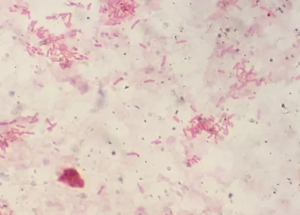 Figure: B. pseudomallei in Gram-stain (image source-Ian Martin, idmic-net).
Sample depends on the site of infection. Sputum and purulent discharge from the lesion are commonly used.
Microscopy and Staining
B. pseudomallei is a Gram-negative bacillus that typically exhibits a bipolar or safety pin appearance, which is better appreciated when stained with methylene blue.
Culture
B. pseudomallei is an aerobe that grows on ordinary media such as nutrient agar, blood agar, and MacConkey agar. Colonies are often dry, rough, and wrinkled (corrugated), which can resemble Pseudomonas stutzeri. Ashdown's agar is the selective medium: on it, B. pseudomallei forms characteristic dry, wrinkled, purple-pink colonies over several days.
An important safety point: because laboratory staff can be infected, the laboratory must be alerted whenever melioidosis is suspected, and the organism is handled under the appropriate biosafety conditions. It is also frequently misidentified by automated systems, so clinical suspicion matters.
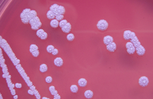 Figure: Burkholderia pseudomallei on Ashdown’s medium(image source: Gavin Koh ref-2)
Important properties that differentiate it from Pseudomonas stutzeri include
- Gelatin liquefaction positive
- Utilizes arginine
- Positive for intracellular poly-β-hydroxybutyrate (PHB).
Latex agglutination test: Culture can be confirmed by latex agglutination test using specific antisera.
Antigen detection
Rapid agglutination tests for detecting B. pseudomallei antigen in urine are available.
Serology
Antibodies against B. pseudomallei can be detected and measured in blood.
Treatment of melioidosis
Two things make treatment distinctive, and both are high-yield.
First, B. pseudomallei is intrinsically resistant to many common antibiotics (including many penicillins, older cephalosporins, gentamicin, and others). This narrows the choices sharply and is why empirical "blind" antibiotics for sepsis can fail if melioidosis is not considered.
Second, treatment has two phases, and skipping the second phase leads to relapse. This two-phase structure is the single most important thing to remember about melioidosis treatment.
- Intensive phase: intravenous therapy to control the acute infection. The drug of choice is ceftazidime or a carbapenem (meropenem or imipenem).
- Eradication phase: a prolonged course of oral therapy to clear organisms hiding inside cells and prevent relapse. The first-choice drug is trimethoprim-sulfamethoxazole (co-trimoxazole), with co-amoxiclav as an alternative (for example, in pregnancy).
The eradication phase must be continued for months. This is because the organism survives inside cells, so a short course leaves hidden bacteria that cause relapse, the reason melioidosis can recur. Prompt, correct treatment greatly reduces what is otherwise a very high mortality in severe disease.
Prevention, especially for people with diabetes in endemic areas, focuses on avoiding exposure: wearing boots and gloves for farming and soil or water contact, and staying out of heavy rain and floodwater aerosols where possible. There is no licensed vaccine, though several are in development.
For the related intracellular Category A/B select agent that also causes pneumonic and disseminated disease, see the article on Francisella tularensis.
How to Remember
| Device | The memory hook |
|---|---|
| Soil, rain, bare feet, diabetes | Melioidosis = a diabetic in the tropics who contacts wet soil, often in the monsoon. Those four together should trigger the diagnosis. |
| Time-bomb | It can stay dormant and reactivate years later, the "Vietnamese time-bomb." Ask about past travel to endemic areas even years ago. |
| The great mimic | It ranges from a skin abscess to pneumonia to fatal sepsis and seeds abscesses in many organs. It imitates many diseases, including tuberculosis. |
| Safety-pin, motile | Bipolar "safety-pin" gram-negative rod, and it is motile (unlike B. mallei). |
| Ashdown's = wrinkled purple | On Ashdown's selective agar it forms dry, wrinkled, purple-pink colonies. |
| Two phases or it comes back | Intensive IV phase (ceftazidime or a carbapenem), then a long oral eradication phase (co-trimoxazole). Skip the second phase and it relapses. |
| Resistant to the usual drugs | It shrugs off many common antibiotics, so you must think of it and choose the right drugs. |
Key exam facts in one table
| Feature | Burkholderia pseudomallei |
|---|---|
| Disease | Melioidosis (Whitmore's disease) |
| Organism | Aerobic, non-fermentative, gram-negative rod; bipolar "safety-pin"; motile |
| Habitat | Soil and water in the tropics (Southeast Asia, northern Australia, South Asia) |
| Transmission | Skin inoculation, inhalation, ingestion; person-to-person rare |
| Main risk factor | Diabetes mellitus (also alcohol use, CKD, immunosuppression) |
| Intracellular | Facultative intracellular; escapes phagosome, spreads cell to cell via actin |
| Key virulence | Capsule, type III secretion system, actin-based motility, tissue enzymes |
| Latency | Can reactivate years later ("time-bomb") |
| Clinical range | Skin abscess, pneumonia, septicemia, chronic disseminated abscesses |
| Most severe form | Septicemic (bloodstream) melioidosis |
| Selective medium | Ashdown's agar (dry, wrinkled, purple-pink colonies) |
| Look-alike | Pseudomonas stutzeri (differentiate: gelatin, arginine, PHB) |
| Resistance | Intrinsically resistant to many antibiotics |
| Treatment | Intensive phase (ceftazidime or carbapenem) then eradication phase (co-trimoxazole) |
| Why prolonged | Intracellular survival → relapse if the eradication phase is short |
| Biothreat | CDC Category B agent |
Where Students Get Confused
| Confusion | The clarification |
|---|---|
| Why is diabetes so central to melioidosis? | The organism survives inside cells, so cell-mediated immunity is needed to control it. Diabetes impairs that response, making diabetics far more likely to develop severe disease. It is the strongest risk factor. |
| Why does treatment have two phases? | The intensive IV phase controls the acute infection; the long oral eradication phase clears organisms hiding inside cells. Skipping or shortening the eradication phase causes relapse. |
| Why can it appear years after exposure? | B. pseudomallei can persist quietly inside cells and reactivate later when immunity drops, which is why it is called a "time-bomb." A travel history, even from years ago, can be relevant. |
| B. pseudomallei vs Pseudomonas stutzeri | Colonies can look similar, but B. pseudomallei is gelatin-liquefaction positive, uses arginine, and stores PHB. It is also motile and has the bipolar safety-pin look. |
| Why do common antibiotics fail? | It is intrinsically resistant to many drugs, including several penicillins, older cephalosporins, and aminoglycosides. Ceftazidime or a carbapenem is needed for the intensive phase. |
| B. pseudomallei vs B. mallei | B. pseudomallei causes melioidosis and is motile and environmental. B. mallei causes glanders (mainly in horses), is non-motile, and is not free-living in soil. |
References and Further Readings
- Tille, P. M. (2022). Bailey & Scott's Diagnostic Microbiology (15th ed.). Elsevier.
- Procop, G. W., & Koneman, E. W. (2017). Koneman's Color Atlas and Textbook of Diagnostic Microbiology (7th ed.). Wolters Kluwer.
- Carroll, K. C., Pfaller, M. A., et al. (2020). Murray's Medical Microbiology (9th ed.). Elsevier.
- Wiersinga, W. J., Virk, H. S., Torres, A. G., et al. (2018). Melioidosis. Nature Reviews Disease Primers, 4, 17107.
- Centers for Disease Control and Prevention. Melioidosis: clinical overview. CDC (current version).
Frequently Asked Questions
Who is most at risk of melioidosis?
Who is most at risk of melioidosis?
People with diabetes are at the highest risk, which is the single most important risk factor. Other risks include heavy alcohol use, chronic kidney disease, chronic lung disease, and weakened immunity. Most people who develop melioidosis have at least one of these, together with exposure to soil and water in an endemic tropical region.
How do people catch melioidosis?
How do people catch melioidosis?
From the environment, not usually from other people. The organism lives in soil and water in the tropics, and infection follows skin contact with contaminated soil or water (often barefoot farming), inhaling contaminated aerosols during heavy rain, or swallowing contaminated water. Cases rise during the monsoon season.
Why is melioidosis called the "Vietnamese time-bomb"?
Why is melioidosis called the "Vietnamese time-bomb"?
Because B. pseudomallei can survive quietly inside the body for a long time and reactivate months or even years after the original exposure, sometimes when the person's immunity later weakens. This is why a travel history from years earlier can be relevant.
Why does melioidosis treatment take so long?
Why does melioidosis treatment take so long?
Because the organism hides and multiplies inside host cells. Treatment has two phases: an intensive intravenous phase to control the acute infection, then a prolonged oral eradication phase lasting months to clear the hidden bacteria. If the eradication phase is cut short, the infection can relapse.
Which antibiotics are used for melioidosis?
Which antibiotics are used for melioidosis?
The intensive phase uses intravenous ceftazidime or a carbapenem such as meropenem. The eradication phase uses oral co-trimoxazole (trimethoprim-sulfamethoxazole), or co-amoxiclav as an alternative. Many common antibiotics do not work, because the organism is intrinsically resistant to them.
How is melioidosis diagnosed in the laboratory?
How is melioidosis diagnosed in the laboratory?
Mainly by culture from blood, sputum, pus, or other samples, using selective Ashdown's agar where it forms wrinkled purple-pink colonies. Because the organism can be misidentified and is a laboratory hazard, it is important to tell the laboratory when melioidosis is suspected so it is handled safely and identified correctly.

Tankeshwar Acharya, MSc (Medical Microbiology)
Tankeshwar Acharya is an Assistant Professor in the Department of Microbiology at Patan Academy of Health Sciences (PAHS), Nepal, where he has been teaching and practicing clinical microbiology for over 14 years. He is the founder of Microbe Online, one of the leading free microbiology education resources on the web, covering bacteriology, mycology, parasitology, immunology, and clinical laboratory diagnostics written from direct experience in both the classroom and the diagnostic laboratory.
Comments
No comments yet. Be the first to share your thoughts.
Leave a comment
All comments are reviewed before they appear.