
Gram Positive Cocci of Medical Importance
Gram positive cocci by arrangement, clusters, chains, pairs, and tetrads, covering Staphylococcus, Streptococcus, Enterococcus, and Micrococcus with key identification tests

On this page
The most common gram-positive cocci of medical importance belong to Staphylococcus, Streptococcus, and Enterococcus genera. etc. Most organisms belonging to these genera are non-motile and do not form spores (exception; Sporosarcina ureae is spore-forming gram-positive cocci). Other gram-positive cocci are Micrococcus, Peptococcus, and Peptostreptococcus. Organisms belonging to these genera are part of the normal flora of the skin and are rare human pathogens.
Before testing anything biochemically, arrangement under the microscope already narrows the field:
| Arrangement | Organism(s) | Quick clue |
|---|---|---|
| Clusters (grape-like) | Staphylococcus (S. aureus, S. epidermidis, S. saprophyticus) | Catalase positive |
| Chains | S. pyogenes, S. agalactiae, viridans streptococci | Catalase negative |
| Pairs (diplococci) | S. pneumoniae (lancet-shaped), Enterococcus | Catalase negative |
| Tetrads | Micrococcus | Catalase positive |
How to Remember
Arrangement first, then catalase, then everything else, that's the same order a working bench microbiologist actually uses, and it's the fastest way to narrow four genera down to one in under a minute:
- Look at the shape: clusters, chains, pairs, or tetrads.
- Run catalase: positive narrows you to Staphylococcus or Micrococcus, negative narrows you to Streptococcus or Enterococcus.
- Everything past that (coagulase, novobiocin, bacitracin, optochin, CAMP, hippurate) is species-level confirmation within whichever pair the first two steps already selected.
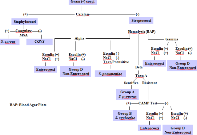 Figure: Overview of Biochemical tests for differentiating Gram positive cocci
Staphylococcus species
Staphylococcus genus contains more than 30 species, two of which, Staphylococcus aureus and S. saprophyticus are commonly pathogenic in humans. S. epidermidis, primarily a normal skin flora of skin can also cause bloodstream infections in neonates and people with prosthetic implants and catheters.
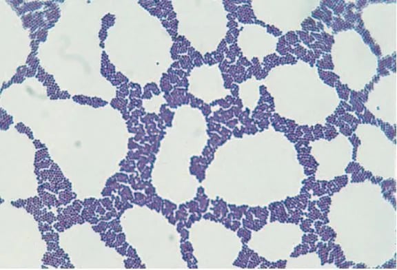 Figure: Staphylococcus in Gram Stain
Staphylococcus aureus
Staphylococcus aureus is a normal flora of the nose and skin but also one of the notorious pathogens. It is catalase and coagulase positive gram-positive cocci. Major diseases caused by S. aureus are sepsis, pneumonia, wound infections, folliculitis, necrotizing fasciitis, scalded skin syndrome, surgical site infection, toxic shock syndrome, and food poisoning.
Major virulence factors of S. aureus are coagulase, collagenase, hyaluronidase, immunoglobulin proteases, protein A, toxic shock syndrome toxin (TSST-1), enterotoxin, exfoliatin, etc.
Read more: Staphylococcus aureus: Properties, Pathogenesis & Lab Diagnosis
Some strains of S. aureus, such as methicillin-resistant Staphylococcus aureus (MRSA), are highly resistant to multiple antibiotics. Bactericidal agent vancomycin is an effective treatment of MRSA infections.
Staphylococcus saprophyticus
Staphylococcus saprophyticus is found primarily on the mucosa of the genital tract in young women and causes urinary tract infections (UTI), especially cystitis in sexually active young women. In this group, it is second to E. coli as a cause of community-acquired UTIs.
Read more about Staphylococcus saprophyticus characteristics and diagnosis.
Staphylococcus epidermidis
Staphylococcus epidermidis are normal flora of the human skin and mucous membranes. They are opportunistic pathogens and can cause neonatal sepsis, prosthetic hip infection, intravascular catheter infection, and endocarditis on prosthetic heart values.
These coagulase-negative staphylococci (CONS) do not produce exotoxins, but some strains may produce glycocalyx, which helps them to adhere to foreign bodies such as prosthetic implants and catheters.
Streptococcus species
Streptococci are spherical gram-positive cocci arranged in pairs or chains. All streptococci are catalase-negative. Traditionally, streptococci are identified based on hemolysis type shown in blood agar cell wall structure and reaction of antibodies to the specific bacterial antigen. S. pneumoniae and viridans streptococci are alpha-hemolytic, S. agalactiae and S. pyogenes are beta-hemolytic.
C-carbohydrate antigen and M protein are two important antigens of beta-hemolytic streptococci. Based on antigenic differences in C carbohydrate, beta-hemolytic streptococci are arranged into groups A-U, also known as Lancefield grouping.
Some other streptococci are non-hemolytic (gamma-hemolysis). Currently, the molecular analysis of the 16S ribosomal ribonucleic acid (rRNA) sequences is helpful to identify the isolates. Streptococci cause various infections, such as pneumonia, sore throat, pyogenic skin infections, and meningitis.
Streptococcus pyogenes
Group A Streptococcus (S. pyogenes), also known as “flesh-eating bacteria,” is the leading bacterial cause of pharyngitis (strep. throat) and cellulitis. It also causes impetigo, necrotizing fasciitis, scarlet fever, and streptococcal toxic shock syndrome. Rheumatic fever and acute post-streptococcal glomerulonephritis (PSGN) are the post-streptococcal sequelae of S. pyogenes infections. It is beta-hemolytic, bacitracin-sensitive, and pyrrolidonyl arylamidase (PYR) test positive.
M proteins, hyaluronic acid capsule, adhesions, hemolysins, pyrogenic exotoxins, and spreading factors (deoxyribonucleases, streptokinase, and hyaluronidase) are the major virulence factors of S. pyogenes.
Read more: Streptococcus pyogenes (GAS): Properties, Pathogenesis, and Lab Diagnosis
Streptococcus pneumoniae
Streptococcus pneumoniae (pneumococcus) is the normal upper respiratory tract flora and the most common cause of community-acquired pneumonia. It also causes bacteremia, meningitis, otitis media, sinusitis, and conjunctivitis. Pneumococci are gram-positive lancet-shaped diplococci. They produce alpha-hemolysis on blood agar, optochin-sensitive, and bile soluble.
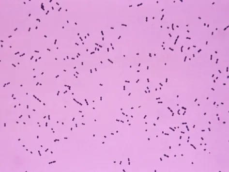 Figure: Gram positive diplococci: Streptococcus spp
Autolysins, IgA1 proteases, pneumolysin, polysaccharide capsules, and teichoic acids are the major virulence factors of S. pneumoniae. There are >90 different capsular serotypes of S. pneumoniae. The capsular serotype of an individual pneumococcal isolate is determined by using quellung reaction.
Read more: Streptococcus pneumoniae: Properties, Pathogenesis, and Diagnosis
Streptococcus agalactiae
Group B Streptococcus (S. agalactiae) colonizes the genital tract of some women. It is the leading cause of neonatal sepsis, pneumonia, and meningitis. The infection is acquired before or during the birthing process. Because of this CDC recommends screening of all pregnant women for GBS carriage between 35 to 37 weeks of gestation.
S. agalactiae is beta-hemolytic streptococci that are resistant to bacitracin and hydrolyze hippurate. CAMP test positive results are the special diagnostic tests for identifying S. agalactiae.
Read more: Streptococcus agalactiae (GBS): Properties, Pathogenesis, Lab Diagnosis
Viridans Streptococci
Viridians group streptococci are also α-hemolytic streptococci, but unlike S. pneumoniae, they are optochin resistant and bile-insoluble. Viridans streptococci are normal flora of the human oral cavity and pharynx but are one of the major causes of bacterial endocarditis.
Streptococcus mutans, Streptococcus mitis, and Streptococcus sanguinis are the medically important species of viridans group of streptococci. S. mutans synthesizes polysaccharide dextran and is responsible for forming dental plaque leading to dental caries.
Read more: Viridans Streptococci: Pathogenesis and Lab Diagnosis
Group D streptococci
These include enterococci (e.g., Enterococcus faecalis and Enterococcus faecium) and non-enterococci (e.g., S. bovis). Group D streptococci are mostly α-hemolytic, but some are β-hemolytic, and others are nonhemolytic.
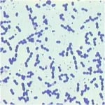 Figure: Enterococcus faecalis in gram stain
Enterococci are members of the normal flora of the colon and are noted for their ability to cause urinary, biliary, and cardiovascular infections. Enterococcus faecalis is an important cause of hospital-acquired UTIs and endocarditis. Strains of E. faecium and E. faecalis are vancomycin-resistant (also known as vancomycin-resistant enterococci-VRE).
Read more: Enterococcus faecalis: Properties, Pathogenesis, Lab Diagnosis
Micrococcus
Micrococci are gram-positive cocci usually arranged in tetrads or pairs and are part of the normal flora of the skin. Micrococci are usually considered contaminants of clinical specimens (blood) and are rarely implicated as a cause of human infections.
The two most common species of micrococci are Micrococcus luteus and Micrococcus roseus. Micrococcus gives non-hemolytic colonies with a wide variety of pigments (white, tan, yellow, orange, or pink) in blood agar. They can be differentiated from staphylococci using microdase test, micrococci are modified oxidase (microdase) positive and susceptible to bacitracin.
Peptococcus
Peptococci are anaerobic gram-positive cocci, resembling staphylococci, found as members of the normal flora of the mouth and colon. They are also isolated from abscesses of various organs, usually from mixed anaerobic infections. Peptococcus niger is the only species of this genus.
Peptostreptococcus
Peptostreptococci are anaerobic gram-positive cocci found as members of the normal flora of the mouth, gut, colon, and genital tract of women. Peptostreptococci are also isolated from abscesses of various organs, usually from mixed anaerobic infections.
These organisms grow well under anaerobic or microaerophilic conditions and produce variable hemolysis. Peptostreptococcus magnus and Peptostreptococcus anaerobius are clinically important species.
References
- Madigan, M., Martinko, J., Stahl, D., & Clark, D. (2012). Brock Biology of Microorganisms (13th ed). Pearson Education
- Bailey & Scott’s Diagnostic Microbiology, Forbes, 11th edition
- Review of Medical Microbiology and Immunology, Lange Medical Books, 13th edition
Frequently Asked Questions
What are the main genera of gram-positive cocci of medical importance?
The most clinically significant genera are Staphylococcus, Streptococcus, and Enterococcus. Micrococcus, Peptococcus, and Peptostreptococcus are also gram-positive cocci but are rare pathogens, mostly normal flora.
How does cell arrangement (clusters, chains, pairs, tetrads) help identify gram-positive cocci?
Arrangement under the microscope narrows identification before any biochemical test is run: clusters suggest Staphylococcus, chains suggest Streptococcus, pairs (diplococci) suggest S. pneumoniae or Enterococcus, and tetrads suggest Micrococcus. This is typically followed by the catalase test to confirm the genus-level call.

Tankeshwar Acharya, MSc (Medical Microbiology)
Tankeshwar Acharya is an Assistant Professor in the Department of Microbiology at Patan Academy of Health Sciences (PAHS), Nepal, where he has been teaching and practicing clinical microbiology for over 14 years. He is the founder of Microbe Online, one of the leading free microbiology education resources on the web, covering bacteriology, mycology, parasitology, immunology, and clinical laboratory diagnostics written from direct experience in both the classroom and the diagnostic laboratory.
Comments
No comments yet. Be the first to share your thoughts.
Leave a comment
All comments are reviewed before they appear.