
Electron Microscope: Principle, Types, Applications
How electron microscopes use electron beams instead of light to reveal detail far below what light microscopy can resolve, and how TEM and SEM differ in what they can show you.
In the 1960s, before smallpox was eradicated, a man walked into a busy London hospital outpatient department with a widespread vesicular rash and a mild fever. He said he'd just returned from East Africa, where smallpox was still endemic, and that he'd had chickenpox as a child but was never vaccinated against smallpox. It was late on a Friday. If this was smallpox, the hospital needed to alert public health authorities and potentially warn everyone who had shared that waiting room. If it was chickenpox, none of that was necessary.
There was no time to wait for a culture or a serology result. An electron microscopist examined fluid from one of his lesions directly under a transmission electron microscope. Within minutes, the answer was visible: poxviruses like variola have a distinctive brick-shaped, complex outer structure, while herpesviruses like varicella-zoster are spherical and enveloped. The two are nothing alike under EM, even though the rash they cause can look nothing alike either.
This is the same principle behind the variola virus micrograph in this article's own figure. Electron microscopy doesn't just make things bigger, it reveals structural detail no light microscope can resolve at all, which is exactly why it remains one of the fastest ways to answer an urgent identification question when speed and specificity both matter.
Electron microscope as the name suggests is a type of microscope that uses electrons instead of visible light to illuminate the object. Electromagnets function as lenses in the electron microscope, and the whole system operates in a vacuum.
Since electrons have a very short wavelength, the resolving power of electron microscopes is very high and produces a high-resolution image on a fluorescent screen, like a television screen.
Electron microscopes are used for detailed investigation of the ultrastructure of a wide range of biological and inorganic specimens including microorganisms, cells, large molecules, biopsy samples, metals, and crystals.
German physicist Ernst Ruska, working with Max Knoll, built the first electron microscope in 1931.
Components of an Electron Microscope
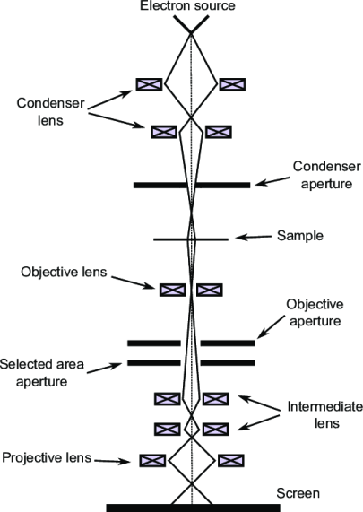 Figure: Schematic diagram of Transmission Electron Microscope
Like any ordinary microscope, the electron microscope also uses a light source, a combination of lenses to produce a magnified image, however, this vary slightly as compared to ordinary light microscope.
- Source of light: The source of light is replaced by a beam of very fast-moving electrons. These electron beams are obtained when the tungsten filament in an electron microscope, is heated by applying a high voltage current, which is used as a light beam.
- Electromagnetic fields (instead of lenses): The lenses that make the specimen seem bigger are replaced by a series of coil-shaped electromagnets through which the electron beam travels also known as electromagnetic lenses. There are 3 sets of electromagnetic lenses in an electron microscope as compared to two in light microscopes. In addition to the condenser and objective lenses, a third projector lens is also present. The magnetic lens of an electron microscope can have different powers (focal lengths and magnification) depending on the amount of current flowing through the electrical coils. In an ordinary microscope, the glass lenses bend (or refract) the light beams passing through them to produce magnification. In an electron microscope, the coils bend the electron beams the same way.
- Image viewing and recording system: The magnified image of the specimen is formed as a photograph (called an electron micrograph) or as an image on a TV screen.
In electron microscope, the entire illuminating imaging system is usually referred to as the microscope column and is constructed upside down as compared to a light microscope.
Types of Electron Microscope
There are two types of electron microscopes with different operating styles: the transmission electron microscope (TEM) and the scanning electron microscope (SEM).
Transmission Electron Microscope (TEM)
As the name suggests, this type uses transmitted electrons as light sources. It provides a detailed view of the internal structure of the prepared test samples. Tissues must be cut in thin sections for viewing under TEM. Its magnifying power is very high as compared to SEM and shows a 2-dimensional image. It is the most commonly used type of electron microscope.
Scanning Electron Microscope (SEM)
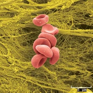 Figure: RBC seen in Scanning Electron Microscope(Image source: University of Iowa)
It uses scattered electrons as the light source. SEM scans the surface of the objects and provides three-dimensional views of the surface structures. In SEM sample is prepared by coating it with a thin layer of metal such as gold or palladium. It has low magnifying power compared to TEM.
The major differences between scanning electron microscope (SEM) and transmission electron microscope (TEM) are available in Differences Between SEM and TEM.
Transmission Electron Microscope (TEM): Specimen Preparation and Principle
The steps below describe specimen preparation and imaging specifically for TEM. SEM preparation is different, and simpler, requiring only a thin conductive metal coating rather than ultra-thin sectioning; see the comparison article above for the full contrast.
Specimen Preparation
The specimen suitable for electron microscopes should be very thin (20-100 nm thickness) so the bacterial cells and any other biopsy materials should be slice into thin layers. The general flow of sample preparation is as follows:
Unlike visible light, electron beams can not penetrate even a single cell.
- Fixation: Cells are fixed by using glutaraldehyde or osmium tetroxide for stabilization
- Dehydration: Specimen is then dehydrated with organic solvents (e.g. acetone or ethanol)
- Embedding: Specimen is embedded in plastic polymer and then, is hardened to form a solid block.
- Slicing: Specimen is cut into thin slices by an ultramicrotome knife. Slices thus prepared are then mounted on a metal slide.
Principle
The electronic beam is obtained from a heated tungsten filament which generates electrons. A large voltage is applied between the cathode (the tungsten filament) and the anode, which excites the electrons and it travels with high velocity towards the condenser lens. The condenser lenses focus the electronic beams through the specimen and electrons are scattered depending upon the thickness or refractive index of different parts of the specimen.
The denser regions in the specimen scatter more electrons and therefore appear darker in the image since fewer electrons strike that area of the screen. In contrast, transparent regions are brighter. The electron beam coming out of the specimen passes to the objective lens, which has high power and forms the intermediate magnified image. The projector throws its image onto a fluorescent screen which may be substituted by a photographic plate to make a permanent record.
Advantages of Electron Microscope
- Magnification and higher resolution: Transmission electron microscopes achieve a resolution of approximately 0.2 nm and magnifications of up to 1,000,000x, compared with a resolution of approximately 0.2 μm and a magnification limit of about 2,000x for light microscopes. Scanning electron microscopes resolve down to approximately 1–20 nm, with magnifications up to about 100,000x.
- High-quality images– Electron microscopes produce highly detailed images of structures which are of high quality, revealing complex and delicate structures.
Limitations of Electron Microscope
Electron microscope is a major capital investment and is not needed for the laboratory diagnosis of most infectious diseases. The major limitations of this microscope are as follows:
- It is expensive, requires a large space in the laboratory, and is highly sensitive equipment.
- Require trained manpower for proper sample preparation to avoid artifacts and proper handling of the microscope.
- Electron microscopes operate in a vacuum; therefore, live specimens cannot be observed.
- Since the penetration power of the electron beam is very low, the object should be ultra-thin. For this, the specimen is dried and cut into ultra-thin sections before observation (for TEM). This could lead to distortions in the structures. This requirement applies specifically to TEM; SEM does not require thin sectioning, since it images the specimen's surface rather than transmitting electrons through it.
- Magnetic fields and vibrations caused by other lab equipment may interfere with their operation.
- Only black and white images can be produced by an electron microscope.
Applications of Electron Microscope
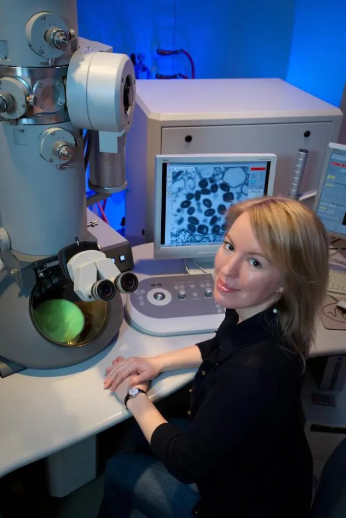 Figure: Scientist observing variola virus in TEM (Image source: CDC/James Gathany)
Electron microscopes are powerful research tools. We are able to discover many new morphological features of bacteria, bacterial components, fungi, viruses, and parasites using electron microscopes.
Apart from Microbiology, electron microscopy have a diverse range of applications in many different fields such as technology, industry, biomedical science, and chemistry.
- In pathology, it is used to examine microscopic features of different diseases including tumors.
- In chemistry, it is used for quality control and assurance, analysis of atomic structures, and drug development.
- In the technology industry, it is used for high-resolution 2D and 3D imaging, semiconductor inspection, computer chip manufacture.
- In forensics SEM is widely used for gunshot residue analysis, firearm identification (bullet markings comparison), investigation of gemstones and jewelry, counterfeit banknotes, examinations of non-conducting materials, etc.
- In geology, SEM is a routine technology employed in the study of rocks and minerals.
How to Remember
- The one-word distinction that explains everything else: TEM is transmission, electrons pass through the specimen, revealing internal structure, as a flat, 2D density shadow. SEM is scanning, electrons bounce off the surface, revealing external structure, as a 3D-looking surface image. Every other difference (sample prep, resolution, magnification) follows from this one choice: through vs. off.
- Why a vacuum is non-negotiable: electrons scatter off air molecules almost immediately, the same way footsteps get lost in fog. Removing the air is what keeps the electron beam a clean, focused beam instead of a scattered blur before it ever reaches the specimen.
- EM images are never actually in color: any color seen in an electron micrograph, including the ones illustrating this cluster, is added afterward. If a question describes a "colorized" EM image, that's simply confirming the image is genuinely black and white at capture.
- The poxvirus-vs-herpesvirus shape distinction, as a fast recall device: brick-shaped and complex means orthopoxvirus (smallpox, vaccinia); spherical and enveloped means herpesvirus (varicella-zoster, herpes simplex). This single visual distinction is what let a 1960s EM lab resolve a potential public health emergency in minutes.
Key exam facts in one table
| Fact | Detail | Why it's tested |
|---|---|---|
| Invention | Electron microscope developed by Ernst Ruska (with Max Knoll), 1931 | Commonly asked "who and when" historical fact |
| Nobel recognition | Ruska received the 1986 Nobel Prize in Physics for the electron microscope | Shared that year with Binnig and Rohrer, for the scanning tunneling microscope |
| Why a vacuum is required | Air molecules scatter the electron beam before it reaches the specimen | Explains why live specimens can never be examined by EM |
| Number of lens sets | Three electromagnetic lens sets (condenser, objective, projector) vs. two glass lens sets in light microscopy | A specific, often-tested structural difference |
| TEM resolution and magnification | ~0.2 nm resolution; up to ~1,000,000x magnification | Standardized across this site's microscopy cluster |
| SEM resolution and magnification | ~1–20 nm resolution; up to ~100,000x magnification | Standardized across this site's microscopy cluster |
| Image color | Always black and white at capture; any color seen is added afterward | A frequently overlooked but testable detail |
Where Students Get Confused
- Treating "electron microscope" as a single instrument rather than a category. TEM and SEM answer fundamentally different questions (internal structure vs. surface structure) and require different specimen preparation; neither is simply a "better" or "worse" version of the other.
- Assuming the TEM-specific specimen preparation and limitations apply to SEM as well. Ultra-thin sectioning, for example, is a TEM requirement; SEM specimens need only a thin conductive metal coating.
- Assuming EM images are naturally in color. Every electron micrograph is fundamentally black and white; any color version has been added digitally afterward, purely for visual clarity.
- Assuming higher magnification always means a "better" image. As with light microscopy, magnification is meaningless without matching resolution, and the specimen preparation required for very high magnification (like TEM's ultra-thin sectioning) is itself a limitation in certain contexts.
References and further readings
- Tille, P. (2017). Bailey & Scott’s Diagnostic Microbiology (14 edition). Mosby.
- Madigan Michael T, Bender, Kelly S, Buckley, Daniel H, Sattley, W. Matthew, & Stahl, David A. (2018). Brock Biology of Microorganisms (15th Edition). Pearson.
Frequently Asked Questions
Who invented the electron microscope, and when?
Why do electron microscopes require a vacuum?
What is the resolution and magnification of a transmission electron microscope compared to a light microscope?
Are electron microscope images ever in color?
How was electron microscopy historically used to distinguish smallpox from chickenpox?

Tankeshwar Acharya, MSc (Medical Microbiology)
Tankeshwar Acharya is an Assistant Professor in the Department of Microbiology at Patan Academy of Health Sciences (PAHS), Nepal, where he has been teaching and practicing clinical microbiology for over 14 years. He is the founder of Microbe Online, one of the leading free microbiology education resources on the web, covering bacteriology, mycology, parasitology, immunology, and clinical laboratory diagnostics written from direct experience in both the classroom and the diagnostic laboratory.