
Bird Seed Agar (Niger Seed Agar): Composition, Uses, and Cryptococcus Identification
Bird seed agar selectively detects Cryptococcus neoformans by its brown-black melanin production from caffeic acid. Learn the principle, composition, colony appearance, and how it differentiates C. neoformans from other Cryptococcus species.

On this page
A 32-year-old HIV-positive patient presents with severe headache, fever, and neck stiffness. CSF shows lymphocytic pleocytosis with elevated protein. India ink reveals yeast cells with large capsular halos. The CSF is inoculated onto bird seed agar (Niger seed agar / Staib medium). After 72 hours at 30°C: brown-black colonies, melanin production from caffeic acid confirming Cryptococcus neoformans. A nearby colony of Candida albicans (contaminant) remains white.
In resource-limited settings where molecular identification is unavailable, bird seed agar provides a rapid, inexpensive, and specific presumptive identification of Cryptococcus neoformans from CSF, blood, and respiratory specimens. The melanin reaction distinguishes C. neoformans and C. gattii from all other yeasts.
Bird Seed Agar, also known as Staib medium, is a selective and differential medium for isolation of Cryptococcus neoformans from clinical specimens and differentiation of it from other Cryptococcus species. It is also known as caffeic acid agar or niger seed agar. Due to the presence of phenoloxidase enzyme, Cryptococcus species can utilize various phenolic compounds as their substrate and produce dark brown, melanin-like pigments, thus presumptively identified.
In 1966, Shields and Ajello modified Staib’s bird seed agar by making the medium selective with an antimicrobial additive.
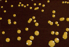 Figure: Colonies of Cryptococcus neoformans in birdseed agar
Principle
The extract of Guizotia abyssinica seeds contains caffeic acid. Phenoloxidase enzyme produced by Cryptococcus neoformans utilizes caffeic acid as a substrate and produces melanin which in turn is absorbed by the yeast cell wall forming a tan to reddish-brown pigmentation. Glucose is the energy source in the medium. Creatinine enhances melanization of some strains of Cryptococcus neoformans. Agar is the solidifying agent. Chloramphenicol is added to inhibit the growth of bacteria and fast-growing fungi.
The melanin mechanism in detail: Cryptococcus neoformans possesses the enzyme laccase (phenol oxidase), which oxidizes diphenolic compounds, particularly caffeic acid (present in the Niger seed/bird seed extract) to melanin. This melanin deposits in the cell wall, producing the distinctive brown-black colony color. Most other pathogenic yeasts, including Candida species, Saccharomyces, and non-neoformans Cryptococcus species (except C. gattii), lack laccase and therefore remain white or cream-colored on bird seed agar.
This melanin production is also a virulence factor, as discussed in the pigments article.
Composition
| Ingredients | Gm / Litre |
|---|---|
| Guizotia abyssinica seeds | 70g |
| Creatinine | 0.780g |
| Dextrose | 10g |
| Chloramphenicol | 0.050g |
| Agar | 20g |
| Final pH ( at 25°C) 6.7±0.2 |
Procedure for preparation of media
- Suspend required quantity (as per manufacturer’s instruction) of powder media in 1 liter of distilled water.
- Heat to boiling to dissolve the medium completely.
- Sterilize by autoclaving at 15 lbs pressure (121°C) for 15 minutes.
- Cool to 45°C and add 100 mcg diphenyl per ml of medium (1 ml of sterile 1% w/v aqueous solution of diphenyl).
- Mix well and pour into sterile Petri plates.
NOTE: The composition and method for media preparation vary according to the manufacturer. Some media contain pre-added phosphate and other antibiotics like penicillin G, gentamicin, while others need supplements to be added.
Interpretation of results
Plates inoculated with suspected samples are observed after incubation at 30°C for 2 weeks. The presence of golden brown to black pigmented smooth colonies is indicative of Cryptococcus neoformans. Other species like Cryptococcus laurentii, Saccharomyces cerevisiae, etc produce non-pigmented colonies. Candida appears as white colonies.
Uses
Bird Seed Agar is used for the selective isolation of Cryptococcus neoformans and Cryptococcus gattii.
Cryptococcus Species on Bird Seed Agar
| Species | Colony Colour on BSA | Clinical Significance |
|---|---|---|
| C. neoformans var. grubii (serotype A) | Brown-black | Most common; AIDS-associated cryptococcal meningitis worldwide |
| C. neoformans var. neoformans (serotype D) | Brown-black | Less common; immunocompromised patients |
| C. gattii (serotypes B and C) | Brown-black | Affects immunocompetent hosts; tree-associated (eucalyptus); Pacific Northwest and tropics |
| C. laurentii | White/cream | Non-pathogenic; no laccase |
| C. albidus | White/cream | Non-pathogenic; no laccase |
| Candida spp. | White/cream | Negative on BSA |
| Rhodotorula spp. | Salmon/orange (own pigment) | Non-melanin pigment; distinct from Cryptococcus brown |
Key point: Both C. neoformans and C. gattii produce brown-black colonies on bird seed agar, they cannot be differentiated from each other by this medium alone. CanaVanine-Glycine-Bromothymol (CGB) agar or molecular methods are needed to distinguish these two species.
Quality control
- Cryptococcus neoformans ATCC 32045 can be used as a positive control that shows brown to black pigmented colonies.
- Escherichia coli ATCC 25922 can be used as a negative control in which growth is inhibited partially or completely.
Key Exam Facts in One Table
| Feature | Detail |
|---|---|
| Also called | Niger seed agar, Staib medium, birdseed agar |
| Named after | Walter Staib (developed the medium) |
| Key substrate | Caffeic acid (from Guizotia abyssinica Niger seeds) |
| Reaction | Laccase (phenol oxidase) oxidises caffeic acid → melanin |
| C. neoformans appearance | Brown-black colonies at 72h–5 days |
| C. gattii appearance | Brown-black (same as C. neoformans) |
| Candida appearance | White/cream, no melanin |
| Rhodotorula appearance | Salmon/orange (own pigment), not melanin |
| Distinguishes | C. neoformans/gattii from all other clinically important yeasts |
| Does NOT distinguish | C. neoformans from C. gattii — need CGB agar or molecular |
| Incubation | 25–30°C; 72h–5 days |
| Clinical use | CSF, blood, respiratory specimens in Cryptococcus-suspected cases |
| Melanin as virulence factor | Laccase protects from oxidative killing; may reduce amphotericin B efficacy |
References and further readings
- Acharya T., Hare J. (2022) Sabouraud Agar, and Other Fungal Growth Media. In: Gupta V.K., Tuohy M. (eds) Laboratory Protocols in Fungal Biology. Fungal Biology. Springer, Cham. https://doi.org/10.1007/978-3-030-83749-5_2
- Staib F. Cryptococcus neoformans und Guizotia abyssinica (syn. G. oleifera DC). Z Hyg Infektionskr. 1962;148:466–475. (Original description of the medium.)
- Kwon-Chung KJ, Bennett JE. Medical Mycology. Lea & Febiger; 1992.
- Larone DH. Larone's Medically Important Fungi: A Guide to Identification. 6th ed. ASM Press; 2018.
- Perfect JR, Dismukes WE, Dromer F, et al. Clinical practice guidelines for the management of cryptococcal disease: 2010 update by the Infectious Diseases Society of America. Clin Infect Dis. 2010;50(3):291–322. https://doi.org/10.1086/649858
- Murray PR, Rosenthal KS, Pfaller MA. Medical Microbiology. 9th ed. Elsevier; 2020.
Frequently Asked Questions
How does bird seed agar identify Cryptococcus neoformans?
How does bird seed agar identify Cryptococcus neoformans?
Bird seed agar (Niger seed/Staib medium) contains caffeic acid derived from Guizotia abyssinica (Niger seeds). Cryptococcus neoformans possesses the enzyme laccase (phenol oxidase), which oxidises caffeic acid to melanin. This melanin deposits in the cell wall, producing distinctive brown-black colonies within 72 hours to 5 days at 30°C. Most other pathogenic yeasts, including all Candida species, lack laccase and remain white or cream colored. The brown-black colony color on bird seed agar is essentially diagnostic for Cryptococcus neoformans or C. gattii (both possess laccase) in the clinical laboratory.
Can bird seed agar differentiate Cryptococcus neoformans from Cryptococcus gattii?
Can bird seed agar differentiate Cryptococcus neoformans from Cryptococcus gattii?
No. Both C. neoformans and C. gattii produce laccase and give identical brown-black colonies on bird seed agar. They cannot be differentiated by this medium alone. Differentiation requires CanaVanine-Glycine-Bromothymol blue (CGB) agar: C. gattii grows on CGB agar and turns the medium blue (produces ammonia from glycine), while C. neoformans does not grow on CGB. This distinction matters clinically because C. gattii primarily infects immunocompetent hosts (unlike C. neoformans which predominantly causes disease in immunocompromised patients), affects different geographic areas, and may respond differently to antifungal therapy.

Tankeshwar Acharya, MSc (Medical Microbiology)
Tankeshwar Acharya is an Assistant Professor in the Department of Microbiology at Patan Academy of Health Sciences (PAHS), Nepal, where he has been teaching and practicing clinical microbiology for over 14 years. He is the founder of Microbe Online, one of the leading free microbiology education resources on the web, covering bacteriology, mycology, parasitology, immunology, and clinical laboratory diagnostics written from direct experience in both the classroom and the diagnostic laboratory.
Comments
No comments yet. Be the first to share your thoughts.
Leave a comment
All comments are reviewed before they appear.