

A 35-year-old man presents with three weeks of productive cough, night sweats, and weight loss. A chest X-ray shows an upper lobe cavity. The clinician requests a sputum smear for AFB.
In many parts of the world including Nepal, India, and most of sub-Saharan Africa, the Ziehl-Neelsen smear result is the first and sometimes only microbiological evidence available before treatment decisions are made. Culture may take 6–8 weeks on Löwenstein-Jensen medium. Molecular tests (GeneXpert) may not be accessible. The ZN smear, available within hours, guides whether to start anti-TB therapy in a patient who cannot wait.
Learning ZN staining is not an academic exercise. In high-burden settings, it is a clinical skill with direct impact on patient outcomes.
Ziehl-Neelsen (ZN) acid-fast staining technique is used to stain Mycobacterium species, including M. tuberculosis, M. ulcerans, M. leprae, and nontuberculous mycobacteria (NTM). Detection of acid-fast bacilli (AFB) in stained and acid-washed smears examined microscopically may provide the initial bacteriologic evidence of the presence of mycobacteria in a clinical specimen. Smear microscopy is the quickest and easiest procedure that can be performed.
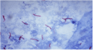 Figure: Acid-fast bacillus (AFB) in Z.N smear
Mycobacteria’s cell wall contains high lipid concentrations, making them waxy, hydrophobic, and impermeable to routine stains such as the Gram Stain. They are also resistant to acid and alcohol and are described as acid-fast bacilli (AFB) or acid alcohol fast bacilli (AAFB).
There are two procedures commonly used for acid-fast staining:
- Carbolfuchsin methods which include the Ziehl-Neelsen and Kinyoun methods (light/bright field microscope)
- Fluorochrome procedure using auramine-O or auramine-rhodamine dyes (fluorescent microscope).
Principle of Ziehl-Neelsen Method of Acid-Fast Staining
Mycobacteria, which do not stain well by Gram stain, stains well with carbol fuchsin combined with phenol.
- In the ‘hot’ ZN technique, the phenol-carbol fuchsin stain is heated to enable the dye to penetrate the waxy mycobacterial cell wall.
- In the ‘cold’ technique known as Kinyoun Method, stains are not heated but the penetration is achieved by increasing the concentration of basic fuchsin and phenol and incorporating a ‘wetting agent’ chemical.
The stain binds to the mycolic acid in the mycobacterial cell wall. After staining, an acid decolorizing solution is applied. This removes the red dye from the background cells, tissue fibers, and any organisms in the smear except mycobacteria which retain (hold fast to) the dye and are therefore referred to as acid-fast bacilli (AFB).
Following decolorization, the sputum smear is counterstained with malachite green, or methylene blue which stains the background material, providing a contrast color against which the red AFB can be seen.
Among the Mycobacterium species, M. tuberculosis and M. ulcerans are strongly acid-fast. When staining specimens for these species, a 3% v/v acid alcohol is used to decolorize the smear, whereas M. leprae is only weakly acid-fast. 0.5-1% v/v decolorizing solution is therefore used for M. leprae smears and also different staining and decolorizing time.
Note: Weakly or partially acid-fast organisms such as M. leprae and Nocardia species are decolorized more readily than M. tuberculosis. A milder decolorizer, typically 0.5 to 1% acid alcohol or 5% sulfuric acid, is used for these smears, with shorter decolorization times. M. leprae is not a nontuberculous mycobacterium; it is a distinct species that happens to be only weakly acid-fast.
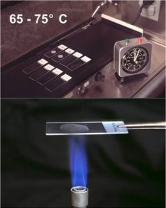 Figure: Heat Fixation of smear (Upper: using an electric heater, lower: using burner)
ZN vs Fluorochrome Staining: Which to Choose?
Two methods are used for acid-fast staining in clinical laboratories. The choice depends on workload, available equipment, and the clinical urgency.
| Feature | Ziehl-Neelsen (Carbolfuchsin) | Auramine-Rhodamine (Fluorochrome) |
|---|---|---|
| Microscope required | Ordinary bright-field microscope | Fluorescence microscope (UV light source) |
| AFB appearance | Red rods on blue/green background | Bright yellow-orange rods on dark background |
| Scanning magnification | 100x oil immersion (entire examination) | 25x or 40x dry objective (scan); 100x to confirm |
| Time to screen one slide | 15–20 minutes | 3–5 minutes |
| Sensitivity vs ZN | Reference standard | Higher, detects paucibacillary specimens better |
| Specificity | High | Slightly lower, artefacts can fluoresce |
| Cost | Low reagent cost | Higher (fluorescence microscope) |
| WHO recommendation | Acceptable | Preferred where fluorescence microscope available |
| Confirmation of positive | Not required | Confirm with ZN if uncertain |
Key point: Auramine-rhodamine is more sensitive than ZN for direct smear examination because slides can be scanned at lower magnification, covering more area per unit time and detecting fewer bacilli. A specimen with 100 AFB/mL may be missed on ZN but detected on auramine-rhodamine. WHO recommends fluorochrome staining as the preferred method where fluorescence microscopy is available.
A positive auramine-rhodamine result that is clinically unexpected should be confirmed by ZN staining of the same slide. Fluorescence artefacts (dust, fibers) can occasionally give false-positive results.
For the full procedure for auramine-rhodamine staining, see: Auramine-Rhodamine Fluorochrome Staining
Sample Collection & Preparation
Due to overnight accumulation of secretions, first morning specimens are more likely to yield better recovery of AFB.
- Direct Smear: Smear prepared directly from a patient specimen prior to processing.
- Indirect Smear: Smear prepared from a processed specimen after centrifugation (is used to concentrate the material)
Assessing Sputum Quality Before Staining
The sensitivity of ZN smear microscopy depends critically on specimen quality. A saliva sample labelled as sputum will give a false-negative result regardless of how perfectly the staining is performed.
Before preparing a smear, assess the specimen macroscopically:
| Specimen Appearance | Assessment | Action |
|---|---|---|
| Mucopurulent, thick, opaque, yellow/green | Acceptable | Proceed with smear preparation |
| Blood-stained, rust or red tinged | Acceptable | Note blood-staining; proceed |
| Mucoid, slightly opaque, stringy | Acceptable | Select the most opaque portion |
| Salivary, thin, watery, frothy | Unacceptable | Request repeat specimen |
| Very small volume (<1 mL) | Borderline | Process but note volume; request repeat |
For smear preparation: Always select the most purulent, caseous, or blood-stained portion of the specimen: this is where AFB are most concentrated. Spreading too thin misses AFB; spreading too thick prevents proper decolorization.
Smear size: Approximately 20 mm by 10 mm. When dry, the smear should be just readable through (you should be able to read newsprint through it). Too opaque = too thick.
Reagents required
- Carbol fuchsin stain (filtered)
- Acid alcohol 3% v/v (or 20% sulfuric acid)
- Malachite green 5 g/l (0.5% w/v) or Methylene blue, 5g/l
Ziehl-Neelsen Staining Procedure
Time needed: 45 minutes
- Prepare the sputum smear. Spread the sputum evenly over the central area of the slide using a continuous rotational movement. The recommended size of the smear is about 20 mm by 10 mm.
- Air dry. Place slides on the dryer with the smeared surface upwards and air dry for about 30 minutes.
- Heat fix the dried smear. Heat fixation of untreated specimens may not kill M. tuberculosis, so continue to handle slides with care. Alcohol fixation is bactericidal.
- Apply the primary stain. Cover the smear with carbol fuchsin stain.
- Heat the smear until vapor just begins to rise, at about 60°C. Do not overheat, boil, or allow the slide to dry. Add more stain if necessary. Leave the heated stain on the slide for 5 minutes.
- Wash off the stain with clean water.
- Apply the decolorizer. Cover the smear with 3% v/v acid alcohol for 2 to 5 minutes, or with 20% sulfuric acid, until the smear is sufficiently decolorized and appears pale pink. Check that no more red color runs off the surface when the slide is tipped. Add a little more decolorizer for very thick slides or those that continue to bleed red dye.
- Wash well with clean water.
- Counterstain. Cover the smear with malachite green stain for 1 to 2 minutes.
- Wash off the stain with clean water.
- Dry and examine. Wipe the back of the slide clean and place it in a draining rack for the smear to air dry. Do not blot dry. Examine the smear microscopically using the 100x oil immersion objective with a 10x eyepiece, for a total magnification of 1000x, and scan the smear systematically.
Procedural note
- Acid alcohol is flammable; therefore, use it with care.
- Take great care while heating carbol fuchsin (as the staining rack may contain volatile chemicals) to reduce the fire risk.
- Slides must not touch each other when placed on a staining rack to prevent the transfer of material from one slide to another.
Results of Acid Fast Staining
- AFB: red, straight, or slightly curved rods, occurring singly or in small groups, may appear beaded.
- Cells: green
- Background material: green
| Reagent | Acid Fast | Non-Acid Fast |
|---|---|---|
| Carbol fuchsin with heat | Red (hot pink) | Red (hot pink) |
| Acid alcohol | Red | Colorless |
| Methylene blue/malachite green | Red | Blue/green |
Troubleshooting ZN Staining
| Problem | Likely Cause | Action |
|---|---|---|
| No red bacilli seen but TB strongly suspected | Paucibacillary specimen; inadequate decolorization time | Request early morning specimen × 3 consecutive days; use sodium hypochlorite concentration technique; consider fluorochrome staining |
| Background too dark (heavy blue/green staining) | Counterstain applied too long; smear too thick | Reduce counterstain time to 1 minute; prepare thinner smear |
| AFB appear faded/pale pink rather than red | Carbol fuchsin too dilute or expired; heating insufficient | Replace carbol fuchsin; ensure steam rises during heating without boiling |
| Red artefacts appearing (non-AFB structures) | Food particles, vegetable fibers in sputum; scratch on slide | Select purulent portion of specimen; use scratch-free slides; examine morphology carefully (AFB are straight or slightly curved rods, not irregular) |
| Smear washing off during staining | Inadequate heat fixation | Ensure complete air-drying before fixation; increase heat fixation passes; consider methanol fixation |
| Carbol fuchsin does not steam during hot technique | Flame too low; slide holder interference | Adjust flame; ensure slide is not resting on an insulating surface |
The 1-9 AFB/100 fields dilemma: When scanty AFB are found (1–9 per 100 fields), the grading guidelines recommend reporting the exact number and requesting a repeat specimen. This is because saprophytic NTM from tap water, glass slide scratches, or contaminated staining reagents can occasionally produce a false-positive result at very low counts. Always correlate with clinical findings: a patient with a classic TB presentation and 2 AFB/100 fields is a different situation from an asymptomatic person with 1 AFB/100 fields found incidentally.
Grading of sputum smear for Mycobacterium tuberculosis
Sputum smear is graded as scanty, 1+, 2+, and 3+.
| Observation | Grading |
|---|---|
| No AFB seen while observing more than 300 fields | No AFB seen |
| 1-9 AFB/100 fields | Scanty; report the exact number seen |
| 10-99 AFB/ 100 fields | Report as 1+ |
| 1-10 AFB/field at least in 50 fields | Report as 2 + |
| More than 10 AFB/field at least in 20 fields | Report as 3+ |
- When no AFB is seen after examining 300 fields, report the smear as ‘No AFB seen’.
- When very few AFB are seen i.e. when only 1 or 2 AFB are seen after examining 100 fields, request a further specimen. Such AFB may come from tap water containing saprophytic mycobacteria, from a scratch on the glass slide, or from carryover when the same piece of blotting paper is reused during drying.
- When any red bacilli are seen, report the smear as ‘AFB positive’ and give an indication of the number of bacteria present using the grading scale above. The greater the number, the more infectious the patient.
Important: The grading scale shown above follows WHO/IUATLD recommendations. Some laboratories use slight variations (always follow your national TB program guidelines). The key principle across all systems is that higher grades correlate with greater infectiousness and higher organism burden.
Grading is not just administrative, it has clinical implications:
- A 3+ smear-positive patient is highly infectious and requires immediate isolation precautions
- A patient converting from 2+ to negative on treatment can be used to monitor treatment response
- Smear grades should be documented at treatment initiation (month 0) and at months 2, 5, and 6 to track bacteriological response per national TB program guidelines
Partial and modified acid-fast organisms (Nocardia, Cryptosporidium, Cystoisospora) require modified decolorization, typically 0.5 to 1% acid alcohol or 5% sulfuric acid rather than 3% acid alcohol, and should not be reported on the standard TB smear report. If these organisms are clinically suspected, a modified ZN preparation should be specifically requested and clearly labelled.
Advantages
- Microscopy of sputum smears is simple and inexpensive, quickly detecting infectious cases of pulmonary TB;
- Sputum specimens from patients with pulmonary TB – especially those with the cavitary disease – often contain sufficiently large numbers of acid-fast bacilli to be readily detected by microscopy.
Limitation of AFB Microscopy
- Does not distinguish between viable and dead organisms
Follow-up specimens from patients on treatment may be smear-positive yet culture negative - Microscopy for acid-fast bacilli (AFB) cannot distinguish drug-susceptible from drug-resistant strains.
- Limited sensitivity: A high bacterial load, roughly 5,000 to 10,000 AFB/mL, is required for smear detection. Culture is far more sensitive, detecting on the order of 10 to 100 viable bacilli per mL. Smear sensitivity is further reduced in patients with extra-pulmonary TB, HIV-co-infection, and those with disease due to nontuberculous mycobacteria (NTM). Many TB patients have negative AFB smears with a subsequent positive culture. Negative smears do not exclude TB disease.
- Limited specificity: All mycobacteria are acid-fast, so ZN does not provide species identification. The local prevalence of M. tuberculosis and NTM determines the predictive value of a positive smear for TB.
List of Acid Fast Organisms
- Mycobacterium tuberculosis
- Mycobacterium leprae (weakly acid-fast)
- Other Mycobacteria
- Nocardia spp: Partial acid-fast
- Rhodococcus spp: Partial acid-fast
- Legionella micdadei: Partially acid-fast in tissue
- Cryptosporidium oocysts: acid-fast (modified ZN, cold method)
- Cystoisospora belli oocysts, formerly Isospora: acid-fast (modified ZN, cold method)
- Cyclospora cayetanensis oocysts: variably acid-fast
- Taenia saginata eggs: acid-fast
Sodium Hypochlorite Centrifugation Technique to Concentrate AFB
This technique is used to concentrate the AFB present in sputum, as it increases the chances of detecting AFB in sputum smears. Sodium hypochlorite (NaOCl) liquefies sputum and greatly reduces the viability of M. tuberculosis, which lowers the risk to laboratory staff. It does not replace standard TB biosafety precautions, and specimens must still be handled with appropriate PPE and containment.
Procedure
- Transfer 1-2 ml of the sputum’s purulent part (i.e. containing any caseous materials) to a screw cap universal bottle or other containers of 10-20 ml capacity.
- Add an equal volume of concentrated sodium hypochlorite (bleach) solution and mix well.
- Leave at room temperature for 10-15 minutes, shaking at intervals to break down the mucus in the sputum.
- Add about 8 ml of distilled water and mix well.
- Centrifuge at 3000 g for 15 minutes. When centrifugation is not possible, leave NaOCl-treated sputum to sediment overnight.
- Remove and discard supernatant fluid using a glass Pasteur or plastic bulb pipette. Mix the sediment.
- Transfer a drop of well-mixed sediment to a clean scratch-free glass slide and spread the sediment to make a thin preparation and allow air-drying.
- Heat fix the smear, stain it using the Ziehl-Neelsen technique, and examine it microscopically.
How to Remember: ZN Staining
Why mycobacteria are acid-fast?
Mycobacteria have a waxy cell wall rich in mycolic acid (long-chain fatty acids that make the wall hydrophobic and impermeable). The carbol fuchsin dye penetrates this waxy wall only with heat (hot ZN) or increased dye concentration (cold/Kinyoun method). Once inside, the dye is held so tightly by the mycolic acid that even acid-alcohol (one of the strongest decolorizing agents used in microbiology) cannot remove it. Everything else in the smear decolorizes; the AFB holds fast. That is where "acid-fast" comes from.
Reagent sequence "Can Anyone Make Mycobacteria Colorful?"
- Carbol fuchsin: primary stain (all cells red)
- Acid-alcohol: decolorizer (removes red from everything except AFB)
- Malachite green / Methylene blue: counterstain (background turns green/blue)
Grading memory:
- Scanty = 1–9 per 100 fields (report exact count)
- 1+ = 10–99 per 100 fields
- 2+ = 1–10 per field (50 fields)
- 3+ = >10 per field (20 fields)
The numbers increase with grade: more AFB, more infectious, higher grade number.
Key Exam Facts in One Table
| Feature | Detail |
|---|---|
| Full name | Ziehl-Neelsen acid-fast (carbolfuchsin) staining |
| Principle | Mycolic acid in mycobacterial cell wall retains carbol fuchsin after acid-alcohol decolorization |
| AFB appearance | Red/pink rods; straight or slightly curved; may appear beaded |
| Background color | Blue (methylene blue) or green (malachite green) |
| Hot technique | Carbol fuchsin heated until vapor rises at about 60°C, held for 5 minutes; standard ZN |
| Cold technique (Kinyoun) | No heat; higher concentrations of fuchsin and phenol + wetting agent |
| Decolorizer for MTB | 3% acid-alcohol (strongly acid-fast) |
| Decolorizer for M. leprae / Nocardia | 0.5–1% acid alcohol or 5% sulfuric acid (weakly/partially acid-fast) |
| Grading: scanty | 1–9 AFB per 100 fields — report exact count, request repeat |
| Grading: 1+ | 10–99 AFB per 100 fields |
| Grading: 2+ | 1–10 AFB per field in 50 fields |
| Grading: 3+ | >10 AFB per field in 20 fields |
| Fluorochrome alternative | Auramine-rhodamine: higher sensitivity, faster screening, requires fluorescence microscope |
| Organisms detected | M. tuberculosis, M. leprae, NTM, Nocardia (partial), Cryptosporidium oocysts (modified ZN) |
| Key limitation | Cannot distinguish viable from dead AFB; cannot identify species; cannot detect drug resistance |
| Specimens | Sputum (early morning × 3), BAL, urine, CSF, tissue biopsy, pus |
| Sensitivity threshold | ~5,000–10,000 AFB/mL required for positive smear (vs 10–100 for culture) |
Where Students Get Confused
- A negative smear does not rule out TB. This is the single most consequential misreading of a ZN report. Smear microscopy needs roughly 5,000 to 10,000 bacilli per mL to turn positive, while culture detects 10 to 100. A patient with early disease, extrapulmonary TB, or HIV coinfection can be genuinely infected and reliably smear-negative. The report says no acid-fast bacilli were seen in the fields examined. It does not say the patient does not have tuberculosis. Treatment decisions in a strongly suspicious case are made on clinical grounds, not on the smear alone.
- A smear-positive result during treatment does not mean the treatment is failing. ZN stains mycolic acid, and mycolic acid is still there in a dead bacillus. Patients on effective therapy can remain smear-positive for weeks while culture has already turned negative, because the stain is counting bodies rather than survivors. Culture, not smear, answers whether the organism is still alive. This is why the same limitation that makes ZN fast also makes it the wrong test for monitoring cure.
- Acid-fast is not a stronger version of Gram-positive. Students often assume mycobacteria are simply hard-to-stain Gram-positives. Mycobacteria have a peptidoglycan layer, so structurally they sit on the Gram-positive side, but they do not stain reliably by Gram stain at all, because the waxy mycolic acid layer blocks dye entry. Requesting a Gram stain on a specimen where TB is suspected wastes a specimen. Acid-fastness is a separate property requiring a separate stain, not a subcategory of the Gram reaction.
- Not everything that is acid-fast is a mycobacterium. Nocardia, Rhodococcus, Cryptosporidium, Cystoisospora, and Cyclospora all retain carbol fuchsin to some degree. A positive ZN on a stool or tissue specimen means something quite different from a positive ZN on sputum. Partially acid-fast organisms are also decolorized by the standard 3% acid alcohol used for TB smears, which is why they need a modified method and should never be reported on a routine TB smear form.
- All mycobacteria look identical on a ZN smear. A positive result says acid-fast bacilli are present. It does not say M. tuberculosis. Nontuberculous mycobacteria stain the same way, and in a low-TB-prevalence setting or in a specimen prone to environmental contamination, a positive smear is considerably less likely to mean tuberculosis than the same result in a high-burden clinic. Species identification requires culture or a molecular test.
- Scanty results are a request for more information, not a diagnosis. Finding 1 to 9 bacilli per 100 fields sits in a zone where saprophytic mycobacteria from tap water, a scratch on the slide, or carryover between slides can produce the same picture. Reporting the exact count and requesting a repeat specimen is the correct response. The count matters more than the label at this level, which is why scanty smears are reported as a number rather than a plus grade.
- Heat fixation does not sterilize the slide. A fixed smear is not a safe smear. Heat fixation of an untreated specimen may leave viable M. tuberculosis on the slide, which is why the procedure specifies continued care in handling and why alcohol fixation is preferred where the bactericidal effect matters. Students who have learned heat fixation on routine bacteriology slides carry over an assumption of safety that does not hold for TB work.
References
- Van Deun, A., Hossain, M. A., Gumusboga, M., & Rieder, H. L. (2008). Ziehl-Neelsen staining: theory and practice. The international journal of tuberculosis and lung disease : the official journal of the International Union against Tuberculosis and Lung Disease, 12(1), 108–110.
- Chen, P., Shi, M., Feng, G. D., Liu, J. Y., Wang, B. J., Shi, X. D., Ma, L., Liu, X. D., Yang, Y. N., Dai, W., Liu, T. T., He, Y., Li, J. G., Hao, X. K., & Zhao, G. (2012). A highly efficient Ziehl-Neelsen stain: identifying de novo intracellular Mycobacterium tuberculosis and improving detection of extracellular M. tuberculosis in cerebrospinal fluid. Journal of clinical microbiology, 50(4), 1166–1170. https://doi.org/10.1128/JCM.05756-11
- Laifangbam, S., Singh, H. L., Singh, N. B., Devi, K. M., & Singh, N. T. (2009). A comparative study of fluorescent microscopy with Ziehl-Neelsen staining and culture for the diagnosis of pulmonary tuberculosis. Kathmandu University medical journal (KUMJ), 7(27), 226–230. https://doi.org/10.3126/kumj.v7i3.2728
- World Health Organization. Tuberculosis Diagnostics Technology and Market Landscape. 5th ed. Geneva: WHO; 2021.
- World Health Organization. Implementing Tuberculosis Diagnostics: A Policy Framework. Geneva: WHO; 2015.
- Steingart KR, Henry M, Ng V, et al. Fluorescence versus conventional sputum smear microscopy for tuberculosis: a systematic review. Lancet Infect Dis. 2006;6(9):570–581. https://doi.org/10.1016/S1473-3099(06)70578-3

Comments
No comments yet. Be the first to share your thoughts.
Leave a comment
All comments are reviewed before they appear.