Working Mechanism of the Light Microscope: Resolution, Numerical Aperture, and Oil Immersion
The physics behind a light microscope's resolving power, why magnification alone can't reveal more detail, and why oil immersion is required at 100X.
A student examines a Gram-stained smear under the 40X dry objective, looking for the organism behind a patient's wound infection. Nothing conclusive shows up, just a faint, blurry haze where bacteria should be. Cranking the focus knob back and forth doesn't help. Neither would switching to a stronger eyepiece.
The fix isn't more magnification. It's a drop of oil and the 100X objective. Suddenly, individual purple cocci in clusters resolve clearly enough to report gram-positive cocci in clusters, consistent with Staphylococcus species.
Nothing about the specimen changed between those two views. What changed is how much of the light scattered by the bacteria the lens was actually able to collect. That's the real subject of this article: not how the parts of a microscope are arranged, but why some arrangements let you see detail that others physically cannot, no matter how hard you try to magnify your way there.
Principle of Light Microscope
A light microscope forms an image by passing light through a specimen and magnifying it in two stages: the objective lens first produces a magnified primary image, and the eyepiece magnifies that image again before it reaches the eye.
| Objective magnfication | Eyepiece magnification | Total magnification |
|---|---|---|
| 10X | 10X | 100 diameters |
| 40X | 10X | 400 diameters |
| 100X | 10X | 1,000 diameters |
Total magnification is magnification by the objective lens × magnification by the ocular lens.
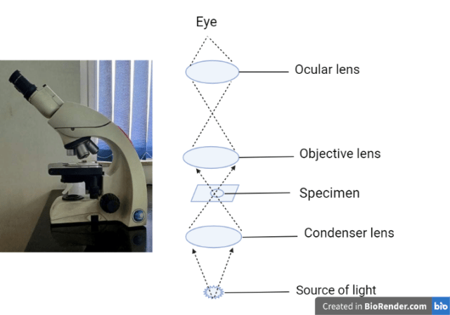 Figure: Working mechanism of Light microscope
For the full path light takes through every part of the microscope, illuminator, condenser, diaphragm, stage, objective, body tube, and eyepiece, see Parts of a Microscope and Their Functions.
Resolution and Numerical Aperture
Resolution or Resolving power is the ability of a lens to distinguish two adjacent points as distinct and separate. The shorter the wavelength of light used in the instrument, the greater the resolution. The white light (wavelength of 450-550 nm) used in a compound light microscope has a relatively long wavelength and cannot resolve structures smaller than about 0.2 µm.
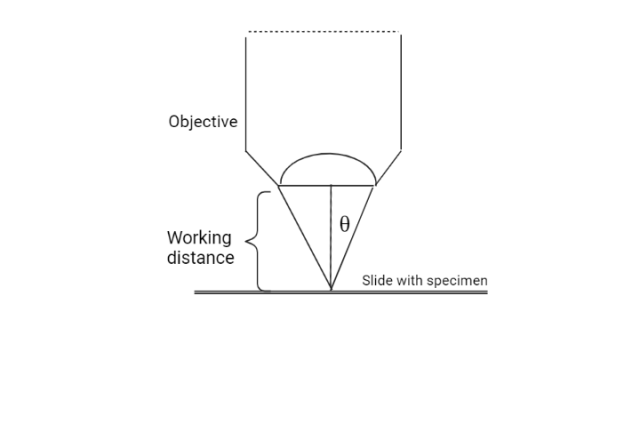
The numerical aperture (NA) is the widest cone of light that can enter the lens. The Abbe equation gives the minimum distance (d or resolution) between two objects that reveal them as separate entities.
 Lambda (λ): wavelength of light used to illuminate the specimen.
n sinθ: the numerical aperture (NA)
The angular aperture θ is ½the angle of the cone of light that enters a lens from a specimen.
d = λ / (2 × NA) = 0.5λ / (n sinθ), where n is refractive index
When d becomes smaller, the resolution increases, and finer detail can be identified in a specimen. To make d smaller, the value of λshould be smaller, and the value of NA should be greater. Thus the greatest resolution is obtained with the light of the shortest wavelength and an objective with the maximum NA.
This equation is the reason no amount of magnification can rescue a blurry image: d is a physical floor set by wavelength and numerical aperture alone. For a standard light microscope, that floor works out to roughly 0.2 μm, enough to resolve bacteria (1–10 μm), but nowhere near enough to resolve a virus (20–300 nm). Raising the numerical aperture, which is exactly what immersion oil does, is one of the only two ways to push that floor lower.
Oil Immersion in the 100X Objective
When a beam of light passes from air into a glass, it is bent towards the normal, and when it passes back from glass to air, it is bent far away from the normal. This has little effect on low power objectives, but with high power lenses, this bending limits the amount of light that can enter the lens and affects the objective’s NA and consequently its resolving power.
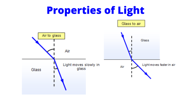 Figure: Properties of light
The bending effect and its limitations on the objective lens can be avoided by replacing the air between the specimen and the lens with an oil that has the same optical properties as glass, i.e., immersion oil.
Immersion oil is a colorless transparent liquid. Oil and glass slide has the same refractive index, i.e., 1.5. Light projected from the illuminator passes the oil without deviation and is projected upwards to the objective lens.
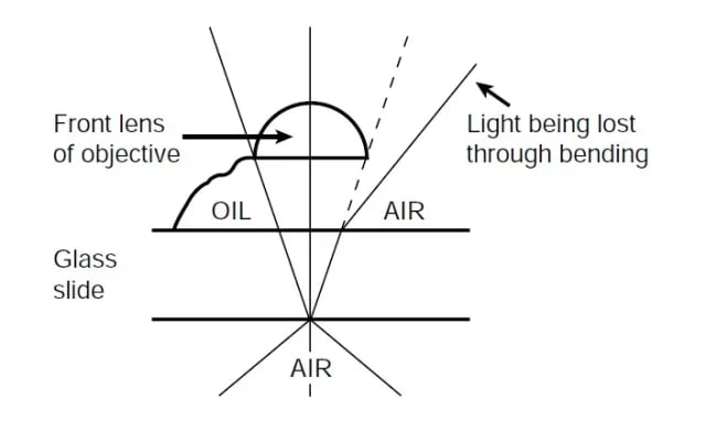 Figure: Working principle of an oil immersion objective
With immersion oil, the light passes in a straight line from glass through the oil and back to glass as though it were passing through glass all the way. Thus oil helps to collect extra oblique light and provides better resolution and a brighter image. Some 50X objectives and all 100X objectives are immersed in oil to observe objects as small as bacteria.
This is exactly what happened in the Gram stain example above. At 40X dry, light scattered by the bacteria at wide angles was lost at the air-glass interface before it could reach the objective, lowering the effective numerical aperture and, by the Abbe equation, raising the resolvable distance d past the point where individual bacteria could be distinguished. Oil immersion doesn't magnify anything further; it recovers that lost light, raises the numerical aperture back up, and lowers d enough to resolve the organism.
How to Remember
- What actually shrinks d: Only two things lower the resolvable distance in the Abbe equation, shorter wavelength (bluer light) or higher numerical aperture (a fatter cone of light entering the lens). Magnification isn't one of them. If a question describes "increasing magnification to see more detail" as the fix for a blurry image, that's the wrong answer, resolution has a physical floor magnification can't cross.
- Oil doesn't magnify, it rescues: Immersion oil doesn't add any magnifying power of its own. It matches glass's refractive index (1.515) so light stops bending and scattering at the slide-to-lens interface, recovering light that would otherwise miss the objective entirely. Think of it as recovering lost signal, not amplifying it.
- The counterintuitive direction of "better": In the Abbe equation, a smaller d means better resolution, closer together points can still be told apart. If a question phrases it as "higher resolution," check whether it means a smaller d value or a colloquial "sees more detail"; both point the same direction, but reading the equation with "smaller is better" firmly in mind avoids a common sign-reversal mistake.
- Only 100X (and some 50X) gets oil, never 40X or below: The bending effect that oil corrects is negligible at low power and becomes significant only at high power, which is why oil is reserved for the highest-magnification objectives and never applied to 40X or lower.
Key exam facts in one table
| Concept | Detail | Why it's tested |
|---|---|---|
| Abbe equation | d = λ / (2 × NA) | The formula connecting wavelength and numerical aperture to resolving power; a frequent short-answer or fill-in-the-blank item |
| What lowers d (improves resolution) | Shorter wavelength, higher numerical aperture | Magnification is deliberately absent from this list, the most commonly tested distinction in this topic |
| Light microscope resolving limit | ~0.2 μm, using white light (450–550 nm) | Explains why bacteria (1–10 μm) are visible but viruses (20–300 nm) are not, regardless of magnification |
| Refractive index of immersion oil and glass | Both 1.515 | The reason light passes from glass through oil back to glass without bending at either interface |
| Objectives requiring oil | 100X always; some 50X objectives | 40X and below are always used dry; using oil on them is a technique error, not an enhancement |
| Why oil improves the image | Recovers wide-angle light otherwise lost to refraction at the glass-air interface, raising effective NA | Not magnification, a common wrong answer on exams |
Where Students Get Confused
- Assuming more magnification fixes a blurry image. Past the resolution limit set by wavelength and numerical aperture, additional magnification only produces a bigger blur, not more detail. The Abbe equation has no magnification term in it for exactly this reason.
- Thinking immersion oil magnifies the specimen. It doesn't add magnifying power. It matches the refractive index of glass so that light which would otherwise scatter and miss the objective at wide angles is recovered, effectively raising the numerical aperture.
- Misreading which direction is "better" in the Abbe equation. A smaller value of d means finer resolution (two closer points can still be told apart), which can feel backwards if "resolution" is read as something that should get numerically bigger to improve.
- Applying oil to the wrong objective. Oil is used only with the 100X objective (and some 50X objectives), never with 40X or lower; the refraction effect oil corrects for is negligible at those lower powers, so oil there does nothing but cause a mess.
- Treating resolution and magnification as the same property. Magnification is how much bigger an image appears; resolution is whether two close points can still be told apart at all. A microscope can have very high magnification and still show nothing more than a large, blurry, uninformative image if its resolving power is exceeded.
References
Madigan, M. T., Martinko, J. M., Stahl, D. A., & Clark, D. P. (2011). BROCK Biology of Microorganisms (13thedition). Benjamin Cumming.
Prescott, L. M. (2002). Microbiology (5th edition). The McGraw-Hill Companies.
Frequently Asked Questions
Why can't increasing magnification reveal more detail once the resolution limit is reached?
Does immersion oil magnify the image at 100X?
What is the resolving power of a standard light microscope, and why does it matter?
Which objective lenses require immersion oil?
What is numerical aperture, and how does it relate to resolution?

Tankeshwar Acharya, MSc (Medical Microbiology)
Tankeshwar Acharya is an Assistant Professor in the Department of Microbiology at Patan Academy of Health Sciences (PAHS), Nepal, where he has been teaching and practicing clinical microbiology for over 14 years. He is the founder of Microbe Online, one of the leading free microbiology education resources on the web, covering bacteriology, mycology, parasitology, immunology, and clinical laboratory diagnostics written from direct experience in both the classroom and the diagnostic laboratory.