
Types of Microscopes: Classification, Comparison, and Clinical Uses
A complete guide to types of microscopes — bright-field, dark-field, phase-contrast, fluorescence, confocal, electron, and more. Includes a master comparison table, resolution limits, and clinical microbiology applications.
A microscope is an optical or electronic instrument that produces magnified images of objects too small to be seen with the naked eye. Microscopes are among the most important tools in biology, medicine, and materials science; enabling discoveries from the identification of bacteria by Antonie van Leeuwenhoek in 1676 to the visualisation of individual atoms by modern scanning probe microscopes.
Choosing the right type of microscope depends on three questions:
- What is the size of the object? (bacteria at 1–10 μm require a different instrument than viruses at 20–300 nm)
- Does the specimen need to be alive or fixed? (electron microscopes require a vacuum; living specimens cannot be examined)
- What information is needed? (surface structure, internal structure, chemical composition, or motility?)
Classification of Microscopes
Microscopes are classified into three major groups based on their illumination source:
Microscopes
├── Light (Optical) Microscopes
│   ├── Simple microscope
│   └── Compound microscope
│       ├── Bright-field microscope (standard)
│       ├── Dark-field microscope
│       ├── Phase-contrast microscope
│       ├── Fluorescence microscope
│       ├── Confocal microscope
│       ├── Inverted microscope
│       ├── Polarizing microscope
│       └── Stereo (dissecting) microscope
├── Electron Microscopes
│   ├── Transmission electron microscope (TEM)
│   └── Scanning electron microscope (SEM)
└── Scanning Probe Microscopes
    ├── Atomic force microscope (AFM)
    ├── Scanning tunnelling microscope (STM)
    └── Magnetic force microscope (MFM)
Master Comparison Table
| Microscope type | Resolution | Max magnification | Living specimens | Image | Primary use | Clinical microbiology use |
|---|---|---|---|---|---|---|
| Bright-field | ~0.2 μm | ~2,000× | Yes (unstained) / No (stained) | Colour (stained) | Routine lab work | Gram stains, blood films, wet preparations |
| Dark-field | ~0.2 μm | ~1,000× | Yes | Bright on dark background | Motile organisms, unstained spirochetes | Treponema pallidum detection in syphilis chancre fluid |
| Phase-contrast | ~0.2 μm | ~1,500× | Yes | Contrast without staining | Living cell observation | Motility studies, endospore and inclusion body examination |
| Fluorescence | ~0.2 μm | ~1,000× | Yes/No | Fluorescent bright on dark | Pathogen identification, immunofluorescence | Auramine-rhodamine for TB; DFA for Legionella, Chlamydia, Bordetella |
| Confocal | ~0.2 μm | ~1,500× | Yes | 3D optical sections | Cell biology, biofilm imaging | Research — biofilm structure, intracellular pathogens |
| Inverted | ~0.2 μm | ~1,500× | Yes | Colour (stained) | Cell culture observation | Mycoplasma detection in cell cultures |
| Polarizing | ~0.2 μm | ~1,000× | No | Birefringent objects bright | Crystals, fibres, parasites | Schistosome eggs, helminth larvae identification |
| Stereo (dissecting) | ~10 μm | 5–45× | Yes | 3D, colour | Gross dissection, macroscopic | Macroscopic parasite identification, colony morphology |
| TEM | ~0.2 nm | ~1,000,000× | No | Black and white | Ultrastructure, viruses, molecules | Research — virus morphology, cell ultrastructure |
| SEM | ~1–20 nm | ~100,000× | No | 3D black and white (surface) | Surface topology | Research — biofilm surface, tick identification, parasite morphology |
| AFM/STM/SPM | ~0.1 nm | Atomic | No | 3D surface maps | Atomic/molecular surface | Research — DNA, protein structure |
Light Microscopes
Light (optical) microscopes use visible light — or in the case of fluorescence microscopes, ultraviolet or blue light — as the illumination source. They use glass lenses to focus and magnify the image. The maximum resolution of a light microscope is approximately 0.2 μm (200 nm), which is sufficient to visualise bacteria (1–10 μm), fungi (2–200 μm), most parasites, and all mammalian cells, but insufficient for viruses (20–300 nm).
Simple Microscope
A simple microscope uses a single magnifying lens — essentially an advanced magnifying glass. It uses natural or reflected light and has no condenser. Maximum magnification is approximately 10–300×. Simple microscopes are used in basic educational settings, field botany, and gemology. They have no role in diagnostic microbiology.
Compound Microscope
The compound microscope is the standard instrument in clinical and research microbiology laboratories. It uses two sets of lenses — the objective lens (close to the specimen) and the ocular lens or eyepiece (close to the eye) — to produce magnifications of up to 2,000×. The total magnification is calculated by multiplying the eyepiece magnification (usually 10×) by the objective magnification (4×, 10×, 40×, or 100×).
The compound microscope exists in several variants, each designed for specific applications:
1. Bright-Field Microscope
The bright-field microscope is the most widely used microscope in diagnostic and teaching laboratories worldwide. Light passes directly through the specimen — the image appears dark against a brighter background (hence "bright-field"). Unstained biological specimens have little contrast and are difficult to visualise; most clinical applications therefore require staining.
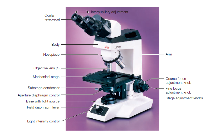 Figure: A bright-field microscope
Key specifications:
- Resolution: ~0.2 μm
- Maximum useful magnification: ~1,000–2,000×
- Light source: tungsten-halogen lamp (white light) or LED
- Specimens: fixed and stained (gram stain, Ziehl-Neelsen, Giemsa) or unstained wet preparations
Clinical applications:
- Gram-stained smears for bacterial morphology and arrangement
- Ziehl-Neelsen acid-fast stained smears for Mycobacterium tuberculosis
- Giemsa-stained blood films for malaria parasites
- Wet preparations for intestinal parasites
- Urine microscopy for cells, casts, and bacteria
- Blood cell differential counts
→ Parts of a Microscope and Their Functions
→ Working Mechanism of the Light Microscope
2. Dark-Field Microscope
The dark-field microscope uses a specialised dark-field condenser containing an opaque disc that blocks all direct light from reaching the objective lens. Only light that is reflected or refracted (scattered) by the specimen enters the objective. The result is a bright image of the specimen against a completely dark background — the reverse of the bright-field image.
Key advantage: Organisms too transparent or too thin to visualise by bright-field microscopy become clearly visible as bright, self-luminous objects against the dark background. No staining is required.
Key specifications:
- Resolution: ~0.2 μm
- Specimen: wet, unstained, living or fresh
- Cannot be used with oil immersion condenser unless a special oil-immersion dark-field condenser is available
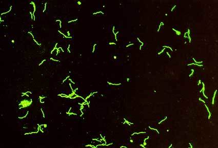 Figure: Positive FTA-Abs test result showing Treponema pallidum coated with host anti-treponemal antibodies.
Clinical applications:
- Detection of Treponema pallidum in chancre fluid from primary syphilis lesions; the spirochetes appear as bright, corkscrew-shaped, motile organisms against the dark background. This remains the most sensitive test for primary syphilis before antibodies develop
- Detection of Leptospira interrogans in urine or blood during acute leptospirosis
- Detection of Borrelia spp. in blood during febrile episodes of relapsing fever
- Examination of motile organisms in fresh preparations
→ Dark-Field Microscopy: Principle, Procedure, Uses
→ Demonstration of Treponema pallidum using Dark-Field Microscopy
3. Phase-Contrast Microscope
Living cells and microorganisms are largely transparent — they have very similar refractive indices to the surrounding medium, making them nearly invisible in bright-field microscopy without staining. The phase-contrast microscope exploits subtle differences in the refractive index and thickness of different cellular structures to produce contrast without staining.
When light passes through structures of different refractive indices, it changes phase (the timing of its wave cycle). The phase-contrast microscope uses a special phase plate in the objective and an annular diaphragm in the condenser to convert these invisible phase differences into visible differences in brightness. Structures with higher refractive index appear darker; those with lower refractive index appear brighter — all without any staining.
Key advantage: Living cells can be observed in their natural state without fixation or staining — biological processes, motility, and cell division can be followed in real time.
Clinical applications:
- Observation of bacterial motility without staining
- Visualisation of bacterial endospores and intracellular inclusion bodies (polyhydroxybutyrate, polyphosphate, sulfur granules)
- Examination of living protozoa and fungi
- Observation of living mammalian cells in tissue culture
→ Phase-Contrast Microscope: Principle, Parts, Applications
4. Fluorescence Microscope
The fluorescence microscope uses ultraviolet (UV) or short-wavelength visible light (blue light, wavelength 330–500 nm) as its illumination source. When this high-energy light strikes a fluorochrome (fluorescent dye) attached to the specimen, the fluorochrome absorbs the short-wavelength light and immediately re-emits light of a longer wavelength — producing visible fluorescence.
A barrier filter between the objective and the eyepiece blocks the excitation light while allowing only the emitted fluorescent light to pass through. The result is a brightly fluorescent specimen against a completely dark background — achieving excellent contrast and sensitivity.
Two types of fluorescent labelling:
- Direct fluorescence — the fluorochrome is attached directly to the target organism or molecule
- Indirect fluorescence (immunofluorescence) — a fluorochrome-labelled antibody is used to detect a specific antigen. Direct fluorescent antibody (DFA) and indirect fluorescent antibody (IFA) tests are widely used in clinical diagnostics
Key specifications:
- Light source: mercury vapour lamp, xenon lamp, or LED
- Resolution: ~0.2 μm (same as bright-field, but much higher sensitivity for specific targets)
- Requires: fluorochrome stains or fluorescent-labelled antibodies
Clinical applications:
- Auramine-rhodamine staining for Mycobacterium tuberculosis in sputum — more sensitive than Ziehl-Neelsen (ZN) staining; positive organisms fluoresce bright yellow-orange against a dark background
- Direct fluorescent antibody (DFA) tests for Legionella pneumophila, Chlamydia trachomatis, Bordetella pertussis, Rabies virus (brain impression smear)
- Acridine orange staining for detecting bacteria and fungi in blood culture bottles and clinical specimens
- Calcofluor white staining for fungi — cell walls of fungi fluoresce bright blue-white, allowing rapid detection of fungal elements in clinical specimens
- FITC-labelled antibodies in immunofluorescence assays (IFA) for virus antibody detection
- Distinction of live bacteria (green fluorescence) from dead bacteria (red fluorescence) using LIVE/DEAD staining kits
→ Fluorescence Microscope: Principle, Types, Applications
5. Confocal Microscope
The confocal microscope is an advanced fluorescence microscope that uses point illumination (a focused laser beam) and a pinhole aperture in front of the detector to eliminate out-of-focus fluorescent light from above and below the focal plane. Only light from the precise focal plane of interest reaches the detector.
By systematically scanning the laser across the specimen and collecting images at multiple focal depths, a confocal microscope can produce a series of optical sections that are reconstructed computationally into a three-dimensional image of the specimen.
Key advantage over standard fluorescence: Eliminates background blur from out-of-focus structures, producing sharper images with better contrast — and allows true 3D reconstruction of cells and tissues.
Applications:
- Imaging biofilm architecture in three dimensions
- Studying intracellular pathogen localisation (e.g. Chlamydia inclusions, Mycobacterium survival within macrophages)
- Cell biology research — tracking protein localisation, cell signalling
- Not used in routine diagnostic microbiology — primarily a research tool
6. Inverted Microscope
The inverted microscope has its objectives positioned below the stage rather than above it. The light source and condenser are above the stage. This reversed configuration allows specimens to be examined from underneath in their containers (petri dishes, flasks, multiwell plates) without disturbing or removing them.
Key advantage: Cells growing on the bottom of tissue culture dishes or flasks can be observed without removing the container lid, maintaining sterility and allowing long-term observation.
Applications:
- Monitoring cell culture growth and morphology
- Detection of cytopathic effects (CPE) in viral cultures
- Detection of Mycoplasma contamination in cell cultures
- Tissue culture work in diagnostic virology and research laboratories
7. Polarizing Microscope
The polarizing microscope incorporates two polarising filters — a polariser below the specimen and an analyser above it. Light is polarised (restricted to vibrating in one plane) before it reaches the specimen. When it passes through birefringent (double-refracting) materials — substances with different refractive indices in different orientations; the polarisation is altered and these structures appear brightly illuminated between crossed polarisers.
Applications in microbiology and medicine:
- Identification of helminth eggs (Schistosoma, Ascaris): shells are birefringent
- Identification of asbestos fibres and silica crystals in lung specimens
- Examination of gout crystals (monosodium urate needle-shaped, negatively birefringent) and pseudogout crystals (calcium pyrophosphate, rhomboid, positively birefringent) in joint fluid
- Examination of birefringent granules in fungi (Sporothrix asteroid bodies)
- Identification of some antiparasitic drug crystals in urine
8. Stereo Microscope (Dissecting Microscope)
The stereo microscope (also called the dissecting microscope) is a low-magnification optical microscope designed to provide a three-dimensional view of specimens by using two separate optical paths angled slightly apart — one for each eye. It uses light reflected from the surface of an object rather than transmitted through it.
Key specifications:
- Magnification: 5–45× (some models up to 250×)
- Resolution: ~10 μm (insufficient for bacteria or fine cellular detail)
- Three-dimensional image (allows depth perception)
- Specimens can be large, intact, and non-transparent
Applications:
- Macroscopic examination of parasites (worms, ectoparasites, arthropods)
- Examination of colony morphology on agar plates at low magnification
- Dissection of biological specimens
- Microsurgery and ophthalmic surgery
- Watchmaking, circuit board assembly and inspection
- Entomology and archaeology
Electron Microscopes
Electron microscopes use a beam of electrons instead of light as the illumination source. Since electrons have wavelengths approximately 100,000 times shorter than visible light (~0.005 nm vs ~500 nm), the resolving power of electron microscopes is dramatically superior to light microscopes approximately 0.2 nm for TEM, compared to 0.2 μm for light microscopes. This is a 1,000-fold improvement in resolution.
Critical limitation: Electron microscopes require a vacuum because electrons are scattered by air molecules. Living specimens cannot be examined. All specimens must be fixed, dehydrated, and specially prepared a process that takes hours to days. Electron microscopes are expensive (US$100,000–$10,000,000), require specialist operation and maintenance, and are not used in routine diagnostic microbiology.
9. Transmission Electron Microscope (TEM)
In TEM, a beam of electrons is transmitted through an ultra-thin specimen (20–60 nm thick sections). Different regions of the specimen scatter electrons to different degrees depending on their density and composition — dense structures scatter more electrons and appear darker in the image; less dense structures appear lighter.
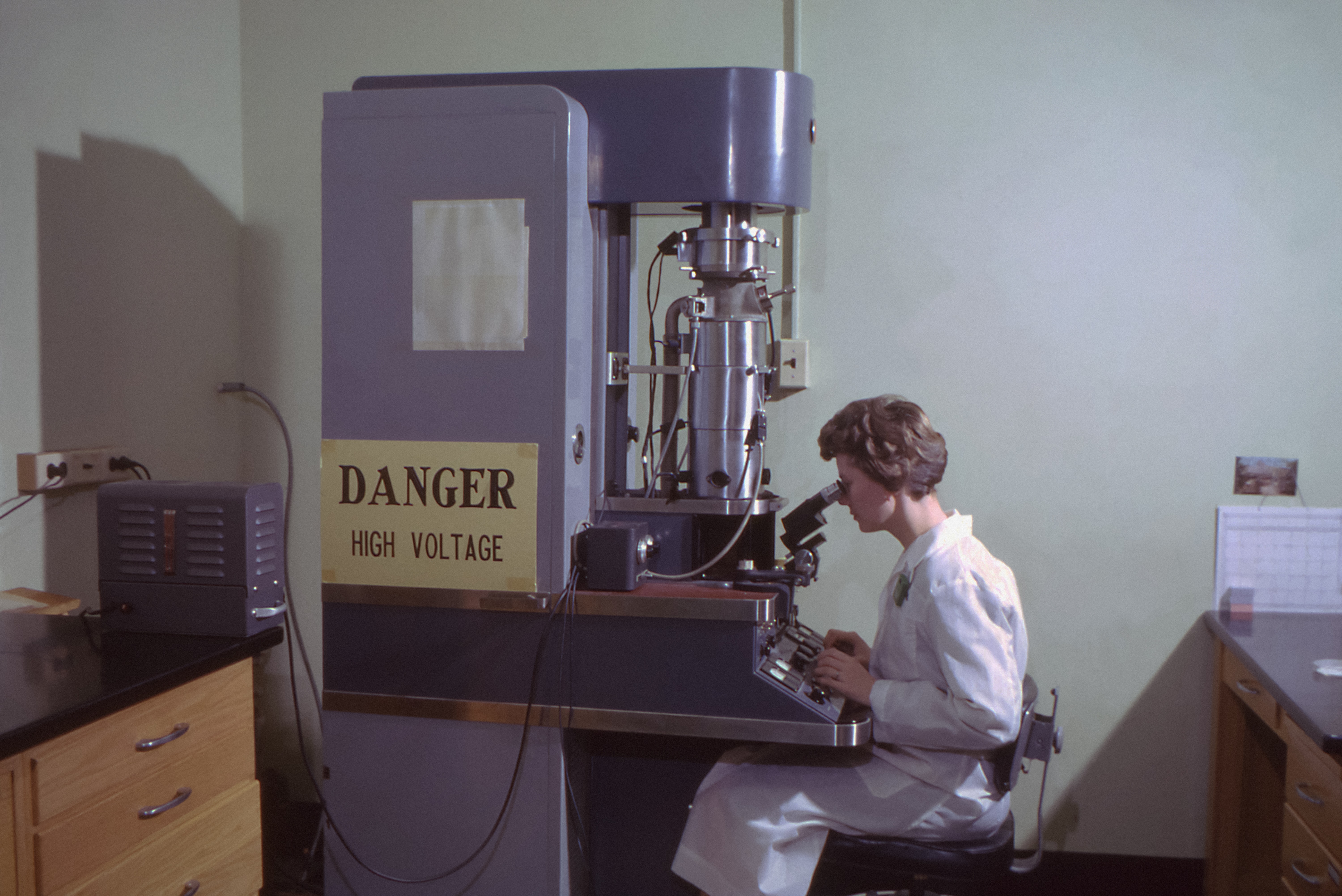 Figure: Transmission electron microscope
Key specifications:
- Resolution: ~0.2 nm
- Maximum magnification: ~1,000,000×
- Specimen preparation: ultra-thin sectioning, heavy metal staining (osmium, uranium, lead)
- Image: black and white (false colour added artificially)
- Reveals internal ultrastructure
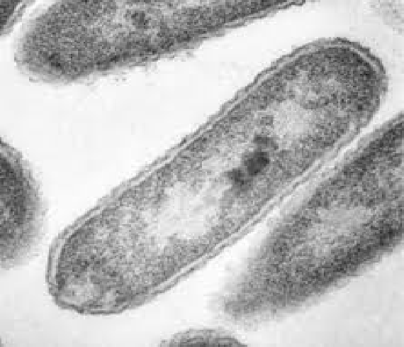 Figure: Rod-shaped bacteria as seen by Transmission Electron Microscope (TEM).
Applications:
- Visualisation and morphological identification of viruses — critical in outbreak investigation before molecular methods are available
- Examination of cell ultrastructure — mitochondria, ribosomes, nuclear membranes
- Visualisation of individual protein and nucleic acid molecules
- Negative staining technique for rapid virus identification in clinical specimens (e.g. rotavirus wheel-shaped particles in diarrhoea outbreaks)
→ Electron Microscope: Principle, Types, Applications
→ Differences between SEM and TEM
10. Scanning Electron Microscope (SEM)
In SEM, a narrow electron beam scans back and forth across the surface of a specimen coated with a thin film of a heavy metal (typically gold or platinum). The specimen does not need to be sectioned. Electrons scattered from the metal coating are collected and used to build a three-dimensional image of the specimen's surface.
Key specifications:
- Resolution: ~1–20 nm
- Magnification: 15× to 100,000×
- Specimen preparation: fixation, dehydration, metal coating; specimen is destroyed in the process
- Image: three-dimensional surface image, black and white (false colour commonly added)
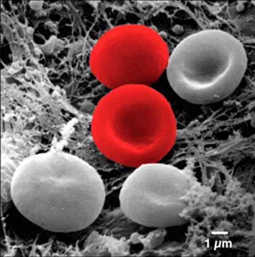 Figure: Image of RBCs obtained by SEM after artificial coloring. Images provided by the SEM are black and white.
Applications:
- Examination of the three-dimensional surface morphology of microorganisms, cells, and tissues
- Characterisation of biofilm architecture and surface colonisation
- Identification of ectoparasites (ticks, mites, lice) and arthropod vectors
- Examination of the external surface features of parasites and eggs
- Research — investigation of host-pathogen surface interactions
- Materials science — examination of metal surfaces, crystals, and nanostructures
Scanning Probe Microscopes
Scanning probe microscopes (SPMs) work on an entirely different principle from both light and electron microscopes. A physical probe with an extremely sharp tip (often just a single atom wide) is scanned across the specimen surface at atomic proximity. The interaction between the probe tip and the specimen surface; electrostatic force, magnetic force, quantum tunnelling current, or physical contact is measured and converted into a surface map.
Key advantage: Achieves atomic-level resolution can image individual atoms and molecules. Does not require a vacuum for some types.
Key limitation: Can only map surfaces no internal structure is revealed. Very slow scanning speed. Not used in biology or clinical medicine except in research.
Types of scanning probe microscopes
Atomic Force Microscope (AFM): The probe tip physically contacts or approaches the specimen surface. The bending (deflection) of the cantilever arm holding the tip is measured using a laser. AFM can image biological molecules (DNA, proteins, cell membranes) in near-physiological aqueous conditions. It is unique among high-resolution microscopes in not requiring vacuum or heavy metal staining.
Scanning Tunnelling Microscope (STM): Measures the quantum tunnelling current between the probe and a conducting specimen surface under a known voltage. Achieves true atomic resolution — individual atoms can be imaged and even manipulated. Restricted to conducting surfaces.
Magnetic Force Microscope (MFM): Uses a magnetised probe tip to map magnetic forces on the specimen surface. Used in materials science for magnetic storage media and magnetic nanoparticle research.
Clinical Uses Quick Reference
Which microscope is used for which diagnostic application?
| Clinical situation | Microscope type | Key stain/technique |
|---|---|---|
| Gram stain of clinical specimen | Bright-field | Crystal violet, iodine, safranin |
| Suspected pulmonary tuberculosis | Fluorescence (preferred) or Bright-field | Auramine-rhodamine (fluorescence) or Ziehl-Neelsen (bright-field) |
| Suspected syphilis — primary chancre | Dark-field | Fresh, unstained — no stain needed |
| Malaria diagnosis — peripheral blood | Bright-field | Giemsa thick and thin blood smear |
| Intestinal parasite examination | Bright-field | Saline/iodine wet preparation |
| Cryptococcus in CSF | Bright-field | India ink (capsule detection) |
| Suspected fungal infection in tissue/BAL | Fluorescence | Calcofluor white |
| Suspected Legionella in respiratory specimen | Fluorescence | Direct fluorescent antibody (DFA) |
| Bacterial motility | Phase-contrast or Dark-field | Wet preparation, no stain |
| Virus identification — outbreak investigation | TEM | Negative staining |
| Biofilm structure in research | Confocal | Fluorescent dyes |
| Macroparasite identification | Stereo (dissecting) | No stain required |
| Cell culture monitoring | Inverted | Unstained, transmitted light |
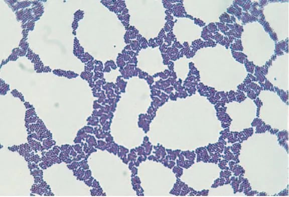 Figure: Gram positive cocci in clusters
Portable Microscopes
Modern advances in optical engineering have produced compact, portable microscopes suitable for field use, point-of-care testing, and resource-limited settings:
Pocket microscope: Battery-operated, magnification 20–250×. Fits in a pocket. Useful for preliminary field examination but insufficient magnification for bacteria. Suitable for examining large parasites, crystals, and surface structures.
Read more: Pocket Microscope: Parts, Working Principle, and Uses
Handheld digital microscope (USB microscope): Connects to a smartphone or computer via USB or Bluetooth. LED-illuminated. Images are viewed on a screen rather than through an eyepiece, making them accessible to multiple viewers simultaneously. Used in education, quality control, forensic science, and field biology.
Read more: Handheld Digital Microscope: Parts, Principle, and Uses
Foldscope: A paper-based microscope costing less than one dollar to manufacture, designed by researchers at Stanford University. Uses a single ball lens and LED for illumination. Achieves magnification of 2,000× with a resolution of 2 μm; sufficient to visualise malaria parasites and other microorganisms. Developed specifically for low-resource settings in developing countries.
References and Further Reading
- Madigan, M. T., Bender, K. S., Buckley, D. H., Sattley, W. M., & Stahl, D. A. (2018). Brock Biology of Microorganisms (15th ed.). Pearson.
- Tille, P. M. (2017). Bailey & Scott's Diagnostic Microbiology (14th ed.). Mosby Elsevier.
- Murphy, D. B., & Davidson, M. W. (2013). Fundamentals of Light Microscopy and Electronic Imaging (2nd ed.). Wiley-Blackwell.
- Abramowitz, M., & Davidson, M. W. Introduction to Microscopy. Olympus Life Science Microscopy Resource Center. Retrieved from https://www.olympus-lifescience.com/en/microscope-resource/
- World Health Organization. (2014). Fluorescence Microscopy for Tuberculosis: Technical Guide. WHO Press.
- Garcia, L. S. (Ed.). (2016). Clinical Microbiology Procedures Handbook (4th ed.). ASM Press.
Frequently Asked Questions
What is the most commonly used microscope in clinical microbiology?
Why can viruses not be seen with a light microscope?
What is the difference between TEM and SEM?
What is the advantage of fluorescence microscopy for TB diagnosis?
What is the difference between dark-field and phase-contrast microscopy?
What does numerical aperture (NA) mean?
What is Köhler illumination?
Which microscope is used to diagnose syphilis in a primary chancre?
: Is a higher magnification microscope always the better choice?
What's the difference in appearance between bright-field and dark-field or fluorescence microscopy?

Tankeshwar Acharya, MSc (Medical Microbiology)
Tankeshwar Acharya is an Assistant Professor in the Department of Microbiology at Patan Academy of Health Sciences (PAHS), Nepal, where he has been teaching and practicing clinical microbiology for over 14 years. He is the founder of Microbe Online, one of the leading free microbiology education resources on the web, covering bacteriology, mycology, parasitology, immunology, and clinical laboratory diagnostics written from direct experience in both the classroom and the diagnostic laboratory.