
Isolation and Enumeration of Bacteria: Techniques and Clinical Significance
How bacteria are isolated as pure colonies and enumerated as CFU/mL, and why both steps matter for diagnosing infection, food safety, and water testing.
A midstream urine sample arrives in the lab from a patient with dysuria and frequency. The clinician needs two different answers from this one specimen, and neither comes from the same step.
First: which organism is causing this? For that, the lab needs a single, pure, well-separated colony it can Gram stain, subculture, and run through identification and susceptibility testing. Mixed growth is useless for this. This is isolation.
Second: how much of it is actually there? A few stray colonies from skin or perineal contamination during collection can look identical to a genuine bladder infection on the plate. The number that separates the two is roughly 10⁵ colony-forming units per mL, the classic Kass threshold still used in labs today. Getting that number requires counting colonies against a known dilution. This is enumeration.
Same specimen, same agar plate in some techniques, but two different questions and two different technique families. Confuse them, and either you never isolate a pure colony to identify, or you never get a number, you just get "growth" or "no growth." Both halves are needed to turn a urine sample into a diagnosis.
Why Isolate and Enumerate Bacteria?
Isolation and enumeration answer two separate but equally practical questions:
- Isolation asks: what is this organism? It separates one species from a mixed population (a clinical specimen, soil, food, water) into a pure colony that can be identified, stained, and tested.
- Enumeration asks: how many are there? It quantifies bacterial load, as either a total count (living and dead) or a viable count (only cells capable of forming a colony), and expresses it as CFU/mL or cells/mL.
Clinically, diagnostic microbiology, food and water safety testing, and pharmaceutical quality control all depend on this pairing. A urine or wound culture needs isolation to name the pathogen and enumeration to judge whether the count is clinically significant or represents contamination. Water and food testing use enumeration methods almost exclusively (MPN, membrane filtration) because the regulatory question is a number against a safety limit, not species identity.
Enumeration of microorganisms is especially important in dairy microbiology, food microbiology, pharmaceutical microbiology, and water microbiology. The number of microorganisms in a culture, sample, or specimen is measured to assess the levels of microbial contamination in raw material or manufactured products (such as medicine). Enumeration is also done to evaluate the effects of antimicrobial agents or the decontamination processes.
Techniques of Isolation
The primary technique for isolating a pure bacterial colony from a mixed specimen is the streak plate technique. It works by progressively thinning out a mixed sample across the surface of an agar plate in a series of streaks, so that by the final streaks, individual cells land far enough apart to grow into separate, well-isolated colonies.
Streak plate isolation is qualitative, not quantitative. It answers "what organisms are present and can I get a pure colony of each?" not "how many organisms were in the original sample?" For the full stepwise procedure, quadrant method, and interpretation of isolated colonies, see the dedicated Streak Plate Technique article.
Techniques of Enumeration
In some cases, it is necessary to know the total number of (both living and dead) microorganisms. For example, in endotoxin (pyrogen) testing, dead and alive cells induce fever when injected into the body. However, in many cases, the number or concentration of living cells is required.
Traditional and rapid methods of enumerating microorganisms
| Viable counts (traditional method) | Total counts (traditional method) | Rapid methods (indirect viable counts) |
|---|---|---|
| Pour plate method | Direct microscopic counting (using Helber or haemocytometer counting chambers) | Epifluorescence often coupled with image analysis |
| Surface spread or spread plate method | Turbidity methods | ATP testing |
| Membrane filter method | Dry weight determination | Impedance method |
| MPN Method | Nitrogen, protein, or nucleic acid determinations | Manometric methods |
Total Count Technique
Total count is a counting procedure enumerating both living and dead cells.
Direct Microscopic count
Direct microscopic counts are possible using special slides known as counting chambers, consisting of a ruled slide and a coverslip. It is constructed so that the coverslip, slide, and ruled lines delimit a known volume. The number of bacteria in a small known volume is microscopically counted, and the number of bacteria in the larger original sample is determined by extrapolation. Dead cells cannot be distinguished from living ones. Only dense suspensions can be counted.

Bacteria can be counted efficiently and accurately with the Petroff-Hausser counting chamber. This is a special slide accurately ruled into squares that are 1/400 mm² in the area; a glass coverslip rests 1/50 mm above the slide so that the volume over a square is 1/20,000 mm² i.e. 1/20, 000, 000 cm². If, for example, an average of five bacteria is present in each ruled square, there are 5 x 20,000,000 or 10^8 bacteria per milliliter. A suspension of unstained bacteria can be counted in the chamber using a phase-contrast microscope.
The formula used for the direct microscopic count is: The number of bacteria per cc = The average numbers of bacteria per large double-lined square X The dilution factors of the large square (1,250,000) X The dilution factor of any dilutions made before placing the sample in the counting chamber, e.g., mixing the bacteria with dye
Advantages of Direct Microscopic count
- Rapid, simple, and easy method requiring minimum equipment.
- Morphology of the bacteria can be observed as they are counted.
- Very dense suspensions can be counted if they are diluted appropriately.
Limitations of Direct Microscopic count
- Dead cells are not distinguished from living cells.
- Small cells are difficult to see under the microscope, and some cells are probably missed.
- Precision is difficult to achieve
- A phase-contrast microscope is required when the sample is not stained.
- The method is not usually suitable for cell suspensions of low density i.e., < 107 Cells per ml, but samples can be concentrated by centrifugation or filtration to increase sensitivity.
Turbidity Method
Turbidity measurements are the most common means of estimating the total number of bacteria present in a sample. Measuring the turbidity using a spectrophotometer or colorimeter and reading the concentration from a calibration plot is a simple means of standardizing cell suspensions for inocula in antibiotic assays or other tests of antimicrobial chemicals.
Viable Count Technique
Viable count records living cells alone. The viable count is more common than the total count. A viable cell is defined as one that can divide and form offspring. Most pharmacopeias and regulatory agencies use a total viable count (TVC), i.e., a viable count that records all the different species or types of microorganisms (e.g., bacteria plus fungi) present in the sample
The usual way to perform a viable count is to determine the number of cells in the sample capable of forming colonies on a suitable agar medium. For this reason, the viable count is often called the plate count or colony count. This method is used to enumerate bacteria in milk, water, foods, soils, cultures, etc., and the number of bacteria is expressed as colony-forming units (CFU) per ml.

With both the spread plate and pour plate method, it is essential that the number of colonies developing on the plates not be too many because, on crowded plates, some cells may not form colonies, and some colonies may fuse, leading to erroneous measurements. It is also essential that the number of colonies not be too few, or the statistical significance of the calculated count will be low. The usual practice, which is the most valid statistically, is to count colonies only on plates that have between 30 and 300 colonies.
The number of bacteria in a given sample may usually be too many to be counted (TMTC). The sample must be diluted to obtain the appropriate colony number so that visible isolated colonies appear. The number of colonies can be used to measure the number of viable cells in that known dilution. Several 10-fold dilutions of the sample are commonly used. Usually, serial dilutions are employed to reach the final desired dilution.
However, if the organism typically forms multiple cell arrangements, such as chains, the colony-forming unit may consist of a chain of bacteria rather than a single bacterium. In addition, some of the bacteria may be clumped together. The development of one colony can occur even when the cells are in aggregates. i.e., cocci in clusters (staphylococci), chains (streptococci), or pairs (diplococci). In such cases, the resulting counts will be lower than the number of individual cells.
Each colony that can be counted is called a colony-forming unit (CFU).By extrapolation, this number can be used to calculate the number of CFUs in the original sample rather than the number of bacteria per milliliter. The assumption made in this type of counting procedure is that each viable cell can yield one colony. There are two ways of performing a plate count: the spread plate method and the pour plate method.
Generally, one wants to determine the number of CFUs per milliliter (ml) sample. To find this, the number of colonies (on a plate having 30-300 colonies) is multiplied by the number of times the original ml of bacteria was diluted (the dilution factor of the plate counted).
For example, suppose a plate containing a 1/1,000,000 dilution of the original ml of the sample shows 150 colonies. In that case, the number of CFUs per ml in the original sample is found by multiplying 150 x 1,000,000, as shown in the formula below:
The number of CFUs per ml of sample = The number of colonies (30-300 plate) X The dilution factor of the plate counted
In the case of the example above 150 x 1,000,000 = 150,000,000 CFUs per ml
Advantage of viable count method
This method is used routinely and with satisfactory results for estimating bacterial populations in milk, water, foods, and many other materials.
- It is easy to perform and can be adapted to measuring populations of any magnitude.
- It is a sensitive method(theoretically, a single viable cell can be detected). For example, if a specimen contains as few as one bacterium per ml, one colony should develop upon the plating of 1 ml.
- It allows for inspection and identification of the organism counted.
Limitation of viable count technique
- Only living cells develop colonies
- Clumps or chains of cells develop into a single colony
- Colonies develop only from those organisms for which the cultural conditions are suitable for growth.
Pour Plate Technique
In the pour plate method, a known volume of diluted sample is mixed into molten agar (cooled to ~45°C) before it sets, so colonies form throughout the depth of the medium rather than only on the surface. This tolerates a larger sample volume than spread plating but exposes the organism briefly to 45°C, so heat-sensitive organisms are better suited to spread plating.
Full procedure, advantages, disadvantages: see dedicated Pour Plate article

Spread Plate Technique
In the spread plate technique, a small volume (≤0.1 mL) of diluted sample is spread across the surface of an already-solidified agar plate using a sterile spreader. Because the sample never touches molten agar, this avoids the 45°C exposure of the pour plate method, at the cost of a smaller usable sample volume.

Full procedure and notes: see dedicated spread plate technique article
Membrane Filter Method
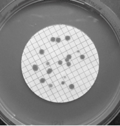 Figure: Membrane filtration technique
A large known volume of sample is passed through the membrane, and then the membrane is placed, without inversion, on the agar surface. Nutrients diffuse through the membrane and allow retained cells to grow into colonies. Membrane filtration detects lower concentrations of microorganisms than other methods, but it can not be used to test microbial loads of viscous samples.
Find more about membrane filtration techniques in this post.
Most Probable Number (MPN) Method
Most probable number (MPN) counts may be used when the anticipated count is relatively low, i.e., from <1 to 100 microorganisms per ml. The procedure involves inoculating multiple tubes of culture medium, usually three or five, with three different volumes of the sample (0.1 ml, 0.001 ml, and 0.001 ml).
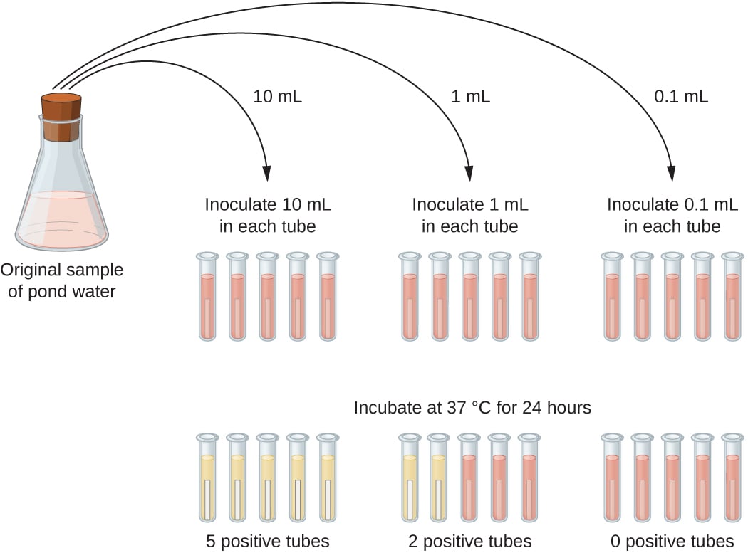 Figure: MPN Technique
The proportions of positive tubes are recorded for each sample volume, and the results are compared with standard tables showing the MPN of organisms per ml of the original sample. MPN technique is commonly used in the water, food, and dairy industries.
Isolation vs. Enumeration: Which Technique Answers Which Question?
| Question being asked | Technique family | Example techniques |
|---|---|---|
| What organism is this? (need a pure colony) | Isolation | Streak plate |
| How many organisms per mL? (moderate-to-high expected count) | Enumeration, direct plating | Pour plate, spread plate |
| How many organisms per mL? (low expected count, <100/mL) | Enumeration, statistical estimate | MPN |
| How many organisms in a large volume of dilute sample? (e.g., water testing) | Enumeration, concentration-based | Membrane filtration |
| How many total cells, dead or alive? | Enumeration, total count | Direct microscopic count, turbidity |
Rapid Methods
Rapid methods enumerate viable microorganisms, usually bacteria and yeasts, within a matter of hours and eliminate traditional methods’ 24-48 hour (or longer) incubation periods. They employ various means for indirect detection of living cells.
Rapid methods are fast, readily automated, and eliminate the need for numerous Petri dishes and incubators but may require the purchase of expensive equipment. Detection limits may vary based on the equipment types, test types, and manufacturers.
Epifluorescent Technique
It uses fluorescent dyes that either exhibit different colors in living and dead cells (e.g., acridine orange) or appear colorless outside the cell but become fluorescent when absorbed and subjected to cellular metabolism (e.g., fluorescein diacetate).
ATP Testing
Living cells generate adenosine triphosphate (ATP) that can readily be detected by enzyme assays, e.g., luciferin emits light when exposed to firefly luciferase in the presence of ATP; light emission can be measured and related to bacterial concentration.
Read details about ATP Testing
Impedance Technique
The resistance, capacitance, or impedance of a culture medium changes due to bacterial or yeast growth and metabolism, and these electrical properties vary in proportion to cell concentration.
Manometric Technique
Manometric techniques are appropriate for monitoring the growth of organisms that consume or produce significant quantities of gas during their metabolism, e.g., yeasts or molds producing carbon dioxide from fermentation.
How to Remember
- Isolation vs. enumeration, in one line: Isolation asks "who is it?" Enumeration asks "how many?" A streak plate answers the first question and cannot answer the second; a pour or spread plate answers the second and is a poor tool for the first, because it is not designed to spatially separate every colony type present.
- Total count vs. viable count: Total count counts everyone, dead or alive, the way a census counts every resident regardless of whether they're home. Viable count only counts those who can still get up and multiply, the way a roll call only counts who answers back.
- Why MPN exists: MPN is used precisely when direct plate counts would fail, because the organism is too sparse (<100/mL) to reliably land 30–300 colonies on a plate. Ask yourself: would this sample even produce a countable plate? If the honest answer is "probably not enough colonies," that's the MPN or membrane filtration territory, not pour or spread plating.
- The CFU trap: A colony-forming unit is not a promise of one cell. Streptococci in chains or staphylococci in clusters can produce one colony from many cells, so a viable count is always a floor, not an exact cell count. If a question describes an organism that grows in clusters or chains and asks whether the CFU count equals the true cell number, the answer is no, it underestimates it.
Key exam facts in one table
| Technique | Category | What it actually measures | Key exam/clinical fact |
|---|---|---|---|
| Streak plate | Isolation (qualitative) | Presence of distinct species as pure colonies | Not used for quantification; answers "what," not "how many." Think of it as sorting mail into named piles, not counting the pile heights. |
| Pour plate | Enumeration (viable count) | CFU/mL, embedded through agar depth | Organism briefly exposed to 45°C molten agar; supports growth of microaerophiles because colonies are partly oxygen-shielded within the agar. |
| Spread plate | Enumeration (viable count) | CFU/mL, surface colonies only | Avoids 45°C exposure entirely; smaller usable volume (≤0.1 mL) than pour plate. |
| Membrane filtration | Enumeration (viable count) | CFU per large filtered volume | Detects lower concentrations than plate methods; not usable for viscous samples that won't pass through the membrane. |
| MPN | Enumeration (statistical estimate) | Probable count when expected count is <1–100/mL | Not an exact count, a statistical estimate read off standard MPN tables from the pattern of positive/negative tubes. |
| Direct microscopic count | Total count | Every cell in view, dead and alive | Petroff-Hausser chamber; the classic exam trap is forgetting this method cannot distinguish live from dead cells. |
| Turbidity | Total count (indirect, rapid) | Optical density as a proxy for total biomass | Fast and simple, used to standardize inoculum density, but says nothing about viability. |
| 30–300 colony rule | Statistical validity rule for viable counts | N/A | Below 30, the count is not statistically reliable; above 300, colonies crowd and merge, undercounting. This range, not an exact number, is the exam-testable fact. |
| ~10⁵ CFU/mL | Clinical correlation (Kass criterion) | Bladder infection vs. contamination in urine culture | The classic threshold separating a significant urine culture from likely contamination; still the reference cutoff cited in clinical practice today. |
Where Students Get Confused
- Treating isolation and enumeration as the same goal. A streak plate isolates a pure colony; it does not, and is not designed to, give a CFU/mL figure. Conversely, a pour or spread plate gives a count but is a poor way to fish a single organism out of dense mixed growth, because colonies from different species are not deliberately spatially separated the way a streak plate separates them.
- Assuming total count and viable count give the same number. Total count includes dead cells; viable (plate) count does not. On a sample with significant cell death, these two numbers can differ substantially, and a question that asks "why might total count exceed viable count on the same sample" is testing exactly this gap.
- Forgetting that a colony can come from more than one cell. For organisms that grow in chains or clusters, one CFU can represent several original cells. Students sometimes treat CFU/mL as a literal cell count; it is a floor estimate, not an exact one.
- Not knowing when to reach for MPN or membrane filtration instead of plate counts. These exist specifically for low-concentration or large-volume-dilute samples (like treated water) where a direct plate count would produce too few colonies to be statistically valid. Students sometimes default to pour/spread plate regardless of expected concentration.
- Missing why pour plate tolerates microaerophiles better than spread plate. Embedding the sample within the agar (pour plate) creates a partially oxygen-limited microenvironment; spread plate colonies sit exposed on the surface. This is a common "why" question, not just a "how" question.
References
- Buss da Silva, N., Mattar Carciofi, B. A., Ellouze, M., & Baranyi, J. (2019). Optimization of turbidity experiments to estimate the probability of growth for individual bacterial cells. Food Microbiology, 83, 109–112. https://doi.org/10.1016/j.fm.2019.05.003
- Clais, S., Boulet, G., Van Kerckhoven, M., Lanckacker, E., Delputte, P., Maes, L., & Cos, P. (2015). Comparison of viable plate count, turbidity measurement and real-time PCR for quantification of Porphyromonas gingivalis. Letters in Applied Microbiology, 60(1), 79–84. https://doi.org/10.1111/lam.12341
- Kass, E. H. (1957). Bacteriuria and the diagnosis of infections of the urinary tract; with observations on the use of methionine as a urinary antiseptic. AMA Archives of Internal Medicine, 100(5), 709–714.
Frequently Asked Questions
What is the difference between a total count and a viable count, and when does the distinction matter clinically?
Why is the membrane filtration method preferred over plate counting for detecting low numbers of bacteria in water?
How does the MPN method estimate bacterial concentration without directly counting colonies?

Tankeshwar Acharya, MSc (Medical Microbiology)
Tankeshwar Acharya is an Assistant Professor in the Department of Microbiology at Patan Academy of Health Sciences (PAHS), Nepal, where he has been teaching and practicing clinical microbiology for over 14 years. He is the founder of Microbe Online, one of the leading free microbiology education resources on the web, covering bacteriology, mycology, parasitology, immunology, and clinical laboratory diagnostics written from direct experience in both the classroom and the diagnostic laboratory.