
Structure of Bacteria: Cell Envelope, Cell Interior, and External Structures
Complete guide to bacterial cell structure — cell wall (gram-positive, gram-negative, acid-fast), plasma membrane, cytoplasm, nucleoid, ribosomes, capsule, flagella, pili, and spores — with clinical significance of each component.
Bacteria are prokaryotic unicellular organisms. They lack a membrane-bound nucleus and membrane-enclosed organelles. This fundamental distinction from eukaryotic cells (fungi, parasites, human cells) has profound implications for antimicrobial therapy: many antibiotics specifically target structures unique to prokaryotes (the peptidoglycan cell wall, 70S ribosomes, and bacterial DNA gyrase) without affecting human cells.
Understanding bacterial cell structure is therefore not just academic. Every major class of antibiotic works by targeting a specific bacterial cellular component:
| Antibiotic class | Target bacterial structure | Examples |
|---|---|---|
| Beta-lactams, glycopeptides | Peptidoglycan cell wall synthesis | Penicillin, amoxicillin, vancomycin |
| Polymyxins | Plasma membrane (LPS outer membrane) | Colistin, polymyxin B |
| Aminoglycosides, tetracyclines, macrolides | 30S or 50S ribosomal subunit | Gentamicin, doxycycline, azithromycin |
| Fluoroquinolones | DNA gyrase (topoisomerase II/IV) | Ciprofloxacin, levofloxacin |
| Rifampicin | RNA polymerase | Rifampicin (TB treatment) |
| Sulfonamides, trimethoprim | Folate synthesis pathway | Cotrimoxazole |
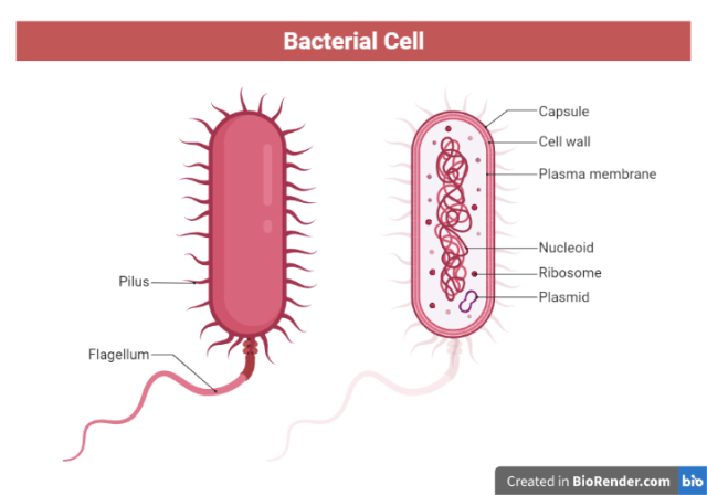 The bacterial cell is organized into three major regions:
- Cell envelope: the outer protective layers (cell wall and plasma membrane)
- Cell interior: the cytoplasm and its contents (nucleoid, ribosomes, inclusions)
- External/surface appendages: structures projecting from the cell surface (capsule, flagella, pili, spores)
The Cell Envelope
The cell envelope is the multilayered boundary between the bacterial cytoplasm and the external environment. It maintains cell shape, provides osmotic protection, and mediates interactions with the host immune system and antibiotics. Its composition differs fundamentally between gram-positive and gram-negative bacteria.
The Cell Wall
The bacterial cell wall is a rigid, chemically complex structure located outside the plasma membrane. It constitutes 20–30% of the dry weight of the bacterial cell and serves two essential functions:
- Mechanical protection: prevents osmotic lysis in hypotonic environments (bacteria are hypertonic relative to their environment; without the wall, osmotic water influx would burst the cell)
- Shape determination: the cell wall is rigid and determines the characteristic shape of the organism (cocci, bacilli, spirilla)
The central component of all bacterial cell walls (except mycoplasma and archaea) is peptidoglycan (also called murein or mucopeptide).
**Peptidoglycan**
Peptidoglycan is a mesh-like polymer consisting of:
- Glycan backbone: alternating units of N-acetylglucosamine (NAG) and N-acetylmuramic acid (NAM) linked by β-1,4 glycosidic bonds
- Short peptide cross-links: tetrapeptide side chains attached to NAM residues, cross-linked by transpeptidase enzymes to form a rigid three-dimensional lattice
The cross-linking transpeptidase reaction is the target of beta-lactam antibiotics (penicillins, cephalosporins, carbapenems) and glycopeptides (vancomycin). By inhibiting this cross-linking step, these antibiotics prevent cell wall synthesis during bacterial division, leading to cell lysis.
Lysozyme — an enzyme present in tears, saliva, nasal secretions, and neutrophil granules — cleaves the β-1,4 glycosidic bond between NAG and NAM, destroying the peptidoglycan backbone. This is one of the first-line innate immune defenses against bacterial infection.
→ Peptidoglycan: Structure and Medical Significance
Gram-positive cell wall
The gram-positive cell wall consists of a thick, multilayered peptidoglycan (20–80 nm; constitutes 40–80% of dry cell wall weight) with no outer lipid membrane. Its key components are:
Teichoic acids: anionic polymers of glycerol phosphate or ribitol phosphate, covalently linked to peptidoglycan. They:
- Regulate peptidoglycan synthesis and autolytic enzymes
- Confer negative charge to the cell surface
- Serve as receptor sites for bacteriophages
- Act as virulence factors, since teichoic acids of S. aureus mediate colonization of nasal epithelium
Lipoteichoic acids (LTAs): teichoic acids anchored to the plasma membrane rather than peptidoglycan. They play a role in immune stimulation similar to LPS in gram-negative bacteria.
Wall teichoic acids (WTAs): covalently bonded to peptidoglycan; important in antibiotic resistance and phage susceptibility.
→ Teichoic Acid: Characteristics and Medical Importance
Gram-negative cell wall
The gram-negative cell wall is structurally more complex with a thin peptidoglycan layer (2–7 nm) sandwiched between the plasma membrane and an outer membrane:
Thin peptidoglycan layer: typically 1 to 3 layers, compared with roughly 20 to 30 layers in gram-positive bacteria; located in the periplasmic space between the inner and outer membranes.
Periplasmic space: the compartment between the plasma membrane and the outer membrane. Contains peptidoglycan, binding proteins (for nutrient uptake), degradative enzymes, and in resistant organisms, beta-lactamases that inactivate beta-lactam antibiotics before they reach their target.
Outer membrane: a bilayer lipid membrane unique to gram-negative bacteria. Its outer leaflet contains:
- Lipopolysaccharide (LPS): a complex molecule consisting of Lipid A (the toxic component or endotoxin), a core oligosaccharide, and the O-antigen polysaccharide chain (used for serotyping)
- Porins: transmembrane protein channels that allow passive diffusion of small hydrophilic molecules (nutrients, some antibiotics) while excluding large or hydrophobic molecules
- Outer membrane proteins (OMPs): structural proteins involved in nutrient uptake, membrane integrity, and virulence
Clinical significance of LPS (endotoxin): When gram-negative bacteria are killed (by antibiotics or host defenses), LPS is released in large quantities. LPS binds to TLR4 (Toll-like receptor 4) on macrophages and monocytes, triggering the massive cytokine release responsible for gram-negative septic shock — fever, hypotension, disseminated intravascular coagulation (DIC), and multi-organ failure. This is why treating gram-negative infections can sometimes initially worsen the clinical picture as bacteria are killed and LPS is released.
→ Lipopolysaccharide (LPS): Characteristics and Functions
Comparison: gram-positive vs gram-negative cell wall
| Feature | Gram-positive | Gram-negative |
|---|---|---|
| Peptidoglycan thickness | Thick (20–80 nm; 40–80% of dry weight) | Thin (2–7 nm; 5–10% of dry weight) |
| Outer membrane | Absent | Present |
| Teichoic acids | Present (wall and lipoteichoic acids) | Absent |
| LPS (endotoxin) | Absent | Present (outer membrane) |
| Periplasmic space | Absent (or minimal) | Present (contains beta-lactamases) |
| Gram stain | Purple (retains crystal violet) | Pink/red (takes up safranin) |
| Susceptibility to lysozyme | More susceptible (peptidoglycan directly exposed) | Less susceptible (outer membrane blocks access to peptidoglycan) |
| Susceptibility to penicillin | Generally more susceptible | Generally less susceptible (outer membrane barrier) |
| Susceptibility to polymyxins | Resistant | Susceptible (target outer membrane) |
Acid-fast cell wall
Acid-fast bacteria (Mycobacterium, Nocardia) have a unique cell wall containing a thick layer of mycolic acids — long-chain (60–90 carbon) fatty acids covalently linked to an arabinogalactan polymer, which in turn is linked to peptidoglycan. This waxy, hydrophobic layer:
- Provides extreme resistance to acid-alcohol decolorization (hence "acid-fast")
- Resists most conventional disinfectants and antibiotics
- Prevents reliable uptake of Gram stain dyes. Mycobacteria are structurally gram-positive but stain poorly and inconsistently, which is why acid-fast staining is used instead
- Requires specialized staining: Ziehl-Neelsen (hot carbol fuchsin) or auramine-rhodamine (fluorescence)
- Is the basis of M. tuberculosis resistance to many antibiotics and host immune killing
→ Cell Wall Composition, Structure and Functions
Cell wall deficient bacteria
Some bacteria naturally lack a cell wall or can lose it under certain conditions:
Mycoplasma: the smallest free-living bacteria; naturally and permanently lack a cell wall. Completely resistant to all beta-lactam antibiotics and glycopeptides. The plasma membrane is stabilized by sterols (unlike all other bacteria). Causes atypical pneumonia (M. pneumoniae) and urogenital infections (M. hominis, Ureaplasma urealyticum).
L-forms: bacteria that have lost their cell wall through antibiotic pressure or mutation. They are pleomorphic, osmotically fragile, and temporarily resistant to beta-lactam antibiotics. May revert to normal cell-walled forms when antibiotic pressure is removed — explaining some treatment failures.
Protoplasts: gram-positive bacteria with the cell wall completely removed by lysozyme in isotonic solution. Osmotically fragile — lyse in hypotonic conditions.
Spheroplasts: gram-negative bacteria with partial cell wall removal. Retain the outer membrane; more osmotically stable than protoplasts.
The Plasma Membrane (Cytoplasmic Membrane)
The plasma membrane is a thin (7–8 nm), flexible phospholipid bilayer lying immediately beneath the cell wall. It is the true selective barrier of the bacterial cell — regulating all transport of materials into and out of the cytoplasm.
Key features distinguishing bacterial plasma membrane from eukaryotic:
- No sterols (cholesterol) in most bacteria except Mycoplasma, which incorporates host cholesterol to stabilize its membrane. This is why antifungal drugs targeting ergosterol (amphotericin B, azoles) have no effect on bacteria.
- Contains respiratory enzymes: the electron transport chain, which in eukaryotes is located in mitochondria, is located in the plasma membrane of bacteria
- Site of ATP synthesis: the proton-motive force driving ATP synthase operates across the plasma membrane
- Contains penicillin-binding proteins (PBPs): the transpeptidase enzymes that cross-link peptidoglycan, and the primary targets of beta-lactam antibiotics
Functions:
- Selective permeability: controls entry of nutrients, exit of waste products
- Energy transduction: site of oxidative phosphorylation (electron transport chain)
- Secretion: Type I to VI secretion systems for virulence factor export
- Contains biosynthetic enzymes for peptidoglycan, LPS, and phospholipid synthesis
The Cell Interior
The Cytoplasm
The bacterial cytoplasm is a gel-like aqueous solution containing all the soluble components needed for cell metabolism — enzymes, metabolites, ions, and small molecules. Unlike eukaryotic cytoplasm, it contains no cytoskeleton (in most bacteria), no membrane-bound organelles, and no endoplasmic reticulum or Golgi apparatus.
The bacterial cytoplasm does contain:
- The nucleoid (bacterial chromosome)
- Ribosomes (70S)
- Plasmids
- Cytoplasmic inclusions (storage granules)
- Occasionally mesosomes (invaginations of plasma membrane — role debated)
The Nucleoid (Bacterial Chromosome)
Bacteria have a single, circular, double-stranded DNA chromosome located in an irregular region of the cytoplasm called the nucleoid (not enclosed in a nuclear membrane). Key features:
- Typically 1–10 Mb in size (human genome ~3,200 Mb for comparison)
- Supercoiled and compacted by nucleoid-associated proteins (NAPs) and DNA gyrase (topoisomerase II)
- DNA gyrase is the target of fluoroquinolone antibiotics (ciprofloxacin, levofloxacin) — inhibiting it prevents DNA replication and causes double-strand breaks
- Contains no introns — all DNA is coding (unlike eukaryotic chromosomes)
- Replication begins at a single origin of replication (oriC)
Ribosomes
Bacterial ribosomes are 70S ribosomes (composed of 30S + 50S subunits), in contrast to eukaryotic 80S ribosomes (40S + 60S subunits). This size difference is clinically crucial — it allows selective targeting of bacterial ribosomes by antibiotics without affecting human ribosomes:
| Ribosomal subunit targeted | Antibiotic class | Effect |
|---|---|---|
| 30S subunit | Aminoglycosides (gentamicin, tobramycin) | Misreading of mRNA → incorrect protein synthesis |
| 30S subunit | Tetracyclines (doxycycline) | Blocks tRNA binding → protein synthesis inhibition |
| 50S subunit | Macrolides (azithromycin, clarithromycin) | Blocks translocation → protein synthesis inhibition |
| 50S subunit | Chloramphenicol | Inhibits peptidyl transferase |
| 50S subunit | Lincosamides (clindamycin) | Blocks translocation |
| 50S subunit | Oxazolidinones (linezolid) | Inhibits formation of initiation complex |
Plasmids
Plasmids are small, circular, extrachromosomal DNA molecules that replicate independently of the main chromosome. They are not essential for basic bacterial survival under normal conditions but confer significant advantages in hostile environments:
- Antibiotic resistance genes: the most clinically significant function; R-plasmids (resistance plasmids) carry genes encoding enzymes that inactivate antibiotics (beta-lactamases, aminoglycoside-modifying enzymes) or efflux pumps that expel antibiotics from the cell
- Virulence factors: toxin genes (e.g. E. coli heat-labile and heat-stable enterotoxins on plasmids), adhesins, iron acquisition systems
- Metabolic capabilities: genes enabling growth on unusual carbon sources
Clinical significance: horizontal gene transfer: Plasmids can be transferred between bacteria of different species through conjugation (plasmid transfer via sex pilus). This is the primary mechanism by which antibiotic resistance spreads rapidly between bacteria — a single plasmid carrying multiple resistance genes can convert a susceptible bacterium into a multi-drug-resistant (MDR) organism in a single transfer event.
Cytoplasmic Inclusions (Storage Granules)
Bacteria store reserve nutrients as cytoplasmic inclusions — concentrated deposits of specific substances that serve as energy and carbon reserves during nutritional deprivation:
| Inclusion type | Composition | Staining | Clinical/diagnostic significance |
|---|---|---|---|
| Metachromatic granules (volutin granules, Babes-Ernst bodies) | Polymerized inorganic phosphate (polyphosphate) | Deep reddish-purple with methylene blue (metachromasia) | Characteristic of Corynebacterium diphtheriae — best seen with Albert or Loeffler's methylene blue stain |
| Polysaccharide granules (glycogen) | Polymerized glucose | Brown with iodine | Energy reserve; found in Bacillus, Clostridium, some gram-negatives |
| Poly-β-hydroxybutyrate (PHB) | Lipid polymer | Black/dark with Sudan Black B; fluoresces with Nile Blue | Carbon and energy reserve; abundant in Bacillus, Pseudomonas |
| Sulfur granules | Elemental sulfur | Refractile under phase contrast | Seen in sulfur-oxidizing environmental bacteria; not in clinical pathogens |
| Magnetosomes | Magnetite (Fe₃O₄) crystals in membrane vesicles | Not routinely stained | Found in magnetotactic bacteria; rare; not clinically significant |
External Structures and Surface Appendages
Bacterial Capsule
The capsule is a thick, well-defined polysaccharide (occasionally polypeptide) layer surrounding the entire cell, located outside the cell wall. It is a major virulence factor in many clinically important bacteria.
Functions:
- Anti-phagocytic: the smooth, negatively charged polysaccharide surface prevents phagocyte adhesion and inhibits phagocytosis, allowing encapsulated bacteria to survive in the bloodstream
- Complement evasion: prevents deposition of C3b opsonin on the bacterial surface, blocking complement-mediated killing
- Biofilm formation: capsular material facilitates adherence to surfaces and to other bacteria in biofilm communities
Clinically important encapsulated organisms:
| Organism | Capsule composition | Disease | Vaccine available? |
|---|---|---|---|
| Streptococcus pneumoniae | Polysaccharide (84 serotypes) | Pneumonia, meningitis, otitis media | Yes — PCV13, PPSV23 |
| Klebsiella pneumoniae | Polysaccharide | Pneumonia, UTI, bacteremia | No |
| Haemophilus influenzae type b | Polyribosylribitol phosphate (PRP) | Meningitis, epiglottitis | Yes — Hib vaccine |
| Neisseria meningitidis | Polysaccharide (ACWY, B) | Bacterial meningitis, septicemia | Yes — MenACWY, MenB |
| Bacillus anthracis | Poly-D-glutamic acid (polypeptide) | Anthrax | Yes (limited availability) |
| Cryptococcus neoformans | Glucuronoxylomannan (fungal) | Cryptococcal meningitis | No |
Laboratory detection:
- India ink preparation: capsule appears as a clear halo around the cell against the dark background (used for Cryptococcus neoformans in CSF)
- Quellung reaction: specific anticapsular antibody causes the capsule to swell visibly — used for S. pneumoniae serotyping
- Mucoidy on culture plates: encapsulated bacteria form mucoid colonies; capsule-less variants form rough colonies
Read more→ Bacterial Capsule: Structure and Importance
Flagella
Flagella are long, whip-like protein filaments that rotate to propel bacteria through liquid environments. They are composed of three structural components: the filament (extracellular, composed of flagellin protein subunits), the hook (flexible coupling between filament and motor), and the basal body (embedded in the cell wall and membrane — the rotary motor).
Flagellar arrangements and clinical examples:
| Type | Description | Examples |
|---|---|---|
| Atrichous | No flagella | Staphylococcus aureus, Klebsiella pneumoniae |
| Monotrichous | Single polar flagellum | Vibrio cholerae (darting motility), Pseudomonas aeruginosa, Campylobacter jejuni |
| Lophotrichous | Cluster of flagella at one pole | Pseudomonas fluorescens, Helicobacter pylori |
| Amphitrichous | Flagella at both poles | Alcaligenes faecalis |
| Peritrichous | Flagella distributed over entire cell surface | E. coli, Salmonella typhi, Proteus mirabilis |
Clinical significance:
- Pathogenesis: flagella mediate movement toward host tissues (chemotaxis), ascending urinary tract infections (E. coli, Proteus), and gastric mucus penetration (H. pylori)
- Identification: Salmonella H (flagellar) antigens used in Kauffmann-White serotyping; Widal test detects antibodies to Salmonella H antigens
- Motility patterns: used in identification: darting motility (Vibrio), swarming (Proteus), corkscrew motility (Spirochetes)
Read more→ Bacterial Flagella: Structure, Importance and Examples
Pili (Fimbriae)
Pili (singular: pilus) are thin, straight, hair-like protein appendages shorter and thinner than flagella. They are composed of pilin protein subunits arranged in a helical array. Two main types exist:
Fimbriae (common pili): short, numerous appendages distributed over the entire cell surface. Primarily mediate adhesion to host cells and surfaces. This initial adhesion is the essential first step in most bacterial infections:
- Type 1 fimbriae of E. coli: bind mannose residues on uroepithelial cells; mediate UTI
- P fimbriae of uropathogenic E. coli: bind Gal-Gal receptors; important in pyelonephritis
- Neisseria gonorrhoeae pili: mediate attachment to urogenital epithelium; required for infection
Sex pili (conjugative pili): longer, fewer in number (1–4 per cell); form the conjugation tube through which plasmid DNA is transferred from donor to recipient bacterium. Critically important in the horizontal spread of antibiotic resistance plasmids.
→ Bacterial Pili/Fimbriae: Characteristics, Types and Medical Importance
Bacterial Endospores
Endospores are dormant, highly resistant structures formed by certain gram-positive bacteria under unfavorable conditions (nutrient depletion, desiccation, heat, UV radiation). They are not reproductive structures — one vegetative cell forms one spore. Sporulation is a survival mechanism.
Only two clinically important genera form endospores:
- Bacillus (aerobic): B. anthracis (anthrax), B. cereus (food poisoning)
- Clostridium (anaerobic): C. tetani (tetanus), C. perfringens (gas gangrene), C. difficile (antibiotic-associated diarrhea), C. botulinum (botulism)
Spore positions (clinically useful):
- Central: B. anthracis; does not distend the sporangium
- Subterminal: C. perfringens; does not distend the sporangium
- Terminal: C. tetani produces the classic "drumstick" or "tennis racket" appearance because the terminal spore is wider than the sporangium
Resistance properties of endospores:
| Challenge | Vegetative cells | Endospores |
|---|---|---|
| Boiling (100°C) | Killed in minutes | Survive for hours |
| Autoclaving (121°C, 15 min) | Killed | Killed |
| 70% alcohol | Killed | Survive |
| Bleach (0.5% sodium hypochlorite) | Killed | Sporicidal with adequate contact time; the recommended agent for C. difficile environmental decontamination |
| UV radiation | Killed | Survive (DNA protected by small acid-soluble spore proteins, SASPs) |
| Desiccation | Killed | Survive for decades |
This extreme resistance is why endospore-forming organisms (particularly C. difficile and anthrax spores) are so difficult to eradicate from healthcare and environmental settings.
Read more→ Bacterial Spores: Structure and Spore-Forming Bacteria
Comparison: Prokaryotic (Bacterial) vs Eukaryotic Cell
| Feature | Bacterial cell (prokaryote) | Human/animal cell (eukaryote) |
|---|---|---|
| Nucleus | Absent — nucleoid region, no nuclear membrane | Present — membrane-bound nucleus with nuclear pore |
| Chromosome | Single, circular, no histones | Multiple linear chromosomes; associated with histones |
| Cell size | 0.2–10 μm | 10–100 μm |
| Cell wall | Peptidoglycan (most bacteria) | Absent in animal cells; chitin in fungi; cellulose in plants |
| Plasma membrane sterols | Absent (except Mycoplasma) | Cholesterol present |
| Ribosomes | 70S (30S + 50S) | 80S (40S + 60S) |
| Mitochondria | Absent — electron transport in plasma membrane | Present |
| Endoplasmic reticulum | Absent | Present |
| Golgi apparatus | Absent | Present |
| Plasmids | Present | Absent (mitochondria and chloroplasts carry their own circular DNA) |
| Introns in DNA | Absent | Present |
| Capsule | Present in many pathogens | Absent |
| Flagella structure | Composed of flagellin; about 20 nm diameter; rotates like a propeller | Composed of tubulin in a 9+2 microtubule arrangement; about 200 nm diameter; beats rather than rotates |
How to Remember
Work from the outside in. The article's three regions are also the order a drug or an antibody meets the cell: external appendages first (capsule, flagella, pili), then the envelope (outer membrane if present, peptidoglycan, plasma membrane), then the interior (nucleoid, ribosomes, plasmids). Anything that has to reach the cytoplasm must cross everything outside it, which is the whole reason gram-negatives are harder to treat.
Every structure has a drug that targets it. This is the single most useful way to hold the whole article:
- Cell wall, so beta-lactams and vancomycin
- Outer membrane, so polymyxins and colistin
- 70S ribosome, so aminoglycosides, tetracyclines, macrolides
- DNA gyrase, so fluoroquinolones
- RNA polymerase, so rifampicin
If a structure is unique to bacteria, it is a drug target. If a structure is shared with human cells, it is not. That one rule explains why there are no useful antibiotics against the plasma membrane phospholipids themselves, and why Mycoplasma, having no wall at all, is untouched by every cell-wall agent.
Thick wall or outer membrane. Gram-positive bacteria chose a thick peptidoglycan coat. Gram-negative bacteria chose a thin one plus an outer membrane. Each solves the same osmotic problem differently, and each has a matching weakness: the exposed thick wall is vulnerable to lysozyme and penicillin, while the outer membrane blocks both but is itself the polymyxin target.
References and further reading
- Madigan MT, Bender KS, Buckley DH, Sattley WM, Stahl DA. Brock Biology of Microorganisms. 16th ed. Pearson; 2021.
- Tille PM. Bailey & Scott's Diagnostic Microbiology. 15th ed. St. Louis: Elsevier; 2022.
- Murray PR, Rosenthal KS, Pfaller MA. Medical Microbiology. 9th ed. Elsevier; 2020.
- Tortora GJ, Funke BR, Case CL. Microbiology: An Introduction. 13th ed. Pearson; 2018.
- Silhavy TJ, Kahne D, Walker S. The bacterial cell envelope. Cold Spring Harb Perspect Biol. 2010;2(5):a000414.
Frequently Asked Questions
What is the difference between a gram-positive and gram-negative bacterial cell wall?
Why do beta-lactam antibiotics not work against Mycoplasma?
What is the clinical significance of bacterial plasmids?
Why are bacterial endospores so resistant to sterilization?
What is the function of LPS (endotoxin) and why is it clinically important?
What is the difference between pili and flagella?
What makes acid-fast bacteria resistant to staining and disinfection?
What is the significance of the periplasmic space in gram-negative antibiotic resistance?

Tankeshwar Acharya, MSc (Medical Microbiology)
Tankeshwar Acharya is an Assistant Professor in the Department of Microbiology at Patan Academy of Health Sciences (PAHS), Nepal, where he has been teaching and practicing clinical microbiology for over 14 years. He is the founder of Microbe Online, one of the leading free microbiology education resources on the web, covering bacteriology, mycology, parasitology, immunology, and clinical laboratory diagnostics written from direct experience in both the classroom and the diagnostic laboratory.