
Phase Contrast Microscope: Principle, Types and Applications
How phase-contrast microscopy makes living, unstained cells visible by amplifying invisible differences in light phase, and why it won Zernike a Nobel Prize.
A lab receives a blood culture bottle flagged positive, gram-positive rods on the smear. One of the first practical questions is whether the organism is motile, since that single observation helps narrow the differential immediately; Listeria monocytogenes, for example, is known for a distinctive tumbling motility at room temperature, while several similar-looking gram-positive rods are not motile at all.
Staining would answer nothing here. Fixing and staining the specimen kills it instantly, freezing any motility forever and destroying the exact clue being looked for. An unstained wet mount under ordinary bright-field microscopy is nearly as unhelpful, living bacterial cells are almost completely transparent, with barely enough contrast against the background to be seen at all, let alone assessed for movement.
A phase-contrast microscope solves both problems at once. It reveals the organisms clearly, without staining, and keeps them alive and moving exactly as they would in the original specimen. The physics behind that trick, converting an invisible property of light into a visible one, is what earned Frits Zernike a Nobel Prize, and it's the reason phase-contrast remains one of the few ways to watch a living organism behave, not just exist, under a microscope.
The phase-contrast microscope is a modified version of the bright-field microscope that helps visualize living cells without affecting the cells’ viability. It is termed phase-contrast because it consists of a unique phase contrast condenser (annular ring) and a phase contrast objective (phase plate). These parts amplify the phase difference of transparent specimens.
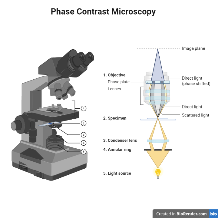 Figure: Phase contrast microscope
Principle of Phase Contrast Microscope
Let us consider light as a wave. Two identical beams of light releases in synchronization from a source. The first light beam strikes a material with a higher refractive index than air. The beam of light slows down, and the number of light waves increases in proportion to the refractive index and thickness of the material. But the second beam travels through the air only. Although the light rays were synchrony, a difference establishes in the beams’ phase after traveling through different mediums. It is termed phase difference.
Every specimen can diffract the beam of light. An image is formed due to the phase difference between diffracted and undiffracted beams (direct light). A stained or colored object has enough phase difference to create an image.
Since the phase difference between diffracted and direct light is insufficient in transparent specimens. A disk (annular ring) in the condenser separates direct and diffracted light. Likewise a special plate (phase plate) in the back of the focal plane of the objective lens increases the phase difference between diffracted light and direct light. The resulting increase in phase difference helps to form the image of transparent objects without staining.
A physicist, Frits Zernike, discovered the phase-contrast phenomenon in 1930, and first described its application to microscopy in 1933. He built the first phase-contrast microscope by 1938, and was awarded the 1953 Nobel Prize in Physics for this work.
Diffraction: the scattering of waves as they touch the specimen’s edge.
Parts of Phase Contrast Microscope
The phase-contrast microscope has all the parts of the microscope and some additional parts. They are:
Annular ring
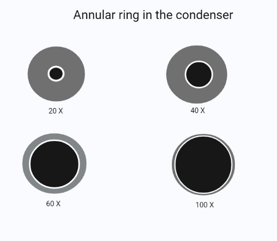 Figure: Annular ring with different power
An annular ring or phase annulus is a clear ring in an opaque disk. The purpose of the apparatus is to produce a circle of light in the front focal plane of the condenser. Thus, the condenser focuses on the specimen plate. The annular ring is different for different objectives.
Phase plate
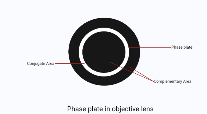 Figure: Phase plate inside an objective lens
A phase plate is a transparent plate with a circular ring in it. The ring of the phase plate is filled with phase advancing and retarding material. The ring of the plate may appear lighter or darker than other parts of the plate. There is an addition of some material that changes light amplitude. The phase plate’s position is complementary to the annular ring. However, the position of the phase ring is over the image of the annulus ring. So the phase plate is a part of objective lens and has conjugate and complementary parts.
Phase telescope
A phase telescope is a special eyepiece that focuses an objective’s back focal image onto a human retina. This procedure is necessary for aligning the annulus ring to the phase plate. Bertrand lens replaces the phase telescope on some microscopes.
Assembling the Apparatus of Phase Contrast Microscope
The proper functioning of the phase microscope requires proper assembling and alignment of the phase apparatus. Follow the steps below to assemble the phase apparatus:
- Kohler illumination is preferred for this type of microscopy because phase-contrast requires the condenser to produce nearly parallel light waves in the specimen.
- After setting set up the microscope on Kohler illumination, open the condenser’s iris fully.
- Then, focus specimen with low power objective with phase plate. And insert the adequately sized annulus ring.
- After that, replace an eyepiece with a phase telescope. And focus the telescope on the annulus ring.
- Align the phase plate with the annulus ring.
- Finally, observe the phase plate and annulus ring through the telescope.
Working mechanism of the Phase Contrast Microscope
- In phase-contrast microscopy, illumination of the specimen occurs by light passing through an annular ring. It produces a hollow cone of light.
- In this microscope, one set of light rays directly comes from the light source (direct light). In contrast, the other set comes from light reflected or diffracted from a particular structure in the specimen.
- Direct light rays strike a phase ring in the phase plate within the objective to give a bright background. At the same time, the diffracted beams miss the ring and pass through the rest of the plate to provide dark and well-defined structures.
- When the two sets of light rays, direct and diffracted beams, are brought together, they form an image of the specimen on the ocular lens with a bright background and different dark areas. This configuration, dark structures against a light background, is what the next section describes as positive phase contrast, the most commonly used form of this technique
Types of Phase Contrast
Depending upon the construction of the phase plates, there are several types of phase-contrast microscopy. The most common types are
Positive phase contrast
Positive phase contrast advances direct light by ¼ wave, producing destructive interference and dark details in a light background. It is the most commonly used form.
Negative phase contrast
Negative phase contrast retards direct light by ¼ wave, producing constructive interference and light details on a dark background.
Either positive or negative phase contrast
The phase plate in this form is of two types. Either the phase plate absorbs direct light, or it can absorb diffracted light.
Applications of Phase Contrast Microscope
A phase-contrast microscope is applied in various biological researches to visualize transparent samples like living cells in culture, pieces of tissue, microorganisms, fibers, subcellular particles, glass fragments, and latex dispersions.
This is a related but distinct problem from the one solved by dark-field microscopy: dark-field detects extremely thin structures like spirochetes through scattered light against total darkness, while phase-contrast amplifies subtle refractive index differences within larger, otherwise transparent structures, such as the internal organization of a living cell.
Limitations of Phase Contrast Microscope
There is some limitation to the phase-contrast microscope. These are:
- It works excellent for observing thin, colorless, and transparent specimens. However, a confusing phase image will be seen if the sample is thick.
- The assembly of the microscope parts makes the whole procedure more expensive.
- The phase plate limits the objective’s NA (numerical aperture).
How to Remember
- Which part lives where: the annular ring lives in the condenser, shaping the incoming light before it reaches the specimen. The phase plate lives in the objective, shaping the light after it leaves the specimen. Incoming shapes the beam; outgoing shapes the image.
- Positive vs. negative, by the word itself: positive phase contrast is the default, dark details on a light background, the same way a normal photograph looks. Negative phase contrast flips it, light details on a dark background, like a photographic negative. The name hints at the inversion.
- Zernike's real achievement, in one line: he found a way to turn an invisible property of light (phase, essentially timing) into a visible one (brightness), without touching the specimen chemically at all. That's the entire principle in a sentence.
- Aligning the apparatus, the one thing to get right first: every alignment step in the assembly procedure depends on Kohler illumination already being correctly set up. If the image looks wrong after aligning the phase plate to the annulus ring, the first thing to re-check isn't the phase apparatus, it's whether Kohler illumination was actually achieved before starting.
Key exam facts in one table
| Concept | Detail | Why it's tested |
|---|---|---|
| Inventor and history | Frits Zernike discovered the phase-contrast phenomenon in 1930, first described its application to microscopy in 1933, and built the first phase-contrast microscope by 1938 | Commonly tested "who and when" historical fact |
| Nobel recognition | Zernike received the 1953 Nobel Prize in Physics for this work | Often the specific detail an exam question asks for |
| Annular ring location | Condenser | Shapes incoming light into a hollow cone before it reaches the specimen |
| Phase plate location | Objective | Amplifies the phase difference between direct and diffracted light after it leaves the specimen |
| Positive phase contrast | Dark specimen details on a light background; the most commonly used type | The default configuration on most commercial phase-contrast microscopes |
| Negative phase contrast | Light specimen details on a dark background | The less common, inverted alternative |
| Key limitation | Reduces the objective's numerical aperture; produces a confusing image on thick specimens | Frequently tested trade-off for this technique |
Where Students Get Confused
- Mixing up which part does what. The annular ring is in the condenser and shapes the light going into the specimen; the phase plate is in the objective and shapes the light coming out. Getting the location backwards is a common exam trap.
- Confusing phase-contrast microscopy with dark-field microscopy. Both are used for living, unstained specimens, but they solve different problems: dark-field reveals extremely thin structures (like spirochetes) via scattered light against total darkness, while phase-contrast reveals subtle internal structure within larger transparent specimens by amplifying phase differences.
- Reversing positive and negative phase contrast. Positive shows dark details on a light background and is the standard, commonly used configuration; negative is the inverted, less common alternative.
- Assuming any alignment problem is a phase-plate problem. In practice, most alignment issues trace back to Kohler illumination not being properly set up first, since every later step in the assembly procedure depends on it.
- Assuming phase-contrast requires some kind of stain or dye. It specifically doesn't; the entire point of the technique is converting an optical phase difference into visible contrast without any chemical treatment of the specimen at all.
References
- A Guide to Phase Contrast: Principles, Applications and Setup. (2022). Scientifica. https://www.scientifica.uk.com/learning-zone/a-guide-to-phase-contrast
- Oldenbourg, R., & Shribak, M. (2010). Microscopes.
- Bagnell, R. Phase Contrast Microscopy. University of North Carolina Microscopy Services Laboratory. https://www.med.unc.edu/microscopy/wp-content/uploads/sites/742/2018/06/lm-ch-10-phase-contrast.pdf
- Madigan, M. T., Bender, K. S., Buckley, D. H., Sattley, W. M., & Stahl, D. A. (2018). Brock Biology of Microorganisms (15th ed.). Pearson.
Frequently Asked Questions
What is the difference between positive and negative phase contrast?
Where are the annular ring and phase plate located in a phase-contrast microscope?
What is the difference between phase-contrast and dark-field microscopy?
What are the main limitations of phase-contrast microscopy?

Tankeshwar Acharya, MSc (Medical Microbiology)
Tankeshwar Acharya is an Assistant Professor in the Department of Microbiology at Patan Academy of Health Sciences (PAHS), Nepal, where he has been teaching and practicing clinical microbiology for over 14 years. He is the founder of Microbe Online, one of the leading free microbiology education resources on the web, covering bacteriology, mycology, parasitology, immunology, and clinical laboratory diagnostics written from direct experience in both the classroom and the diagnostic laboratory.