
Differences Between SEM and TEM

On this page
A research lab is studying a catheter-associated biofilm from a patient with a recurrent, antibiotic-resistant infection. Two very different questions need answering. First: how are the bacteria physically arranged on the catheter surface, are they in a thick, structured layer, and does the surface texture explain how they're resisting removal? Second: what is a candidate antimicrobial peptide actually doing to those bacteria at the cellular level, are membranes rupturing, are internal structures breaking down?
Both questions are legitimate. Neither can be answered by the same instrument. The first is a surface question, answered by scanning electron microscopy, which images the biofilm's three-dimensional architecture in detail. The second is an internal question, answered by transmission electron microscopy, which requires thin-sectioning the bacterial cells to see what's happening inside them. The table below exists to make sure that choice gets made correctly, before the sample prep, not after.
Both scanning electron microscopy (SEM) and transmission electron microscopy (TEM) use a focused beam of electrons instead of light to image a specimen, but they answer fundamentally different questions.
For the shared general principles, invention history, and components common to all electron microscopes, see Electron Microscope: Principle, Types, Applications.
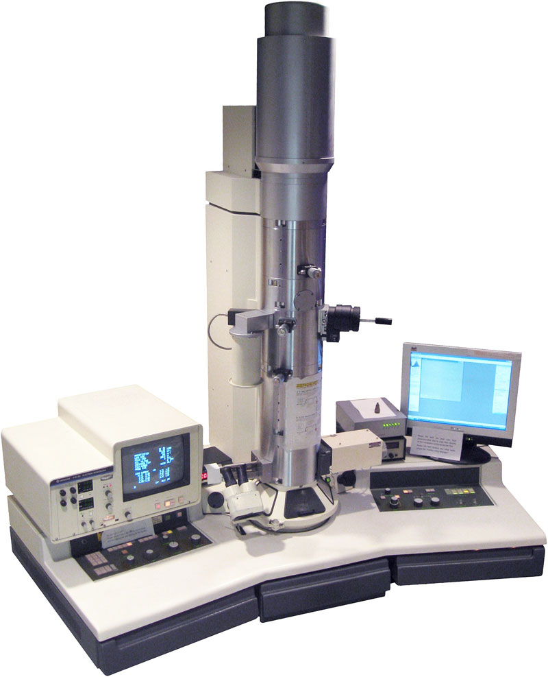 Figure: Electron Microscope (image source)
There are several types of electron microscopes, classified according to their final use. Transmission electron microscope and scanning electron microscope are the two most common types of electron microscope.
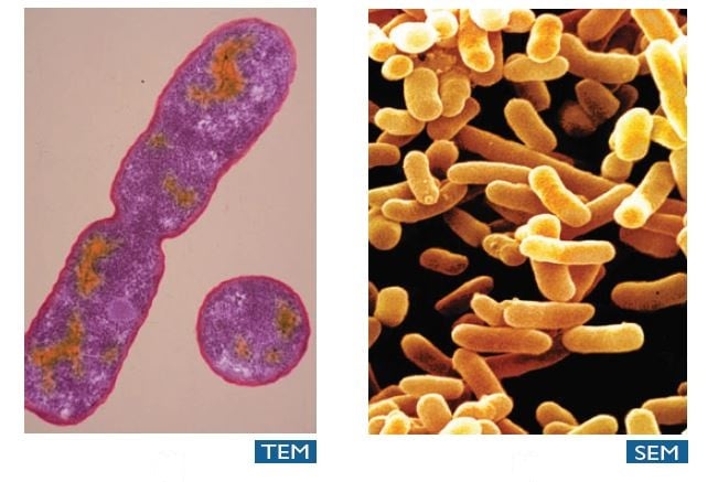 Figure: Colorized electron micrographs of E.coli produced by TEM and SEM (Source: TEM-Dennis kunkel/phototake, TEM-David M.Phillips/Visuals Unlimited)
The major differences between SEM and TEM are as follows
| Properties | Scanning Electron Microscopy (SEM) | Transmission Electron Microscopy (TEM) |
|---|---|---|
| Light Source | SEM is based on scattered electrons, i.e. electrons emitted from the surface of a specimen. It is the EM analog of a stereo light microscope. | Electrons are used as “light source”. TEM is based on transmitted electrons and operates on the same basic principles as the light microscope. |
| Purpose | SEM provides detailed images of the surfaces of cells. SEM focuses on the sample’s surface and its composition, so SEM shows only the morphology of samples. | Transmission electron microscope is used to view thin specimens (tissue sections, molecules, etc). TEM can show many characteristics of the sample, such as internal composition, morphology, crystallization, etc. |
| Sample Preparation | Sample is coated with a thin layer of heavy metal such as gold or palladium. | The sample in TEM has to be cut thinner ( 70-90 nm ) because electrons cannot penetrate very far into materials. |
| Resolution | SEM can resolve objects down to approximately 1–20 nm | TEM has a much higher resolution than SEM, approximately 0.2 nm, sufficient to approach near-atomic levels of detail |
| Magnification | The magnifying power of SEM is up to approximately 100,000X. | The magnifying power of TEM is up to approximately 1,000,000X. |
| Processing of sample (s) | SEM allows for a large amount of sample to be analyzed at a time | With TEM only a small amount of samples can be analyzed at a time. |
| Image formation | Secondary or backscattered electrons arising from the interaction of electron beam and metal-coated specimen are collected and the resulting image is displayed on a computer screen. | Transmitted electrons hit a fluorescent screen giving rise to a “shadow image” of the specimen with its different parts displayed in varied darkness according to their density. The image can be studied directly by the operator or photographed with a camera. |
| 3D picture | SEM provides a 3-dimensional image | TEM provides a 2-dimensional picture. |
| Current Applications | To study topography and atomic composition of specimens, process control and also, for example, the surface distribution of immuno-labels | To image the interior of cells (in thin sections), the structure of protein molecules (contrasted by metal shadowing), the organization of molecules in viruses and cytoskeletal filaments (prepared by the negative staining technique), and the arrangement of protein molecules in cell membranes (by freeze-fracture). |
How to Remember
- Surface vs. interior, in two words: SEM shows you the outside; TEM shows you the inside. Every other difference in the table, sample prep, resolution, magnification, 2D vs. 3D, follows from this one distinction.
- Sample prep, as a costume vs. surgery: SEM specimens just need a "costume," a thin conductive metal coating. TEM specimens need "surgery," fixation, dehydration, embedding, and ultra-thin sectioning, before they can be imaged at all.
- Why TEM has far higher resolution than SEM: TEM directly transmits electrons through the specimen and detects density differences at near-atomic scale; SEM only detects electrons scattered back from the surface, which limits how fine a detail it can resolve. If a question asks which technique can approach atomic-level resolution, the answer is TEM, not SEM.
- The 3D/2D pattern matches the surface/interior pattern exactly: SEM's surface-scanning naturally produces a 3D-looking image, since it's imaging a real surface topology. TEM's straight-through transmission produces a flat, 2D density shadow, since it's imaging what light and electrons pass through, not around.
Key exam facts
| Concept | Detail | Why it's tested |
|---|---|---|
| The one deciding question | Do you need to see the specimen's surface, or its internal structure? | This single question determines which instrument is even usable, before considering cost or convenience |
| Resolution figures (standardized) | TEM ~0.2 nm; SEM ~1–20 nm | Consistent with the rest of this site's microscopy cluster |
| Magnification figures (standardized) | TEM up to ~1,000,000x; SEM up to ~100,000x | TEM's far higher magnification follows directly from its much higher resolving power |
| Sample destruction risk | TEM's thin-sectioning destroys surface architecture; SEM's metal coating destroys the possibility of examining internal structure | Explains why the two techniques are complementary, not interchangeable, for a single research question |
| Image color | Both produce black-and-white images natively; any color shown has been added afterward | Applies equally to both techniques, easy to forget for SEM given how often colorized SEM images circulate |
Where Students Get Confused
- Assuming SEM and TEM are interchangeable, with one just being "better." The real distinction is what each can even see: SEM cannot reveal internal cellular structure no matter how it's used, and TEM cannot reveal true surface topology, since its thin-sectioning process removes the specimen's original surface entirely.
- Assuming higher magnification numbers alone tell you which instrument to pick. TEM's magnification advantage only matters if the question being asked is an internal-structure question; for a surface-architecture question, SEM's lower magnification and 3D imaging are the right tool, not a lesser one.
- Forgetting that both produce black-and-white images. SEM images in particular are very often shown colorized (biofilms, cell surfaces, insects), which can create the impression that SEM captures color information directly; it doesn't.
- Treating sample preparation as a minor technical detail rather than a consequence of the imaging principle. TEM's thin-sectioning and SEM's metal coating aren't arbitrary lab steps, they follow directly and necessarily from transmission vs. surface-scattering imaging, respectively.
References
- Black, J. G., & Black, L. J. Microbiology: Principles and Explorations (9th ed.).
- University of Massachusetts Medical School, Center for Electron Microscopy. What is EM? https://www.umassmed.edu/cemf/whatisem/
- U.S. Department of Veterans Affairs. What Is Electron Microscopy and How Does It Work? https://www.va.gov/DIAGNOSTICEM/What_Is_Electron_Microscopy_and_How_Does_It_Work.asp
- The Nobel Prize. Electron Microscopes. https://www.nobelprize.org/educational/physics/microscopes/tem/
Frequently Asked Questions
What is the main difference between SEM and TEM?
Which has higher resolution, SEM or TEM?
Why can't SEM be used to see the inside of a cell?
Do SEM images show true color?
How does sample preparation differ between SEM and TEM?

Tankeshwar Acharya, MSc (Medical Microbiology)
Tankeshwar Acharya is an Assistant Professor in the Department of Microbiology at Patan Academy of Health Sciences (PAHS), Nepal, where he has been teaching and practicing clinical microbiology for over 14 years. He is the founder of Microbe Online, one of the leading free microbiology education resources on the web, covering bacteriology, mycology, parasitology, immunology, and clinical laboratory diagnostics written from direct experience in both the classroom and the diagnostic laboratory.
Comments
No comments yet. Be the first to share your thoughts.
Leave a comment
All comments are reviewed before they appear.