
Complement Fixation Test (CFT): Principle, Procedure, and Why No Hemolysis Is Positive
CFT explained clearly: the two-system principle, step-by-step procedure, and the key point students get wrong, why no hemolysis means a positive result.

On this page
Here is the fact about the complement fixation test that trips up almost everyone who learns it: no hemolysis means the test is positive. When the red cells stay intact, the patient has the antibody. When the red cells burst, the patient does not. It feels backwards, and that single inverted readout is why so many students look this test up more than once.
The reason it works this way is not a trick. It follows logically from how the test is built, from two systems competing for one limited resource: complement. Once you see the logic, the result reads itself. This article builds that logic step by step, then walks through exactly how to read a positive and a negative result.
The complement fixation test (CFT) is a classic laboratory test used to detect a specific antibody (or antigen) in a patient's serum. It has long been used to help diagnose certain infections by showing whether a person has made antibodies against a particular microbe.
The test relies on the complement system, a group of blood proteins that help antibodies destroy their targets. CFT uses several reagents and several preparation steps, and many versions exist. The microtiter version developed at the US Centers for Disease Control and Prevention (the LBCF test) uses rigorous controls and is widely used.
A note on current use: CFT is now largely a historical method. For most purposes it has been replaced by faster, more sensitive tests such as ELISA (enzyme immunoassay), immunofluorescence, and PCR. It is still taught because its logic is a classic teaching model of antibody detection, and it is still used in a few reference and research settings for certain viral and fungal serology. Learn it for the concept and because it still appears in exams, while knowing it is no longer the frontline test in most laboratories.
 Figure: Fixation of Complement
Principle of the complement fixation test
CFT answers one question: does the patient's serum contain a specific antibody? It answers it indirectly, using complement as a shared resource that two systems compete for.
The key fact about complement: complement is a set of blood proteins that, once activated by an antigen-antibody complex, gets used up. A fixed amount of complement can only be consumed once. The whole test hinges on this: if one reaction uses the complement, none is left for a second reaction.
The test uses two systems, added one after the other.
System 1, the test system (added first):
- A known antigen (from the microbe you are testing for)
- The patient's serum (which may or may not contain antibody against that antigen)
- A measured, limited amount of complement
If the patient has the antibody, it binds the antigen, and this antigen-antibody complex activates and consumes (fixes) the complement. If the patient does not have the antibody, no complex forms, and the complement stays free and unused.
At this point, you cannot see anything. Complement being used or not used is invisible. You need a second system to reveal it.
System 2, the indicator system (added second):
- Sheep red blood cells
- Hemolysin (rabbit anti-sheep antibody, also called amboceptor), which coats the sheep cells
Sheep cells coated with hemolysin are "sensitized": they are ready to be lysed, but only if complement is available. The indicator system is a complement detector. It bursts (hemolysis) when complement is present, and stays intact when complement is absent.
Now the two systems interact, and this is where the inverse readout comes from.
Scenario A, the patient HAS the antibody (positive):
In system 1, the antibody bound the antigen and used up all the complement. When the indicator system is added, there is no complement left for it. The sensitized sheep cells cannot be lysed. No hemolysis. The cells stay intact and settle as a red button at the bottom of the well. This is a positive result.
Scenario B, the patient does NOT have the antibody (negative):
In system 1, no antigen-antibody complex formed, so the complement was never used. It is still free. When the indicator system is added, this free complement is available to the sensitized sheep cells and lyses them. Hemolysis. The cells burst and the well turns clear red. This is a negative result.
So the readout is inverted on purpose. The sheep cells are a stand-in that reports on complement, and complement is only left over when the patient's antibody was absent. No hemolysis = antibody present = positive. Hemolysis = antibody absent = negative.
| What you see | What it means for complement | What it means for the patient | Result |
|---|---|---|---|
| No hemolysis (red button, cells intact) | Complement was used up in system 1 | Antibody is present | Positive |
| Hemolysis (clear red, cells lysed) | Complement was still free | Antibody is absent | Negative |
Why sheep cells lyse at all: complement's terminal proteins assemble into the membrane attack complex (C5b-9), which punches pores in the sheep-cell membrane and releases hemoglobin. That release, seen as the red solution clearing, is the visible signal. It only happens when complement was left free, that is, when the patient had no antibody.
Materials and Reagents
- Sheep erythrocytes suspension (5% suspension of washed sheep RBCs)
- Hemolysin, also called amboceptor (rabbit anti-sheep red-cell antibody). When it coats the sheep cells, they are called sensitized sheep cells, the indicator system.
- Guinea pig complement, free of antibodies to the agent of interest (Note: Guinea pig is the commonest source of fresh complement)
- Barbital-buffered diluents
- Plastic microtiter plate
- Centrifuge adapter for microtiter plates
- Water bath for incubation of plates
- Color standards for judging hemolysis (prepared by lysing various concentrations of red cells)
Procedure of Complement Fixation Test
CFT is run in two stages, added in order.
Stage 1: The complement fixation stage (test system)
- Heat the patient's serum at 56°C for 30 minutes first. This inactivates the patient's own complement, which would otherwise interfere with the measured complement added in the test. (This step is called inactivation.)
- Mix the inactivated patient serum with a known antigen and a standardized, limited amount of complement.
- Incubate.
What happens depends on the patient:
- If the serum contains a specific antibody, it binds the antigen, and the antigen-antibody complex fixes (uses up) the complement.
- If there is no specific antibody, no complex forms, and the complement stays free.
At the end of stage 1 you cannot see the outcome yet. Stage 2 reveals it.
Stage 2: The indicator stage
- Add the indicator system: sheep red blood cells coated with hemolysin (sensitized sheep cells).
- Incubate and read.
- If complement was fixed in stage 1 (antibody present), none is left, so the sheep cells are not lysed. No hemolysis. Positive.
- If complement was left free in stage 1 (antibody absent), it now lyses the sheep cells. Hemolysis. Negative.
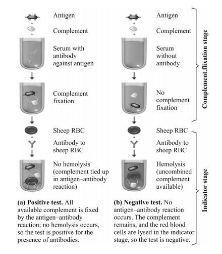 Figure: Complement Fixation Test Procedure/Results
Results and Interpretation
- No lysis of sheep red blood cells: positive CFT. The patient's serum contains the specific antibody. (The antibody used up the complement in stage 1, so none was left to lyse the sheep cells.)
- Lysis of sheep red blood cells: negative CFT. The patient's serum does not contain the specific antibody. (Complement stayed free and lysed the sheep cells.)
Titer and a rising titer. CFT is often run on serial two-fold dilutions of serum to find the titer (the highest dilution still giving a positive, no-hemolysis result). Testing an acute-phase and a convalescent-phase sample together is more useful than a single result: a four-fold or greater rise in titer between the two samples indicates a recent, active infection. A single positive may only reflect past exposure.
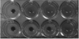 Figure: Complement Fixation Test in Microtiter Plate rows 1 and 2 exhibit complement fixation obtained with acute and convalescent-phase serum specimens, respectively. (2-fold serum dilutions were used). The observed 4-fold increase is significant and indicates infection.
Materials required for Quality Control
- Known positive antibody or antigen
- Known negative antibody or antigen
- Serum control without antigen (to detect anticomplementary activity)
- Antigen controls without serum (to detect anticomplementary activity)
- Tissue control (the cells or tissue in which the antigen was prepared)
- Buffer control without antigen or antibody
- Back titration of complement to document the use of 5CH50 units
Controls should be used along with the test to ensure that
- Antigen and serum are not anticomplementary
- The appropriate amount of complement is used and
- The sheep red blood cells do not undergo autolysis
What the complement fixation test is used for
CFT detects antibodies against a specific microbe, so it has been used to diagnose infection by showing that a patient has mounted an antibody response. Historically it was used for:
- Certain viral infections (for example, some respiratory and arbovirus serology)
- Certain fungal infections (for example, antibody detection in some systemic mycoses)
- Some bacterial and parasitic infections
For most of these, faster and more sensitive tests (ELISA, immunofluorescence, PCR) are now preferred, but the principle of CFT remains a core teaching example of antibody detection.
Advantages and Disadvantages of Complement Fixation Test
Advantages
- Could screen for many viral and bacterial infections using the same basic method
- Inexpensive, needing no specialized instruments.
Disadvantages
- Less sensitive than modern tests such as ELISA, so it can miss low antibody levels
- Time-consuming and labor-intensive
- Often non-specific e.g. cross-reactivity between herpes-simplex virus (HSV) and varicella-zoster virus (VZV).
How to remember
No hemolysis, antibody present. The one line that fixes the whole test. If the red cells survive, the patient's antibody grabbed the complement first. If the red cells die, there was no antibody and complement was free to kill them. "Cells live, antibody's there."
Complement is used once. The entire test rests on this. Whoever gets the complement first (the patient's antibody in system 1, or the sheep cells in system 2) determines the result. If the patient's antibody uses it, the sheep cells get none.
The sheep cells are a detector, not the point. You do not care about sheep cells. They are a visible flag that reports whether complement was left over. Free complement kills them (visible), used-up complement cannot (invisible until you add them).
Two systems, added in order. System 1 (antigen + patient serum + complement) is the real test but is invisible. System 2 (sensitized sheep cells) makes the answer visible. Test first, reveal second.
Key exam facts
| Point | Fact | Memory aid |
|---|---|---|
| Full form | CFT = complement fixation test | Fixation = using up complement |
| What it detects | A specific antibody (or antigen) in serum | Antibody detector |
| Core principle | Complement is used up once; two systems compete for it | Whoever grabs complement first wins |
| Test system (system 1) | Antigen + patient serum + complement | The real test, but invisible |
| Indicator system (system 2) | Sensitized sheep RBCs (sheep cells + hemolysin/amboceptor) | The visible flag |
| Complement source | Guinea pig | Guinea pig gives fresh complement |
| Patient serum step | Heat 56°C for 30 min to inactivate own complement | Inactivate before testing |
| No hemolysis | Positive (antibody present) | Cells live, antibody's there |
| Hemolysis | Negative (antibody absent) | Cells die, no antibody |
| Significant result | Four-fold rise in titer (acute vs convalescent) | Rising titer = active infection |
| Current status | Largely replaced by ELISA/IF/PCR; still taught | Classic, not frontline |
Where students get confused
"Why does no hemolysis mean positive? It feels backwards." Because hemolysis reports on complement, not on antibody directly. If the patient has the antibody, it uses up the complement in the first step, so none is left to burst the sheep cells: no hemolysis. The sheep cells surviving is the signal that the antibody was there. The result is inverted because you are reading complement leftover, and complement is only left over when the antibody was absent.
"What is the sheep red blood cell for?" It is a detector for leftover complement, nothing more. Coated with hemolysin, the sheep cell will burst if, and only if, complement is available. So it converts the invisible "was complement used up?" question into a visible "did the cells burst?" answer.
"What does lysis of the sheep cells tell me about the patient?" That the patient had no specific antibody (a negative result). The complement was never used in step 1, so it was free to lyse the sheep cells in step 2. Lysis means antibody absent.
"Why heat the patient's serum first?" The patient's own serum contains complement. If you leave it in, it adds to the measured complement and throws off the test. Heating to 56°C for 30 minutes inactivates the patient's complement so that only the known, measured complement you add is in play.
"Which animal gives the complement, and which gives the antibody to sheep cells?" Complement comes from guinea pig (a good source of fresh, active complement). The antibody that coats the sheep cells (hemolysin/amboceptor) comes from rabbit (rabbit anti-sheep). Two different animals, two different roles. Students often mix these up.
"Is CFT still used?" Mostly not as the frontline test. ELISA, immunofluorescence, and PCR have replaced it for most purposes. It is still taught because its logic is a classic model, and it is still used in some reference and research settings. Know the concept; know it is largely historical.
Frequently Asked Questions
What is the complement fixation test (CFT)?
What is the complement fixation test (CFT)?
It is a classic laboratory test that detects a specific antibody in a patient's serum, used to help diagnose certain infections. It works by using complement, a group of blood proteins, as a shared resource that reveals whether the antibody is present.
What is the full form of CFT?
What is the full form of CFT?
CFT stands for complement fixation test. "Fixation" refers to complement being used up (fixed) by an antigen-antibody complex.
Why does no hemolysis mean a positive result?
Why does no hemolysis mean a positive result?
Because hemolysis reports on leftover complement, not on the antibody directly. If the patient has the antibody, it binds the antigen and uses up the complement in the first step, leaving none to burst the indicator sheep cells. So no hemolysis means the antibody was present, which is a positive result.
What does hemolysis (lysis of sheep cells) indicate?
What does hemolysis (lysis of sheep cells) indicate?
A negative result. If the sheep cells burst, complement was still free, which means the patient's serum had no specific antibody to use it up in the first step.
What are the two systems in CFT?
What are the two systems in CFT?
The test system (known antigen, patient serum, and a measured amount of complement) and the indicator system (sheep red blood cells coated with hemolysin, called sensitized sheep cells). The test system is the real test but is invisible; the indicator system makes the result visible.
Why is the patient's serum heated before the test?
Why is the patient's serum heated before the test?
Heating to 56°C for 30 minutes inactivates the patient's own complement. If left in, it would add to the measured complement and disturb the test. Only the known, added complement should be active.
Where does the complement come from?
Where does the complement come from?
Guinea pig serum is the usual source, because it provides fresh, active complement. The antibody that coats the sheep cells (hemolysin) comes from rabbit.
What does a four-fold rise in titer mean?
What does a four-fold rise in titer mean?
Comparing an acute-phase and a convalescent-phase serum, a four-fold or greater rise in antibody titer indicates a recent, active infection. A single positive result may only reflect past exposure.
Is the complement fixation test still used?
Is the complement fixation test still used?
It is now largely historical. For most purposes it has been replaced by ELISA, immunofluorescence, and PCR, which are faster and more sensitive. It is still taught as a classic model of antibody detection and is still used in some reference and research settings.
References
- Tille PM. Bailey & Scott's Diagnostic Microbiology. 15th ed. St. Louis: Elsevier; 2022.
- Procop GW, Church DL, Hall GS, et al. Koneman's Color Atlas and Textbook of Diagnostic Microbiology. 7th ed. Philadelphia: Wolters Kluwer; 2017.
- Leber AL, ed. Clinical Microbiology Procedures Handbook. 4th ed. Washington, DC: ASM Press; 2016. doi:10.1128/9781683670438.CMPH
- Murray PR, Rosenthal KS, Pfaller MA. Medical Microbiology. 9th ed. Philadelphia: Elsevier; 2021.
- Abbas AK, Lichtman AH, Pillai S. Cellular and Molecular Immunology. 10th ed. Philadelphia: Elsevier; 2022.

Tankeshwar Acharya, MSc (Medical Microbiology)
Tankeshwar Acharya is an Assistant Professor in the Department of Microbiology at Patan Academy of Health Sciences (PAHS), Nepal, where he has been teaching and practicing clinical microbiology for over 14 years. He is the founder of Microbe Online, one of the leading free microbiology education resources on the web, covering bacteriology, mycology, parasitology, immunology, and clinical laboratory diagnostics written from direct experience in both the classroom and the diagnostic laboratory.
Comments
No comments yet. Be the first to share your thoughts.
Leave a comment
All comments are reviewed before they appear.