
Classification of Bacteria
Classification of bacteria — by cell wall, gram staining, shape, oxygen requirements, temperature, pH, salt, flagella, spore formation, capsule, and nutritional type. Complete guide with Bergey's Manual hierarchy and links to detailed articles.
A Gram stain from a patient's cerebrospinal fluid takes about a minute to read, and it is often the first piece of information a clinician gets. Before any organism grows, before a single biochemical test, the classification of what is on that slide already shapes treatment.
Gram-positive diplococci in a child's CSF suggest Streptococcus pneumoniae. Gram-negative diplococci suggest Neisseria meningitidis. Short Gram-negative coccobacilli raise Haemophilus influenzae. Each points to a different likely organism, a different empiric antibiotic, and a different set of next steps, all from where the organism sits in a classification scheme built on nothing more than cell wall structure and shape.
This is why bacterial classification is not an exercise in memorizing lists. Every scheme in this article, gram reaction, shape, oxygen requirement, capsule, spore formation, is a question the laboratory asks of an unknown organism, and each answer narrows the field. A microbiologist who understands the schemes as a decision tree, rather than a set of tables to recite, can move from an unlabeled slide to a working diagnosis faster than any single test allows.
The sections below build that decision tree one criterion at a time. The formal reference for bacterial classification is Bergey's Manual of Systematic Bacteriology, which organizes bacteria based on phylogenetic relationships derived from 16S ribosomal RNA gene sequencing. In clinical microbiology, however, practical classification systems based on observable properties (gram reaction, shape, oxygen requirements, and biochemical characteristics) are more immediately useful for identifying organisms from patient specimens.
Taxonomic hierarchy of bacteria
Like all living organisms, bacteria are classified using the Linnaean hierarchical system:
| Level | Example (Staphylococcus aureus) |
|---|---|
| Domain | Bacteria |
| Phylum | Firmicutes |
| Class | Bacilli |
| Order | Bacillales |
| Family | Staphylococcaceae |
| Genus | Staphylococcus |
| Species | aureus |
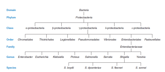 The species is the fundamental unit of bacterial classification. In clinical reporting, bacteria are referred to by their genus and species name (e.g. Staphylococcus aureus, Escherichia coli). A strain is a variant within a species with minor but detectable differences.
1. Classification based on cell wall and gram staining reaction
The nature of the bacterial cell wall is the primary criterion used in clinical bacterial classification. Gram staining — developed by Danish physician Hans Christian Gram in 1884 — divides most bacteria into two major groups based on cell wall composition.
| Group | Cell wall | Gram stain result |
|---|---|---|
| Gram-positive | Thick peptidoglycan layer (20–80 nm); no outer membrane | Purple |
| Gram-negative | Thin peptidoglycan layer (2–7 nm) + lipopolysaccharide outer membrane | Pink/red |
| Acid-fast | Thick waxy mycolic acid layer; resists gram stain | Neither (requires acid-fast stain) |
| Wall-less | No cell wall | Cannot be gram stained (Mycoplasma) |
Gram-positive bacteria
Cocci: Staphylococcus, Streptococcus, Enterococcus, Micrococcus, Peptostreptococcus
Spore-forming rods:
- Aerobic: Bacillus spp.
- Anaerobic: Clostridium spp.
Non-spore-forming rods:
- Non-filamentous: Corynebacterium, Listeria, Erysipelothrix, Lactobacillus
- Filamentous: Actinomyces, Nocardia, Streptomyces
 ### Gram-negative bacteria
Cocci: Neisseria spp., Moraxella catarrhalis, Veillonella (anaerobic)
Coccobacilli: Haemophilus, Bordetella, Brucella, Francisella, Acinetobacter, Pasteurella
Straight rods (Enterobacteriaceae): Escherichia, Klebsiella, Salmonella, Shigella, Proteus, Enterobacter, Serratia, Morganella, Yersinia
Curved and spiral rods: Campylobacter, Helicobacter, Vibrio
Obligate anaerobic rods: Bacteroides, Fusobacterium, Prevotella, Porphyromonas
Special groups
Acid-fast bacteria: Mycobacterium tuberculosis, M. leprae, Nocardia spp. — the waxy mycolic acid cell wall resists both gram stain and decolorization with acid-alcohol; requires Ziehl-Neelsen acid-fast stain.
Wall-less bacteria: Mycoplasma and Ureaplasma — lack a cell wall entirely, making them resistant to all beta-lactam antibiotics and invisible on gram stain.
Spirochetes: Treponema, Borrelia, Leptospira — thin flexible cell walls; too thin to visualize on gram stain; require dark-field microscopy or silver staining.
Check these articles:
→ Gram Staining: Principle, Procedure, Results
→ Peptidoglycan: Structure and Medical Significance
→ Cell Wall Composition, Structure and Functions
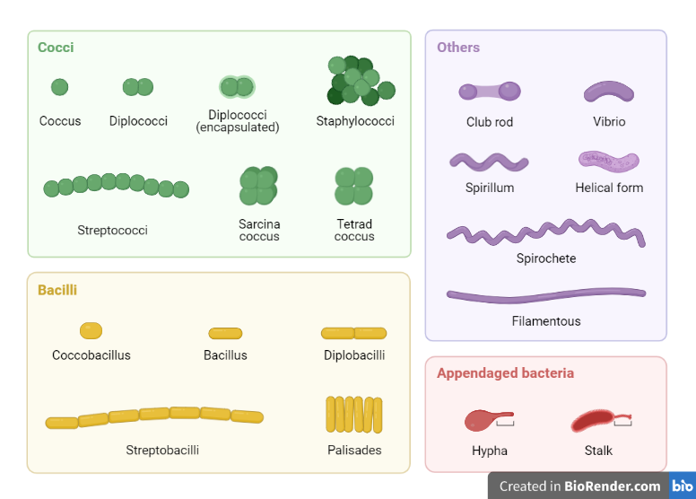
2. Classification based on shape and arrangement
Bacteria are classified into five basic groups based on shape:
| Shape | Name | Examples |
|---|---|---|
| Spherical | Cocci | Staphylococcus, Streptococcus, Neisseria |
| Rod-shaped | Bacilli | E. coli, Bacillus, Clostridium |
| Comma-shaped | Vibrios | Vibrio cholerae |
| Helical/rigid spiral | Spirilla | Spirillum spp. |
| Flexible spiral | Spirochetes | Treponema, Leptospira, Borrelia |
Arrangement (how cells group after division) is equally important diagnostically — grape-like clusters (staphylococci), chains (streptococci), diplococci (pneumococci, gonococci), and palisades (corynebacteria) are all clinically significant.
Check these articles:
→ Characteristics and Shape of Pathogenic Bacteria
3. Classification based on oxygen requirements
The ability to grow in the presence or absence of oxygen is one of the most clinically important bacterial characteristics — it directly determines which culture conditions, media, and incubation systems are required.
| Group | Oxygen relationship | Examples |
|---|---|---|
| Obligate aerobes | Require oxygen; cannot grow without it | Pseudomonas aeruginosa, Mycobacterium tuberculosis, Nocardia, Bacillus |
| Facultative anaerobes | Grow with or without oxygen; prefer oxygen if available | E. coli, Staphylococcus aureus, Klebsiella, Salmonella, Shigella |
| Obligate anaerobes | Cannot tolerate oxygen; killed by exposure | Clostridium tetani, Bacteroides fragilis, Fusobacterium, Prevotella |
| Aerotolerant anaerobes | Do not use oxygen but can survive in its presence | Streptococcus pyogenes, Lactobacillus |
| Microaerophiles | Require reduced oxygen (2–10%); killed by atmospheric O₂ | Campylobacter jejuni, Helicobacter pylori |
| Capnophiles | Require elevated CO₂ (5–10%) for growth | Neisseria gonorrhoeae, Streptococcus pneumoniae, Haemophilus influenzae |
Note: capnophiles are classified by CO₂ need, not O₂, but are grouped here for practical culture reasons.
Check these articles:
→ Oxygen Requirements for Pathogenic Bacteria
→ Cultivation of Aerobic and Anaerobic Bacteria
4. Classification based on temperature requirements
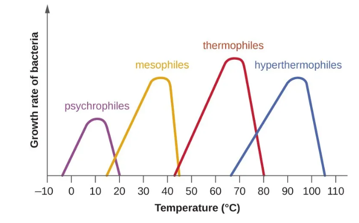 Bacteria are classified into five groups based on their optimum growth temperature. This classification has direct implications in diagnostic microbiology — incubation temperature is selected to favor the target pathogen.
| Group | Min | Optimum | Max | Clinical relevance |
|---|---|---|---|---|
| Psychrophiles | −20°C | 10–15°C | 20°C | Environmental; rarely cause human infection |
| Psychrotrophs | 0°C | 20–30°C | 35°C | Listeria monocytogenes, Yersinia enterocolitica — grow in refrigerators |
| Mesophiles | 10°C | 35–37°C | 45°C | Most human pathogens — optimized for body temperature |
| Thermophiles | 45°C | 50–60°C | 80°C | Geobacillus stearothermophilus — used as autoclave biological indicator. |
| Hyperthermophiles | 60°C | 80–110°C | >121°C | Archaea in hydrothermal vents; no human pathogens |
Check these articles:
5. Classification based on pH requirements
| Group | pH range | Examples |
|---|---|---|
| Acidophiles | 0–5.5 | Sulfolobus, Acidithiobacillus, Helicobacter pylori (tolerates gastric pH) |
| Neutrophiles | 5.5–8.0 | Most human pathogens — E. coli, Staphylococcus, Salmonella |
| Alkaliphiles | 8.0–11.5 | Bacillus alcalophilus, Vibrio cholerae (grows optimally at alkaline pH 8.4–8.6) |
The alkaline pH optimum of Vibrio cholerae is exploited diagnostically — alkaline peptone water (pH 8.4–8.6) is used as an enrichment broth to selectively grow vibrios from stool specimens before plating on TCBS agar.
6. Classification based on salt requirements
| Group | NaCl requirement | Examples |
|---|---|---|
| Non-halophiles | < 1% NaCl | Most human pathogens |
| Halotolerant | Grow best without NaCl but tolerate moderate salt | Staphylococcus aureus (tolerates up to 10% NaCl) — basis of mannitol salt agar selectivity |
| Slight halophiles | 1–5% NaCl optimal | Vibrio parahaemolyticus |
| Moderate halophiles | 5–20% NaCl optimal | Halobacillus, marine organisms |
| Extreme halophiles | 20–30% NaCl optimal | Halobacterium, Haloarcula (archaea, not human pathogens) |
7. Classification based on flagella
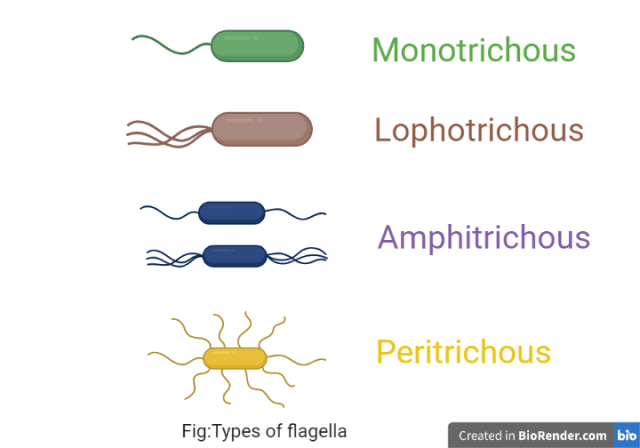 Flagella are protein appendages that provide bacterial motility. Their presence, number, and arrangement are taxonomically significant and are assessed by the Leifson flagella stain or electron microscopy.
| Type | Arrangement | Examples |
|---|---|---|
| Atrichous | No flagella | Staphylococcus aureus, Klebsiella pneumoniae |
| Monotrichous | Single polar flagellum | Vibrio cholerae, Pseudomonas aeruginosa |
| Lophotrichous | Cluster of flagella at one pole | Pseudomonas fluorescens, Helicobacter pylori |
| Amphitrichous | Flagella at both poles (single or cluster) | Alcaligenes faecalis, Aquaspirillum spp. |
| Peritrichous | Flagella distributed all over the cell surface | Salmonella Typhi, E. coli, Proteus mirabilis |
Check these articles:
8. Classification based on spore formation
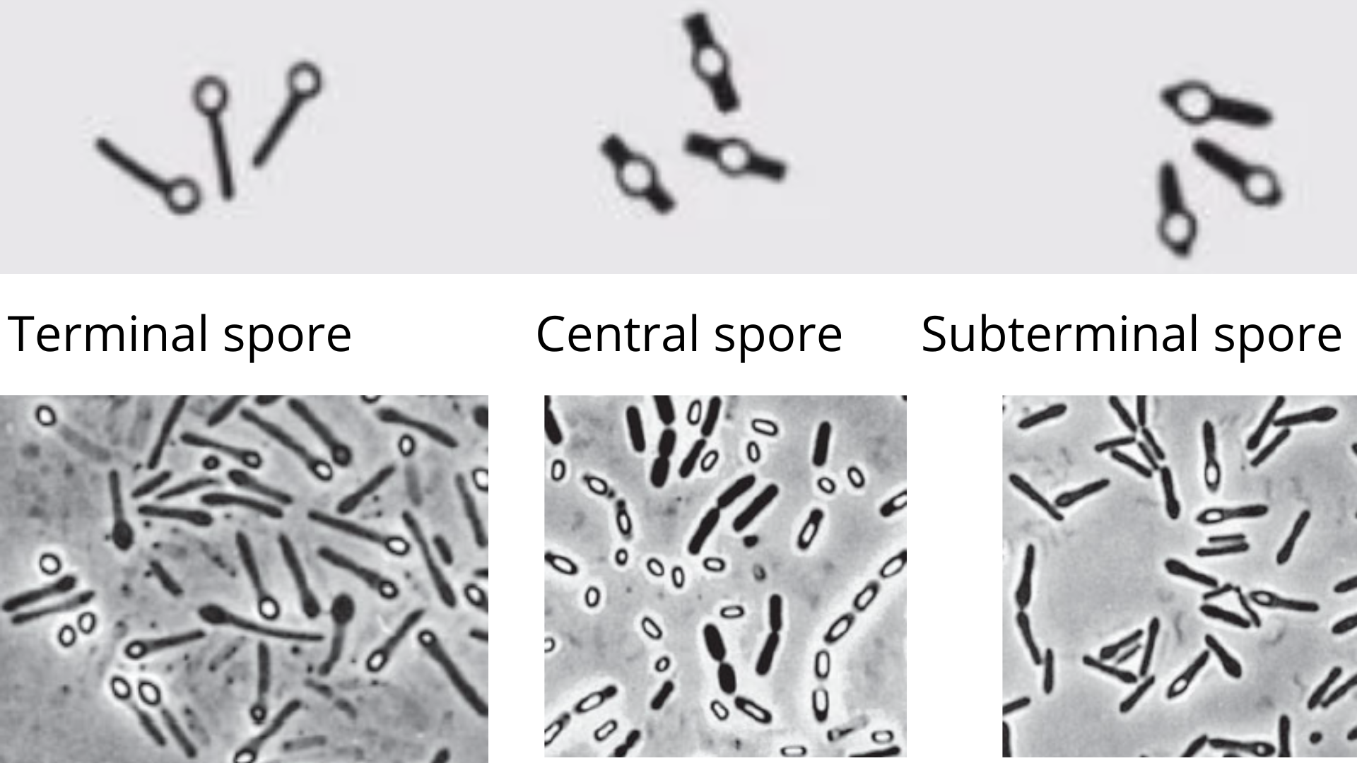 Bacterial endospores are dormant, highly resistant structures formed under adverse conditions (nutrient deprivation, desiccation, extreme temperature). Only gram-positive rods form endospores — this property is clinically significant because endospores resist standard disinfection, boiling, and many sterilization methods.
| Property | Details |
|---|---|
| Spore-forming bacteria | Bacillus spp. (aerobic), Clostridium spp. (anaerobic) |
| Non-spore-forming bacteria | All other bacteria including all gram-negative organisms |
| Spore positions | Central (Bacillus anthracis), subterminal (Clostridium perfringens), terminal (Clostridium tetani — "drumstick") |
| Resistance | Survive boiling (100°C), UV radiation, many disinfectants; killed by autoclaving (121°C, 15 min) |
| Clinical significance | C. tetani (tetanus), C. perfringens (gas gangrene), C. difficile (antibiotic-associated diarrhea), B. anthracis (anthrax) |
Check this article → Bacterial Spores: Structure, Resistance, and Significance
9. Classification based on capsule
A bacterial capsule is a polysaccharide (occasionally polypeptide) layer surrounding the cell wall. Capsule production is an important virulence factor — it protects bacteria from phagocytosis and complement-mediated killing.
| Group | Examples | Clinical significance |
|---|---|---|
| Capsulated bacteria | Streptococcus pneumoniae, Klebsiella pneumoniae, Haemophilus influenzae type b, Neisseria meningitidis, Bacillus anthracis | Enhanced virulence; resist phagocytosis; Quellung reaction for pneumococcus; India ink for Cryptococcus |
| Non-capsulated bacteria | Staphylococcus aureus, Shigella | Virulence achieved by other mechanisms |
Cryptococcus neoformans is a capsulated yeast, not a bacterium, but is included in many capsule discussions because its polysaccharide capsule is demonstrated by the same India ink method.
Check this article → Bacterial Capsule: Structure, Importance and Examples
10. Classification based on nutritional requirements
Bacteria are classified by their sources of carbon, energy, and electrons. While this classification is more relevant to environmental and industrial microbiology, it is tested in microbiology examinations.
| Classification | Criterion | Groups | Examples |
|---|---|---|---|
| Carbon source | Where carbon comes from | Autotrophs — use CO₂ | Cyanobacteria, nitrifying bacteria |
| Heterotrophs — use organic compounds | Most human pathogens | ||
| Energy source | How energy is obtained | Phototrophs — use light | Rhodospirillum, purple bacteria |
| Chemotrophs — use chemical oxidation | Most bacteria including all pathogens | ||
| Electron source | Electron donor | Lithotrophs — use inorganic compounds | Nitrosomonas, Thiobacillus |
| Organotrophs — use organic compounds | Most human pathogens |
Most clinically important human pathogens are chemo-organo-heterotrophs — they obtain energy by oxidizing organic compounds and use organic carbon as their carbon source.
11. Classification based on phylogenetic relationships (Bergey's Manual)
The most scientifically rigorous classification of bacteria is based on 16S ribosomal RNA (16S rRNA) gene sequencing, which reflects evolutionary relationships rather than phenotypic traits. This is the basis of Bergey's Manual of Systematic Bacteriology (5 volumes).
The three domains of life are:
| Domain | Description | Examples |
|---|---|---|
| Bacteria | True bacteria; peptidoglycan cell wall (most); all human bacterial pathogens | E. coli, S. aureus, M. tuberculosis |
| Archaea | Ancient prokaryotes; no peptidoglycan; no human pathogens known | Halobacterium, Methanobacterium, Sulfolobus |
| Eukarya | Eukaryotic organisms | Fungi, parasites, humans |
Major phyla of clinically important bacteria within the domain Bacteria:
| Phylum | Key clinical organisms |
|---|---|
| Firmicutes | Staphylococcus, Streptococcus, Enterococcus, Bacillus, Clostridium, Listeria |
| Proteobacteria | E. coli, Klebsiella, Salmonella, Pseudomonas, Neisseria, Haemophilus, Campylobacter, Helicobacter |
| Actinobacteria | Mycobacterium, Corynebacterium, Nocardia, Actinomyces |
| Bacteroidetes | Bacteroides fragilis, Prevotella, Porphyromonas |
| Spirochaetes | Treponema, Borrelia, Leptospira |
| Tenericutes | Mycoplasma, Ureaplasma (wall-less bacteria) |
| Chlamydiae | Chlamydia trachomatis, Chlamydophila pneumoniae |
Note: The article uses classic names (Firmicutes, Proteobacteria, Actinobacteria, Bacteroidetes, Spirochaetes, Tenericutes) but in 2021 many were formally renamed (Firmicutes → Bacillota, Proteobacteria → Pseudomonadota, Actinobacteria → Actinomycetota, etc.)
Where students get confused
Sorting out the oxygen categories. Aerotolerant vs facultative anaerobe, and microaerophile vs capnophile, are the two most-confused pairs in bacterial classification, and both are questions about oxygen and CO₂ rather than about the broader classification schemes. They are worked through in detail, with an enzyme-ladder memory aid and clinical anchors, in the dedicated article: Oxygen Requirements for Pathogenic Bacteria.
Capsule means virulence, not a taxonomic group. Students treat "capsulated" as a fixed category like Gram-positive. It is not. The capsule is a virulence factor that helps an organism resist phagocytosis, and the same species can have capsulated and non-capsulated strains. Haemophilus influenzae type b is capsulated and invasive; non-typeable H. influenzae lacks the capsule and behaves differently. Classify by capsule to understand virulence, not to assign a permanent identity.
The spore-as-sterilization-indicator paradox. Endospores are described as killed by autoclaving, yet Geobacillus stearothermophilus spores are the standard test used to check whether an autoclave works. Both are true, and that is the point. The spore is the hardest thing in the load to kill, so if the indicator spores are dead, everything easier to kill is dead too. The indicator works precisely because its spores die only when sterilization has genuinely been achieved.
Wall-less does not mean Gram-negative. Mycoplasma takes no Gram stain because it has no cell wall to stain, not because it is Gram-negative. The practical consequence is the one that matters clinically: with no peptidoglycan target, all beta-lactam antibiotics are useless against it, which is why Mycoplasma pneumonia is treated with a macrolide or tetracycline, never a penicillin.
Acid-fast is a third staining category, not a subtype of Gram. The waxy mycolic acid wall of Mycobacterium resists the Gram stain entirely, so a Gram stain of a TB specimen is unreliable. Acid-fast (Ziehl-Neelsen) staining is a separate method for a separate wall chemistry, not a special case of Gram staining.
References and further reading
- Tille, P. M. (2022). Bailey and Scott's Diagnostic Microbiology (15th ed.). St. Louis: Elsevier.
- Madigan, M. T., Bender, K. S., Buckley, D. H., Sattley, W. M., & Stahl, D. A. (2018). Brock Biology of Microorganisms (15th ed.). Pearson.
- Garrity, G. M. (Ed.). (2005). Bergey's Manual of Systematic Bacteriology (2nd ed.). Springer.
- Levinson, W. (2020). Review of Medical Microbiology and Immunology (16th ed.). McGraw-Hill.
- Murray, P. R., Rosenthal, K. S., & Pfaller, M. A. (2020). Medical Microbiology (9th ed.). Elsevier.
Frequently Asked Questions
What are the main criteria used to classify bacteria?
What is the difference between gram-positive and gram-negative bacteria?
What are the major phyla of clinically important bacteria?
What is the difference between obligate aerobes, facultative anaerobes, and obligate anaerobes?
Why are most human pathogens mesophiles?
What is Bergey's Manual?
What is the clinical significance of bacterial capsules?
What is the difference between spirilla and spirochetes?

Tankeshwar Acharya, MSc (Medical Microbiology)
Tankeshwar Acharya is an Assistant Professor in the Department of Microbiology at Patan Academy of Health Sciences (PAHS), Nepal, where he has been teaching and practicing clinical microbiology for over 14 years. He is the founder of Microbe Online, one of the leading free microbiology education resources on the web, covering bacteriology, mycology, parasitology, immunology, and clinical laboratory diagnostics written from direct experience in both the classroom and the diagnostic laboratory.