
Chocolate Agar (CAP): Composition, Preparation, Uses, and Colony Morphology
Chocolate agar is an enriched medium for isolating fastidious pathogens like Haemophilus and Neisseria. Learn its composition, preparation, CO₂ requirement, colony morphology, and key modifications like Thayer-Martin.

On this page
A three-year-old presents with fever, neck stiffness, and a bulging fontanelle. The pediatrician suspects bacterial meningitis and orders a CSF culture. The technician plates the sample onto blood agar and chocolate agar at the same time. By the next morning, the blood agar plate did not grow anything, but the chocolate agar plate shows small, grey, glistening colonies. The organism is Haemophilus influenzae. It cannot grow without two specific nutrients that only chocolate agar reliably provides.
Chocolate agar (CAP or CHOC) is a nonselective, enriched medium used to isolate fastidious pathogens: organisms with exacting nutritional requirements that fail to grow on routine media. The name comes from the color. Heating blood agar to 75–80°C lyzes the red blood cells and turns the medium from red to dark chocolate-brown. That same heating step releases the nutrients that make the medium work.
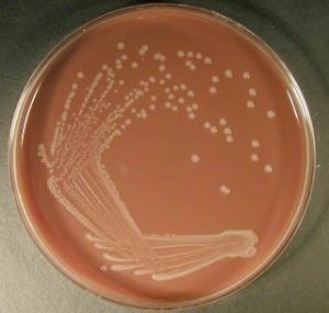 Figure: Haemophilus influenzae on chocolate agar
Principle
Chocolate agar is blood agar that has been heated to lyze the red blood cells. Lysis releases intracellular nutrients into the agar for fastidious bacteria to use: hemoglobin, hemin (X factor), and the coenzyme nicotinamide adenine dinucleotide (NAD, or V factor). Peptone supplies nitrogen and amino acids, sodium chloride maintains osmotic balance, and agar solidifies the medium.
Commercial chocolate agar reaches the same result by a different route and needs no added blood. Casein and animal tissue digest supply nitrogen and amino acids. Cornstarch neutralizes toxic fatty acids, to which Neisseria species are highly sensitive. Potassium phosphate holds the pH steady during growth. A hemoglobin solution supplies X factor (hemin) for Haemophilus species, and IsoVitaleX enrichment supplies V factor (NAD) along with amino acids, vitamins, and other cofactors that support fastidious organisms, including Neisseria species.
Why Chocolate Agar and Not Blood Agar?
This is the central conceptual question about the medium, and it comes down to two growth factors.
Haemophilus influenzae needs both X factor (hemin) and V factor (NAD, nicotinamide adenine dinucleotide) to grow. On blood agar, the red blood cells stay intact. Hemin is locked inside them, and the NAD that is present gets destroyed by NADase enzymes released from the same cells.
Heating blood agar to 75–80°C to make chocolate agar solves both problems at once:
- The red blood cells lyze, releasing hemin (X factor) into the agar.
- The heat inactivates the NADases that would otherwise destroy V factor.
- V factor (NAD) is released and stays available for the bacteria to use.
Neisseria species also need enriched media, and they are additionally sensitive to toxic fatty acids in many base agars. Cornstarch in chocolate agar neutralizes these.
The result is a medium that supports organisms blood agar cannot. This is why the two plates are used together for cerebrospinal fluid, sputum, and genital specimens: blood agar for the majority of organisms, chocolate agar for the fastidious pathogens.
The comparison table below shows the difference directly.
| Feature | Blood Agar | Chocolate Agar |
|---|---|---|
| RBC state | Intact | Lysed by heat |
| X factor (hemin) available | Locked in RBC | Released into agar |
| V factor (NAD) available | Destroyed by NADases | Released; NADases inactivated |
| H. influenzae growth | Poor to none | Good |
| N. gonorrhoeae growth | Poor | Good |
| S. pneumoniae growth | Good (alpha hemolysis visible) | Good (no hemolysis visible) |
| Hemolysis visible | Yes | No (RBCs already lysed) |
Key teaching point: Chocolate agar is not "better" than blood agar. It is used alongside it. Blood agar shows hemolysis patterns that chocolate agar cannot, because the red blood cells are already destroyed. The two media do different jobs.
Composition of Chocolate Agar
The composition of chocolate agar is the same as that of blood agar. The only difference is blood agar is heated in a water bath to prepare chocolate agar. Commercially available chocolate agar medium, however, has a different composition and requires no addition of blood.
Ingredients | Gm/L |
Casein/animal tissue digest | 15.0 |
Cornstarch | 1.0 |
Potassium phosphate, dibasic | 4.0 |
Potassium phosphate, monobasic | 1.0 |
Sodium chloride | 5.0 |
10.0 | |
Hemoglobin solution (2%) | 500.0 ml |
IsoVitaleX enrichment | 10.0 ml |
Preparation of Chocolate Agar
- Prepare the blood agar base as instructed by the manufacturer.
- Sterilize by autoclaving at 121°C for 15 minutes.
- Add 5-7% v/v of defibrinated blood (horse or sheep blood). Then place the media in a water bath of 75 -80°C.Keep swirling gently until the color changes to dark brown.
- Pour into sterile Petri plates under aseptic conditions after the media has cooled to 50-55°C.
- Label the plates with the name of the media, and date of preparation, and store them invertedly at 2-8°C until use.
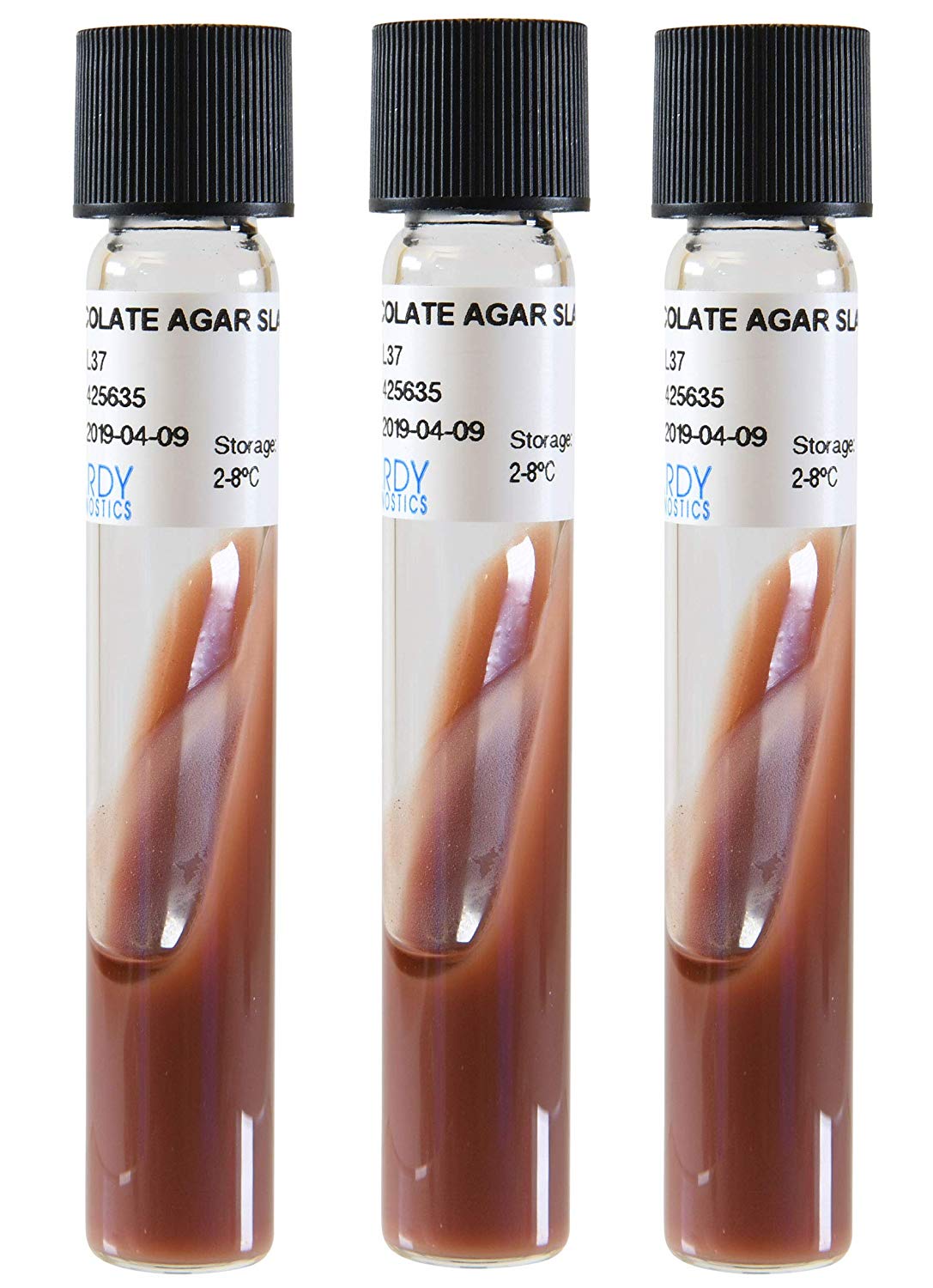 Figure: Chocolate agar slants
Note: Other supplements such as IsoVitaleX could be added during preparation.
Chocolate agar slants
- Dispense 4 mL of the medium into 16X125 mm screw-cap tubes
- Keep the tubes in a slanted position and let them solidify.
This agar slants appear brown to brownish-red color. Store these at 4°C when not in use and warmed to room temperature (25°C) before use.
Inoculation and Incubation
- Allow the plate to reach room temperature and dry the agar surface before use. Inoculate as soon as possible after the specimen is received.
- Inoculate directly from the specimen. Common specimens include cerebrospinal fluid, sputum and lower respiratory samples, blood culture subcultures, genital swabs for Neisseria gonorrhoeae, and eye or ear swabs.
- If culturing from a swab, roll the swab over a small area at the edge of the plate first, then streak out from that area.
- Streak for well-isolated colonies over four quadrants using a sterile loop.
- Incubate at 35 to 37°C in a humidified atmosphere with 5 to 10% CO₂. Haemophilus and Neisseria are capnophilic, so the CO₂ is not optional for reliable recovery. Use a CO₂ incubator, or a candle jar in resource-limited settings, noting that a candle jar provides only about 3% CO₂.
- Read the plate at both 18 to 24 hours and 48 hours. Many fastidious organisms are slow, so do not report the plate negative at 24 hours. For cerebrospinal fluid cultures, extend incubation to 72 hours.
- For respiratory specimens, do not incubate beyond 48 hours, because normal flora overgrow the plate and can mask the pathogen.
- Subculture N. gonorrhoeae within 18 to 24 hours if it is the target organism, because it does not survive well on the primary plate.
Colony Morphology
Almost all organisms grow on this agar giving grey colonies of various sizes. However, it’s use is specifically to isolate fastidious pathogens such as Neisseria and Haemophilus, their colony morphologies are below.
| Organism | Colony on Chocolate Agar | Key distinguishing feature |
|---|---|---|
| Haemophilus influenzae | Small, colorless to grey, moist, non-hemolytic | Characteristic "mousy" or "bleach-like" odor |
| H. parainfluenzae | Similar to H. influenzae; grows without X factor | Requires V factor only (no X factor requirement) |
| Neisseria gonorrhoeae | Pinkish-brown, translucent, smooth, 0.5–1 mm | Small; oxidase positive; confirmed by sugar utilization |
| N. meningitidis | Grey, non-hemolytic, round, convex, smooth, moist, glistening; larger than N. gonorrhoeae | Grows faster than gonococci; confirmed by serogrouping |
| Streptococcus pneumoniae | Small, grey, mucoid colonies; no visible hemolysis (RBCs already lysed) | Hemolysis is not visible in chocolate agar, use blood agar to see alpha hemolysis |
| Moraxella catarrhalis | Grey-white, dry, brittle, dome-shaped; can be slid across agar intact ("hockey puck" sign) | Non-hemolytic; oxidase positive |
Note: Almost all common organisms (staphylococci, streptococci, Enterobacteriaceae) will also grow on chocolate agar, producing grey colonies of varying sizes. The medium is enriched, not selective. Identification of fastidious organisms relies on recognizing their specific colony characteristics within a mixed growth.
Quality Control
Perform sterility testing of the prepared media by incubating 3-5 uninoculated plates of each batch at 35-37°C for 18-24 hours. Regard any growth as positive, and discard the whole lot.
Performance testing of the media should be done by inoculating the following organisms to prepared plates. Then incubating them for 18-24 hours at 35-37°C with 5% CO₂ (or in a candle jar but it can only provide up to 3% CO₂). This helps to determine the growth performance of the completed medium.
Organism | Expected Result |
Neisseria gonorrhoeae ATCC 43069 | Luxuriant growth, small, grey to white colonies |
Haemophilus influenzae ATCC 10211 | Good growth, small, colorless, moist colonies |
Why 5% CO₂?
Haemophilus and Neisseria species are capnophilic, they grow best in elevated carbon dioxide concentrations. CO₂ lowers the pH of the medium slightly and stimulates growth of these organisms. In well-resourced laboratories, a CO₂ incubator set at 5% is standard. In resource-limited settings, a candle jar (a sealed jar with a lit candle that burns until oxygen is consumed, generating approximately 3% CO₂) is an acceptable alternative, though slightly less optimal. Always note which method was used when reporting culture conditions.
Uses of Chocolate Agar
- CSF cultures (bacterial meningitis workup): Chocolate agar is plated alongside blood agar for all cerebrospinal fluid specimens. H. influenzae (especially in unvaccinated children) and N. meningitidis require this medium for primary isolation.
- Respiratory specimens: Sputum, bronchial lavage, and nasopharyngeal swabs are plated on chocolate agar to recover H. influenzae, a common cause of lower respiratory tract infections and COPD exacerbations.
- Genital and urethral specimens (STI workup): Neisseria gonorrhoeae requires enriched media. Chocolate agar supports initial growth; Thayer-Martin medium (a modification) is used when specimens are from non-sterile sites containing mixed flora.
- Eye specimens (conjunctivitis workup): Both H. influenzae and N. gonorrhoeae cause conjunctivitis, including neonatal ophthalmia neonatorum. Chocolate agar is part of the standard eye specimen battery.
- Selective isolation with bacitracin: Chocolate agar supplemented with bacitracin suppresses oral flora (particularly streptococci) and improves primary isolation of H. influenzae from sputum specimens with heavy background flora.
Limitations
- Since this agar is an enriched medium, contamination occurs easily.
- Use of this agar only, makes it difficult to differentiate non-pathogenic organisms from pathogenic ones as the former overgrow pathogenic bacteria.
- It has a reduced concentration of agar, so the surface of the plate is fragile and prone to scratches during streaking.
Modifications of Chocolate Agar
- Thayer-Martin Media: It is a modification of chocolate agar supplemented with vancomycin, nystatin, and colistin. It helps to inhibit the normal flora, including nonpathogenic Neisseria for the selective isolation of N. gonorrhoeae and N. meningitidis.
- Chocolate Agar with bacitracin: This modification is useful as a selective medium to improve the primary isolation of H. influenzae from specimens such as sputum. As they may contain a mixed flora of bacteria and/or fungi.
- Chocolate agar with GC base and growth supplement: This variant of chocolate agar is used to support the special growth requirements (hemin and NAD). It is needed for the isolation of Haemophilus spp. when incubated at 35-37°C in a 5% CO₂ atmosphere.
- Chocolate agar with TSA and growth supplements: It is a modification of chocolate agar that supports the special growth requirements (hemin and NAD) needed for the isolation of fastidious organisms such as H. influenzae when incubated at 35-37°C in a 5% CO₂ atmosphere.
How to Remember
The name tells the story. Heated blood agar turns chocolate-brown because the red blood cells are lysed. That color change is not just cosmetic. It is visual proof that the heating worked and the growth factors have been released. If your chocolate agar still looks red or pink, the blood was not heated enough, and the medium will not support fastidious organisms.
X and V, a simple rule:
- Haemophilus influenzae needs both X and V factors, so it grows on chocolate agar but not on plain blood agar.
- H. parainfluenzae needs V factor only, so it can grow on blood agar as well.
A quick mnemonic: "Influenzae needs eXtra Vitamins" (X = hemin, V = the NAD, a vitamin-like factor).
Satellitism, a bench observation that ties it together:
Streak H. influenzae onto a blood agar plate next to Staphylococcus aureus. The H. influenzae colonies grow only near the S. aureus streak, because S. aureus lyzes the local red blood cells and releases V factor into the surrounding agar. The H. influenzae colonies appear to cluster, or "satellite," around the S. aureus. One plate demonstrates the X and V factor requirement and the principle behind chocolate agar at the same time.
The clinical decision rule:
Suspect Haemophilus or Neisseria? Use chocolate agar. Need to see hemolysis? Use blood agar. For any serious infection workup, plate both.
References
- Tille, P. M. (2022). Bailey and Scott's Diagnostic Microbiology (15th ed.). Elsevier.
- Forbes, B. A., Sahm, D. F., & Weissfeld, A. S. (2007). Bailey and Scott's Diagnostic Microbiology (12th ed.). Mosby Elsevier.
- Mahon, C. R., Lehman, D. C., & Manuselis, G. (2018). Textbook of Diagnostic Microbiology (6th ed.). Elsevier.
- Clinical and Laboratory Standards Institute (CLSI). (2023). M22: Quality Control for Commercially Prepared Microbiological Culture Media (5th ed.). CLSI.
- BD Diagnostics. Chocolate Agar (GC II Agar with IsoVitaleX). Package Insert. Becton, Dickinson and Company.

Tankeshwar Acharya, MSc (Medical Microbiology)
Tankeshwar Acharya is an Assistant Professor in the Department of Microbiology at Patan Academy of Health Sciences (PAHS), Nepal, where he has been teaching and practicing clinical microbiology for over 14 years. He is the founder of Microbe Online, one of the leading free microbiology education resources on the web, covering bacteriology, mycology, parasitology, immunology, and clinical laboratory diagnostics written from direct experience in both the classroom and the diagnostic laboratory.
Comments
No comments yet. Be the first to share your thoughts.
Leave a comment
All comments are reviewed before they appear.