
Calcofluor White Staining: Principle, Procedure, Results, and Clinical Applications

On this page
Calcofluor white staining uses fluorescent dyes to stain the chitin and cellulose in the fungi, plants, and algae cell walls. Calcofluor binds non-specifically to the chitin and cellulose, and exposure to the long-wavelength ultraviolet and short-wavelength visible light helps in colored observation.
Although KOH wet mount is the recommended method for direct examination of fungal stains, it has lower sensitivity and can not differentiate the hyphae from the collagen fibers and other artifacts. So, calcofluor white staining is preferred nowadays.
This technique helps visualize a mixed fungal infection as well. When coupled with the KOH, the calcofluor white stain provides better sensitivity to detect fungi in the clinical specimen. The KOH clears the cell debris. Calcofluor then colors fungus, which appears bright apple-green through the standard barrier filter of a fluorescence microscope.
Principle of Calcofluor White Staining
Calcofluor white (CFW) was originally developed as a textile brightening agent. Its ability to bind cellulose and fluoresce brightly under UV light made it useful for whitening fabric. Microbiologists adopted it for the same reason: fungal cell walls contain chitin and cellulose, and CFW binds both, making fungal elements glow brilliantly under a fluorescence microscope while the surrounding tissue remains relatively dark.
When the CFW comes in contact with clinical specimens, it binds with the 1-3 beta and 1-4 beta polysaccharides on the chitin and cellulose if fungal elements are present. Then it fluoresces into green color when exposed to ultraviolet light. It can be observed under a fluorescent microscope, and the differentiation of infectious compounds is based on color and morphology.
The CFW excites at 380 nm wavelength. The absorption in calcofluor white stain occurs over the 300-412 nm range. Its absorbance peak is 347nm, and its emission peak is 475 nm. Using violet or blue light also gives good results, but one prefers UV light because maximum excitation and fluorescence occur with it. During the fluorescent microscopic observation, fungi and other organisms like a cyst of Pneumocystis fluoresce brilliant apple-green. The green coloration is due to barrier filters in the fluorescence microscope. Other elements present in the sample will fluoresce as reddish-orange. Yellowish-green background fluorescence can also be observed when a tissue sample is used. Observing the slide under the blue light and using various combinations of exciter and barrier filter can diminish background fluorescence.
Evans blue is used as a counterstain and diminishes the background fluorescence by using the blue light excitation (not UV). The addition of 0.1 % of Evans blue minimizes the non-specific background fluorescence. Evans blue counter stain also produces the contrasting orange to ruby-red background and aids in clearly demonstrating the fungi in the surrounding tissue.
Preparation of the Calcofluor White Stain
The preparation of calcofluor white stain requires calcofluor white powder, which is dissolved in distilled water. 1% (w/v) calcofluor white stain is prepared by properly dissolving the 1 g powder of calcofluor white and 100 ml distilled water. The best way to use the solution is to dilute the 1% CFW solution to 0.1%. Storage should be done at room temperature and in the dark helps preserve the solution for years.
Adding 10% KOH solution (10 g KOH powder in 90 ml distilled water and 10 ml glycerol) to the CFW just before use is helpful while handling skin and nail samples.
Likewise, 0.05% to 0.01% Evans blue solution (w/v) can be added to the CFW as counter stain. Evans blue solution can also be used after using a calcofluor white stain.
Calcofluor White Staining Overview
| Method | Calcofluor white staining |
|---|---|
| Use | Detection of fungal pathogens |
| Time required | Approximate 5 minutes |
| Advantages | Clear demonstration of the fungus, rapid method, higher sensitivity |
| Disadvantages | Need fluorescent microscope, which cannot be afforded by every routine laboratory |
Procedure
- Firstly take a clean, grease-free glass slide and place the sample in the center.
- Then add one drop of calcofluor white stain or CFW with Evans blue solution on top of the sample.
- For nail and skin scrapings, add 10% KOH in the slide.
- Then, put the coverslip on top and let it stand for a minute.
- After that, observe the slide under UV light at 100X to 400X magnifications.
Result and Interpretation of Calcofluor White Staining
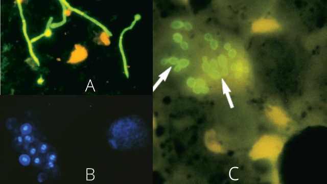 Figure: A. Calcofluor white stain of urine demonstrates Candida albicans B.Calcofluor white staining of Pneumocystis organisms in bronchoalveolar lavage (BAL) specimen C. Calcofluor white stain of sputum showing intracellular yeast cells of Histoplasma capsulatum (arrows), Image source: Bailey and Scott, DOI:10.1177/1753465810380102
- Fungi and the parasites fluoresce apple-green while its other elements fluoresce reddish-orange.
- Cotton fibers fluoresce more intensely than the fungal hyphae, so one must carefully observe them.
- In free-living amoebae such as Acanthamoeba, both trophozoites and cysts fluoresce with CFW however, cysts fluoresce more intensely due to their higher chitin content in the double-walled cyst wall. This differential fluorescence intensity helps distinguish cysts from trophozoites in corneal scraping specimens.
- Cysts of the Pneumocystis are observed as a round cell having a uniform shape with a 5-8 µm diameter. It is differentiated from yeast cells based on budding characteristics and deep internal staining.
- The reaction may be non-specific in the tissue samples.
Application of the Calcofluor White Staining
Calcofluor white staining is one of the methods being used frequently for the rapid detection of fungal infections. Some of the more detailed application of calcofluor white staining are as follows:
- Calcofluor white is a sensitive stain that helps to visualize the hyphae, pseudohyphae, and yeast. Since the chitin concentration is higher in the budding yeasts, calcofluor white stains the bud scars more intensely.
- Calcofluor white stain helps detect non-culturable fungus like Pneumocystis jirovecii.
- Calcofluor stain can be applicable for observing non-fungal agents such as free-living amoebae (Acanthamoeba, Naegleria, and Balamuthia) and larva of Dirofilaria. Calcofluor white stain helps in the rapid diagnosis of Acanthamoeba keratitis from the corneal scrapings and keratectomy specimens.
- Calcofluor white stain can be incorporated into the growth media because it can withstand the autoclave conditions (121°C for 15 minutes). So, a calcofluor white stain can be used as the vital stain growing fungus in the slide culture.
- Calcofluor white stain stains the vegetative cells but not the ascospores. So, it helps in differentiation when incorporated into the ascospore-inducing media.
Vital stain means the process of adding the stain on living cells without killing it.
Specimen-Specific Guide: When and How to Use Calcofluor White
| Specimen Type | Clinical Suspicion | Preparation | What to Look For |
|---|---|---|---|
| Skin scrapings | Dermatophytosis (tinea) | CFW + 10% KOH; let stand 5 min | Septate hyphae fluorescing bright green/blue-white at skin/hair boundary |
| Nail scrapings | Onychomycosis | CFW + 20% KOH (stronger for nail keratin); 10–15 min | Hyphae within nail plate; distinguish from nail fibres (fibres fluoresce more intensely) |
| Hair (scalp, beard) | Tinea capitis | CFW + KOH; examine shaft and root | Hyphae within hair shaft (endothrix) or around shaft (ectothrix) |
| Corneal scraping | Acanthamoeba keratitis | CFW alone or with Evans blue | Double-walled cysts (5–25 μm) fluorescing brightly; trophozoites less prominent |
| BAL fluid | Pneumocystis jirovecii PCP | CFW alone; cytospin preparation | Round oval cysts 4–6 μm; uniform shape; no budding |
| BAL / bronchial washing | Invasive pulmonary fungal infection | CFW + KOH | Septate hyphae (Aspergillus) or aseptate broad hyphae (Mucorales) |
| CSF | Cryptococcal meningitis | CFW + India ink | Yeast cells with narrow-based budding; capsule visible on India ink |
| Sputum | Pulmonary fungal infection | CFW + KOH | Hyphae, yeast cells; note intracellular yeast (Histoplasma within macrophages) |
| Tissue biopsy | Invasive fungal infection | CFW on frozen or paraffin section | Fungal elements in tissue; compare with PAS and GMS |
| Stool | Intestinal protozoa | CFW alone | Cryptosporidium oocysts (4–6 μm); Cyclospora (8–10 μm) |
Combined KOH-CFW method: For skin, nail, and hair specimens, combining 10% KOH with CFW is standard practice. KOH dissolves keratin and host cell debris (clearing the specimen), while CFW simultaneously stains the fungal elements. The combination produces better sensitivity than either method alone.
Limitations of Calcofluor White Staining
Equipment requirement: CFW requires a fluorescence microscope with UV or near-UV light source and appropriate excitation (380–412 nm) and barrier filters. This is the primary limitation in resource-limited settings where KOH wet mount or LPCB are used instead.
Non-specific binding: CFW binds all β-glucan and chitin-containing structures, not just pathogenic fungi:
- Cotton fibers fluoresce more intensely than fungal hyphae and are the most common source of false-positive confusion, particularly in respiratory specimens where cotton-tipped swabs may have shed fibers
- Plant material, cellulose in food particles, and insect cuticle also fluoresce
- The characteristic morphology (branching hyphae, budding yeast) must always be confirmed alongside fluorescence
Cannot distinguish viable from non-viable organisms: CFW stains both live and dead fungal elements, a positive result in a treated patient may represent residual non-viable organisms rather than active infection.
Cannot identify species: Like KOH, CFW reveals fungal morphology but cannot identify genus or species. Culture remains essential for definitive identification and antifungal susceptibility testing.
Background fluorescence in tissue: Collagen, elastin, and some tissue components auto-fluoresce, potentially obscuring fungal elements. Evans blue counterstain reduces this but does not eliminate it entirely.
Troubleshooting Calcofluor White Staining
| Problem | Likely Cause | Action |
|---|---|---|
| Bright fibers obscuring hyphae | Cotton fibers from swab contamination | Use nylon or dacron swabs; examine carefully, fibers are irregular and non-branching; confirm morphology |
| Excessive background fluorescence | Tissue autofluorescence; too much CFW | Add Evans blue counterstain; reduce CFW concentration to 0.1%; use blue light excitation instead of UV |
| Fungal elements not visible despite clinical suspicion | Specimen too sparse; inadequate KOH digestion | Allow KOH to act longer (10–15 min for nail/skin); prepare a second slide with higher inoculum; consider repeat specimen |
| Cysts and hyphae indistinguishable | Morphological overlap at low power | Use 40x objective for morphological detail; look for budding (yeast) vs non-budding (cysts); confirm with culture |
| Fluorescence fading rapidly | High-intensity UV exposure bleaching fluorochrome | Examine promptly; use lower UV intensity; seal coverslip with nail varnish to slow fading |
| Stain solution appears yellow | Degradation due to light exposure | Store in dark container at room temperature; discard yellow-tinged solution; prepare fresh |
How to Remember
"Calcofluor = Chitin Fluoresces": Calcofluor White (CFW) binds to chitin (and cellulose) and fluoresces brightly under UV light. It naturally emits a blue-white glow, but through the standard microscope barrier filter it appears bright apple-green. This is the basic principle. CFW works because it binds to the β-linked polysaccharides found in fungal cell walls, Pneumocystis cyst walls (rich in β-glucan), and Acanthamoeba cyst walls (which contain cellulose and chitin). In simple terms, if a structure contains these β-linked cell wall polysaccharides, CFW will bind to it and make it fluoresce.
The "CFW + KOH = see more" rule: KOH clears the debris; CFW lights up the fungi. Together they outperform either alone for skin, nail, and hair specimens. This is the combined method for dermatophyte diagnosis.
Cotton fibers are the enemy: The biggest practical problem with Calcofluor White (CFW) is that cotton fibers also fluoresce brightly. To an inexperienced observer, they can look like fungal hyphae and cause false-positive results. Before reporting a positive result, always examine the morphology carefully. True fungal hyphae have features such as branching, septation, and a relatively uniform width, whereas cotton fibers do not.
Three non-fungal organisms CFW detects:
- Pneumocystis jirovecii cysts (BAL)
- Acanthamoeba cysts and trophozoites (corneal scraping)
- Cryptosporidium / Cyclospora oocysts (stool)
Key Exam Facts
| Feature | Detail |
|---|---|
| Dye type | Fluorescent brightener; binds β-1,3 and β-1,4 polysaccharides (chitin, cellulose) |
| Excitation wavelength | 380–412 nm (UV/near-UV) |
| Emission colour | Bright apple-green (with barrier filter) |
| Counterstain | Evans blue, reduces background fluorescence; produces orange-red background |
| Combined with KOH | Yes, for skin, nail, hair specimens (KOH clears; CFW stains) |
| Fungi appearance | Bright green/blue-white fluorescence |
| Pneumocystis appearance | Round oval cysts 4–6 μm; uniform; non-budding |
| Acanthamoeba appearance | Double-walled cysts 5–25 μm; intense fluorescence |
| Main advantage over KOH | Higher sensitivity; detects sparse fungal elements; distinguishes hyphae from artefacts |
| Main limitation | Requires fluorescence microscope; non-specific (cotton fibres, plant material also fluoresce) |
| Cannot do | Species identification; distinguish viable from non-viable organisms |
| Storage | Room temperature, in dark; 1% stock diluted to 0.1% working solution |
References
- Monheit, J. E., Cowan, D. F., & Moore, D. G. (1984). Rapid detection of fungi in tissues using calcofluor white and fluorescence microscopy. Archives of pathology & laboratory medicine, 108(8), 616–618.
- Wilhelmus, K. R., Osato, M. S., Font, R. L., Robinson, N. M., & Jones, D. B. (1986). Rapid diagnosis of Acanthamoeba keratitis using calcofluor white. Archives of ophthalmology (Chicago, Ill.: 1960), 104(9), 1309–1312. https://doi.org/10.1001/archopht.1986.01050210063026
- Harrington, B. J., & Hageage, G. J. (2003). Calcofluor White: A Review of its Uses and Applications in Clinical Mycology and Parasitology. Laboratory Medicine, 34(5), 361–367. https://doi.org/10.1309/eph2tdt8335gh0r3
- Chander, J. (2018). Textbook of Medical Mycology (Fourth edition). Jaypee Brothers Medical Publishers Ltd.
- Murray PR, Rosenthal KS, Pfaller MA. Medical Microbiology. 9th ed. Elsevier; 2020.
- Larone DH. Larone's Medically Important Fungi: A Guide to Identification. 6th ed. ASM Press; 2018.
Frequently Asked Questions
What does calcofluor white stain and why does it work on fungi?
Calcofluor White is mainly used to rapidly detect fungi in clinical specimens such as skin scrapings, nail clippings, hair, corneal scrapings, sputum, bronchoalveolar lavage (BAL), tissue, and other body fluids. Because staining takes only a few minutes, it is often used as a quick screening test before culture results are available. The fluorescent stain makes even small numbers of fungal elements easier to see than routine light microscopy.
One important limitation is that CFW is not specific for fungi. Any material containing chitin or cellulose, including cotton fibers, plant material, and some environmental debris, can also fluoresce. For this reason, a positive fluorescent structure should always be identified by its morphology, such as branching, septation, budding, or yeast shape, rather than by fluorescence alone.
What is the combined KOH-CFW method and when is it used?
Cotton fibres are the most common cause of false-positive results with calcofluor white (CFW) staining. They fluoresce very brightly under UV light, sometimes even more brightly than fungal hyphae, and can easily be mistaken for fungi.
To avoid this error:
Use nylon or dacron swabs instead of cotton-tipped swabs for specimen collection.
Do not rely on fluorescence alone. Always examine the morphology of the fluorescent structure.
True fungal hyphae have a uniform width, branching, and septation, whereas cotton fibres are irregular, non-branching, and variable in width. Careful examination under high power helps distinguish cotton fibres from fungal hyphae.

Tankeshwar Acharya, MSc (Medical Microbiology)
Tankeshwar Acharya is an Assistant Professor in the Department of Microbiology at Patan Academy of Health Sciences (PAHS), Nepal, where he has been teaching and practicing clinical microbiology for over 14 years. He is the founder of Microbe Online, one of the leading free microbiology education resources on the web, covering bacteriology, mycology, parasitology, immunology, and clinical laboratory diagnostics written from direct experience in both the classroom and the diagnostic laboratory.
Comments
No comments yet. Be the first to share your thoughts.
Leave a comment
All comments are reviewed before they appear.