
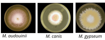
Microsporum: Species, Identification, and the Rice Grain Test
The Microsporum species that matter in the lab: spindle-shaped thick-walled macroconidia, colony appearance of M. canis, M. gypseum, and M. audouinii, Wood's lamp fluorescence, and...
Articles tagged with dermatophytes.
The Microsporum species that matter in the lab: spindle-shaped thick-walled macroconidia, colony appearance of M. canis, M. gypseum, and M. audouinii, Wood's lamp fluorescence, and...
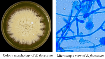
How to identify Epidermophyton floccosum: khaki-green powdery colony, club-shaped (beaver-tail) macroconidia in clusters, and the key feature that it produces macroconidia only and...
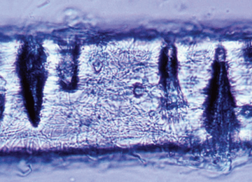
The in vitro hair perforation test for dermatophytes: principle, procedure, and how to read it. Which organisms are positive, and how it separates T. mentagrophytes from T. rubrum...
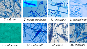
The Trichophyton species that matter in the lab: colony and microscopic appearance, pencil-shaped macroconidia, endothrix vs ectothrix hair invasion, and how to tell T. rubrum from...
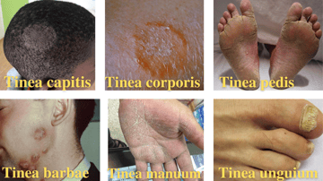
The tinea infections explained by site: tinea capitis, corporis, pedis, cruris, unguium, and more. Common organisms, clinical features, and how the site names work.
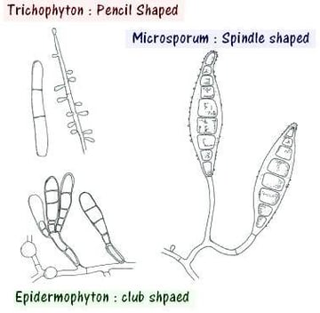
How the laboratory identifies dermatophytes: KOH microscopy, SDA culture, Wood's lamp, and the differential tests that separate look-alike Trichophyton, Microsporum, and Epidermoph...
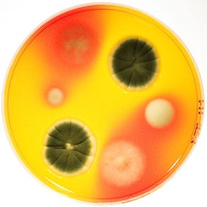
Why a clean, unchanged DTM tube after two weeks doesn't always mean no fungal infection, the color-change logic explained, and the one ingredient that can silently suppress a true...