
Types of Tinea (Ringworm): Sites, Causes, and Clinical Features
The tinea infections explained by site: tinea capitis, corporis, pedis, cruris, unguium, and more. Common organisms, clinical features, and how the site names work.

On this page
A medical student on a dermatology rotation sees three patients in one morning: an itchy ring on a wrestler's shoulder, a boy with a scaly bald patch and broken hairs on his scalp, and an adult with peeling, cracked skin between the toes. All three are ringworm. All three are the same kind of organism, a dermatophyte. Yet each has a different name, a different likely species, and a different treatment.
This is the logic of tinea naming, and once it clicks the whole topic becomes easy. Tinea infections are named for the body site, not the fungus. The species that causes the ring on the shoulder can be the same one causing the peeling between the toes.
This article walks through each tinea infection by site: what it looks like, which organisms usually cause it, and how treatment differs. For how the laboratory identifies the fungus once a specimen arrives, see the companion article: Dermatophytes: Lab Diagnosis and How to Tell the Three Genera Apart.
What "tinea" means
Tinea is the medical name for a dermatophyte infection of the skin, hair, or nails. The everyday word is ringworm, which is misleading because no worm is involved. The name comes from the ring-shaped lesion many of these infections produce: a raised, scaly, advancing border with clearer skin in the center, formed as the fungus spreads outward from where it first landed.
The three genera of dermatophytes, Trichophyton, Microsporum, and Epidermophyton, all live on keratin, the tough protein in skin, hair, and nails. The clinical name of the infection depends only on where on the body it takes hold. This is why the same species, most often Trichophyton rubrum, can appear under several different tinea names.
The tinea infections at a glance
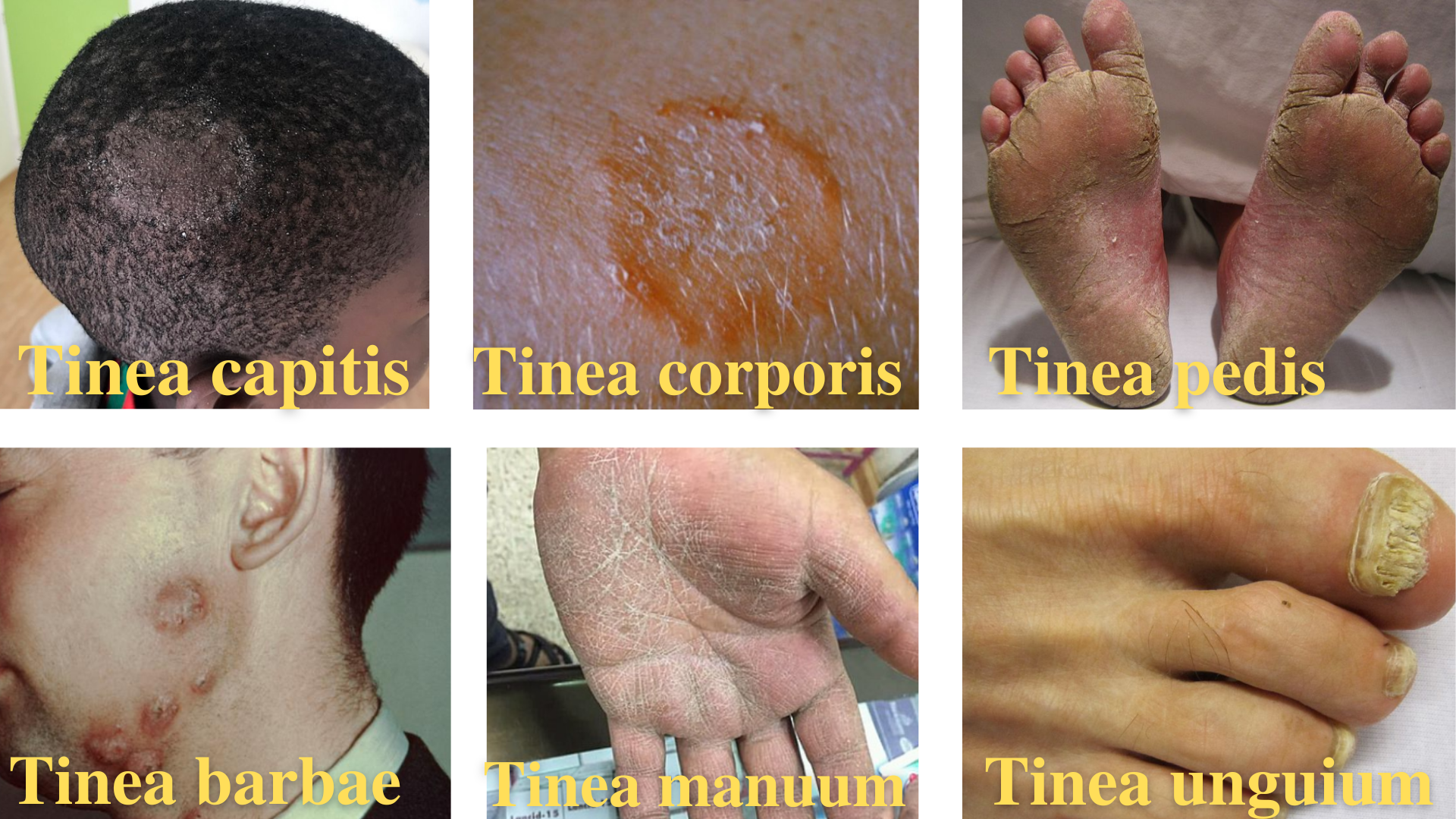 This table is the fastest way to hold the whole topic in your head. Read it site by site: where the infection is, what to call it, which organisms cause it most often, and the one feature that identifies it.
| Site | Name | Common organisms | Key clinical feature |
|---|---|---|---|
| Scalp / hair | Tinea capitis | Trichophyton spp., Microsporum canis | Scaly patches, broken hairs, black dots or bald areas; mostly in children |
| Body (trunk, limbs) | Tinea corporis | Trichophyton rubrum | Classic ring: raised scaly border, central clearing |
| Foot | Tinea pedis (athlete's foot) | T. rubrum, T. mentagrophytes, Epidermophyton floccosum | Scaling and cracking between the toes; itching, burning |
| Groin | Tinea cruris (jock itch) | T. rubrum, E. floccosum | Itchy red patches in the groin folds, spreading to inner thigh |
| Nail | Tinea unguium (onychomycosis) | T. rubrum, T. mentagrophytes, E. floccosum | Thick, discolored, crumbling nail |
| Beard / mustache | Tinea barbae | T. verrucosum, T. mentagrophytes | Scaly patches with pustules in the beard area; adult men |
| Hand / palm | Tinea manuum | T. rubrum, T. mentagrophytes, E. floccosum | Dry scaling of the palm, often with tinea pedis ("two feet, one hand") |
| Face (non-beard) | Tinea faciei | T. rubrum, M. canis | Ring lesions on the face; may lack the classic ring |
A pattern worth noticing right away: Trichophyton rubrum appears in almost every row. It is the single most common dermatophyte worldwide, and it infects skin, nails, and (less often) hair, which is why one species turns up under so many names.
Tinea capitis (scalp ringworm)
Tinea capitis is a dermatophyte infection of the scalp and hair shafts, seen mainly in children. It is caused by Trichophyton species and Microsporum canis. The fungus grows down into the keratinized wall of the hair follicle and along the hair shaft, so the infection damages hair as well as skin. Infected hairs look dull and grey and break easily.
Tinea capitis takes two broad forms, and the difference matters clinically because the inflammatory forms can scar.
Non-inflammatory forms are more common and cause little or no swelling:
- Black dot type. The fungus grows inside the hair shaft (endothrix invasion), which weakens the hair so it snaps off right at the scalp surface. Each broken hair leaves a small black dot in a patch of otherwise smooth scalp. It is usually caused by T. tonsurans and T. violaceum.
- Grey patch type. Round, scaly patches with dull grey hairs, classically caused by Microsporum species. These often fluoresce under a Wood's lamp.
- Seborrheic type. Diffuse scaling that resembles dandruff, which can delay diagnosis.
Inflammatory forms cause swelling and can lead to permanent hair loss:
- Kerion. A severe, boggy, pus-discharging swelling of the scalp, often with tender lymph nodes. It is an inflammatory reaction to the fungus, usually a zoophilic species (T. verrucosum, T. mentagrophytes). Kerion can scar and cause permanent bald patches if not treated promptly. In children, kerion celsi is one of the common severe presentations.
- Favus. A chronic form caused by T. schoenleinii, producing cup-shaped yellow crusts (scutula) around the hair follicles that give a honeycomb appearance (the Latin favus means honeycomb). It can lead to scarring alopecia.
Because tinea capitis involves the hair shaft, it needs oral antifungal treatment; topical creams cannot reach the fungus inside the follicle.
Tinea corporis (body ringworm)
Tinea corporis is the infection of the glabrous (non-hairy) skin of the trunk and limbs. It is most often caused by Trichophyton rubrum. This is the textbook ringworm: one or more ring-shaped lesions with a raised, red, scaly, advancing edge and a clearer center. Lesions may be single, multiple, or run together (confluent).
It spreads readily by skin-to-skin contact, which is why outbreaks are common among wrestlers and other contact-sport athletes (where it is sometimes called tinea corporis gladiatorum). Most cases respond to topical antifungal treatment.
Tinea pedis (athlete's foot)
Tinea pedis is a dermatophyte infection of the feet and toe webs. It is the most common dermatophyte infection in adults, favored by the warm, moist environment inside shoes. The main organisms are T. rubrum, T. mentagrophytes, and Epidermophyton floccosum.
It presents in several patterns:
- Interdigital. The most common form. Scaling, cracking (fissuring), and softening (maceration) of the skin between the toes, with itching and burning. The macerated skin can crack and let bacteria in, causing secondary infection.
- Moccasin (hyperkeratotic). Chronic dry scaling covering the sole and sides of the foot in the distribution of a moccasin. Often caused by T. rubrum. When it also involves one hand, it produces the "two feet, one hand" pattern.
- Vesicular. Fluid-filled blisters, usually on the sole.
- Ulcerative. The most severe form, with breakdown of the skin, often complicated by bacterial infection.
Interdigital and mild cases respond to topical treatment; chronic moccasin-type disease often needs oral antifungals.
Tinea cruris (jock itch)
Tinea cruris is the infection of the groin, upper inner thighs, and skin folds. It is common in adult men, favored by warmth and moisture, and is often accompanied by tinea pedis (the patient reinfects the groin from the feet). The usual organisms are T. rubrum and Epidermophyton floccosum.
It appears as itchy, red-brown patches with a raised scaly border, spreading outward from the groin fold onto the inner thigh. The scrotum is usually spared, which helps distinguish it from candidal infection, where the scrotum is typically involved. Most cases respond to topical treatment, along with keeping the area dry and treating any coexisting tinea pedis.
Tinea unguium (onychomycosis)
Tinea unguium is the dermatophyte infection of the nail, part of the broader term onychomycosis (fungal nail infection from any cause). It is most often caused by T. rubrum, and less often by T. mentagrophytes and Epidermophyton floccosum. Toenails are affected far more often than fingernails.
The infected nail becomes thickened, discolored (yellow-white or brown), and friable, meaning it crumbles easily. The nail may lift away from the nail bed. Because the fungus lives deep within the nail plate, topical treatment alone rarely cures it. Oral antifungals such as terbinafine and itraconazole are the mainstay, and treatment is long because the nail must grow out.
Tinea barbae
Tinea barbae is ringworm of the beard and mustache area, seen in adult men. It is also called barber's itch. The usual organisms are the zoophilic species T. verrucosum and T. mentagrophytes, often acquired from animals, which is why it is more common in farm and rural settings. It produces red, scaly patches with brittle, lusterless hairs and a tendency to pustular folliculitis. Microsporum canis and T. rubrum are found occasionally. Like other hair-involving infections, it usually needs oral treatment.
Tinea manuum
Tinea manuum is ringworm of the hand and palm, caused by T. rubrum, T. mentagrophytes, and Epidermophyton floccosum. It typically shows as dry, fine scaling of the palm and is classically one-sided. It often coexists with tinea pedis in the "two feet, one hand" pattern, where both feet and a single hand are involved.
How treatment differs by site
The single most useful treatment rule follows directly from anatomy: whether the fungus is in skin only, or in hair or nail.
- Skin-only infections (most tinea corporis, cruris, interdigital pedis, manuum) usually respond to topical antifungals: azole creams, terbinafine, ciclopirox, or amorolfine.
- Hair and nail infections (tinea capitis, barbae, unguium, and severe or extensive skin disease) need oral antifungals, because topical agents cannot reach the fungus inside the hair follicle or nail plate. Oral options include terbinafine, itraconazole, and griseofulvin. Terbinafine and itraconazole have largely replaced griseofulvin for nail infections because of better cure rates and shorter courses.
Confirming the organism by laboratory testing matters most before starting a long oral course, since these are given for weeks to months. The diagnostic workup is covered in the dermatophytes lab diagnosis article.
Prevention and control
- Do not share combs, brushes, hats, or towels, and disinfect barbershop instruments (combs, brushes, scissors).
- Disinfect clothing, towels, and bedding after use, as tinea corporis and cruris spread this way.
- Keep skin folds and feet dry; good foot hygiene and dry footwear reduce tinea pedis.
- Treat tinea pedis to prevent it seeding tinea cruris or tinea manuum in the same person.
- Examine and treat infected pets, a common source of Microsporum canis tinea capitis and corporis in children.
How to Remember
The name is the place, not the fungus. Every tinea name is Latin for a body region: capitis (head), corporis (body), pedis (foot), cruris (leg/groin), unguium (nail), barbae (beard), manuum (hand), faciei (face). Learn the sites and the "which organism" question becomes secondary, because T. rubrum causes most of them anyway.
Topical for skin, oral for hair and nail. The one treatment rule that carries the most exam weight. If hair or nail is involved, a cream will not reach the fungus, so the answer is oral.
Black dots mean inside the hair. In tinea capitis, black dots come from endothrix invasion: the fungus grows inside the shaft, weakens it, and the hair snaps at the scalp. "Endo, ends at the scalp."
Two feet, one hand. Tinea pedis on both feet plus tinea manuum on one hand is a recognized pattern, worth remembering because the asymmetry (why one hand?) is a classic exam prompt.
Key exam facts
| Fact | Detail |
|---|---|
| Tinea = | Dermatophyte infection of skin, hair, or nail (= ringworm) |
| Named for | The body site, not the organism |
| Most common cause overall | Trichophyton rubrum |
| Most common cause of tinea capitis | Trichophyton spp. and Microsporum canis |
| Most common cause of tinea unguium | Trichophyton rubrum |
| Kerion | Severe inflammatory tinea capitis; can scar; zoophilic species |
| Favus | T. schoenleinii; honeycomb yellow crusts (scutula) |
| Black dot tinea capitis | Endothrix invasion; T. tonsurans, T. violaceum |
| Two feet, one hand | Tinea pedis (both feet) + tinea manuum (one hand) |
| Topical treatment | Skin-only disease (corporis, cruris, most pedis) |
| Oral treatment | Hair and nail disease (capitis, barbae, unguium) |
| Scrotum spared | Helps distinguish tinea cruris from candidiasis |
Where Students Get Confused
Tinea versicolor is not a true tinea. Despite the name, tinea versicolor (pityriasis versicolor) is not caused by a dermatophyte. It is caused by Malassezia, a yeast, and behaves differently. It is grouped here only to warn against the naming trap: the word "tinea" in "tinea versicolor" is historical and misleading.
The tinea name does not name the fungus. Tinea corporis and tinea pedis can be caused by the same species (T. rubrum). The name tells you the site; only the laboratory tells you the organism.
Ringworm has no worm. The name comes from the ring-shaped lesion with central clearing, not from any parasite. It is entirely fungal.
Onychomycosis and tinea unguium are not exact synonyms. Tinea unguium means a nail infection caused specifically by a dermatophyte. Onychomycosis is the broader term for any fungal nail infection, including those caused by non-dermatophyte molds and yeasts. All tinea unguium is onychomycosis, but not all onychomycosis is tinea unguium.
Kerion is a reaction, not a separate organism. A kerion is the host's strong inflammatory response to a scalp dermatophyte, not a different fungus. It looks like a bacterial abscess but is fungal, so incising and draining it as if it were a bacterial abscess is a mistake.
References
- Chander J. Textbook of Medical Mycology. 4th ed. Jaypee Brothers Medical Publishers; 2018.
- Tille PM. Bailey and Scott's Diagnostic Microbiology. 15th ed. Elsevier; 2022.
- Procop GW, Church DL, Hall GS, et al. Koneman's Color Atlas and Textbook of Diagnostic Microbiology. 7th ed. Wolters Kluwer; 2017.
- Hay RJ. Tinea capitis: current status. Mycopathologia. 2017;182(1-2):87-93. https://doi.org/10.1007/s11046-016-0058-8

Tankeshwar Acharya, MSc (Medical Microbiology)
Tankeshwar Acharya is an Assistant Professor in the Department of Microbiology at Patan Academy of Health Sciences (PAHS), Nepal, where he has been teaching and practicing clinical microbiology for over 14 years. He is the founder of Microbe Online, one of the leading free microbiology education resources on the web, covering bacteriology, mycology, parasitology, immunology, and clinical laboratory diagnostics written from direct experience in both the classroom and the diagnostic laboratory.
Comments
No comments yet. Be the first to share your thoughts.
Leave a comment
All comments are reviewed before they appear.