
Indirect Fluorescent Antibody (IFA) Test: Principle, Procedure, and Uses

On this page
The indirect fluorescent antibody test (IFA) is a semi-quantitative, sensitive test used to detect specific antibodies (or antigens) in a sample. It is the "indirect" arrangement of immunofluorescence: instead of one labeled antibody, it uses two, an unlabeled antibody that binds the target and a second, dye-labeled antibody that binds the first. Its most common job is finding a patient's antibodies in serum, for example when a pathogen is hard to find directly and you look instead for the immune response to it, as in the serodiagnosis of leishmaniasis, syphilis, or toxoplasmosis. The two-antibody design is what separates IFA from the direct (DFA) method, and it is also what makes IFA more sensitive. For how the two methods compare in full, see Immunofluorescence assay.
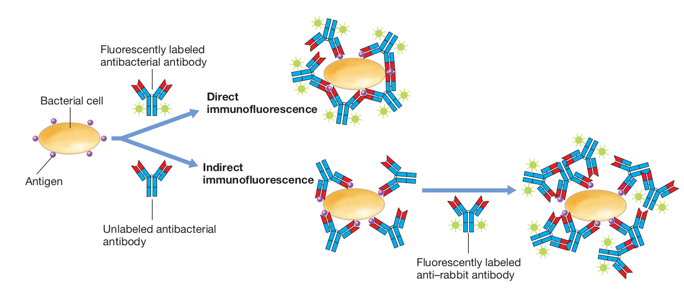 Figure: Direct and indirect immunofluorescence for the detection of antigen (Image source: Brock Biology of Microorganisms)
IFA for the detection of Antibodies
Indirect fluorescent antibody (IFA) test can be used to detect specific antibodies against various etiological agents present in patient serum and cerebral spinal fluid (CSF) samples.
IFA is being used for the diagnosis of:
- Rabies
- Syphilis
- Toxoplasmosis
- Leishmaniasis
- Legionellosis
Principle
Known antigen is immobilized on a glass slide (slides smeared with cells carrying known antigens are commercially available). Test sample (patient serum) is added over the smear. If specific antibodies are present in the serum, the antigen-antibody complex is formed. The serum is washed off and a secondary antihuman immunoglobulin conjugated to a fluorochrome (fluorescein isothiocyanate or rhodamine B) is added.
On examination, the target organism or cell is visible only where the patient's antibodies have bound it and the labeled secondary antibody has attached in turn.
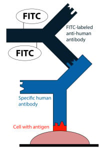 Figure: IFA for antibodies detection
Two fluorophores are commonly used: fluorescein isothiocyanate (FITC), which emits apple-green (sometimes described as yellow-green), and rhodamine, which emits red.
For example, an IFA test for the diagnosis of syphilis uses Treponema pallidum isolated from a lab animal and a smear is prepared on a glass slide. Patient serum is spread over the smear and anti-treponemal antibodies, if present, are allowed to bind. The serum is washed off and a secondary antibody labeled with fluorescein isothiocyanate (FITC) is added. On examination, the T. pallidum bacteria will only be visible. This treponemal IFA, refined with an absorption step to remove cross-reacting antibodies, is the basis of the FTA-ABS test used to confirm syphilis.
Procedure (antibody detection)
- A slide pre-coated with known antigen is used (commercially prepared antigen slides are available).
- Patient serum is applied. If specific antibody is present, it binds the fixed antigen.
- The slide is washed to remove unbound serum proteins.
- A secondary anti-human immunoglobulin antibody, labeled with FITC, is added and binds any patient antibody already attached to the antigen.
- The slide is washed again and examined under a fluorescence microscope. Apple-green fluorescence indicates the patient antibody is present. A titer can be estimated by testing serial serum dilutions.
IFA for detection of Antigens
Principle
Unlike the direct fluorescent antibody test, indirect detection of antigen is a two-step procedure using two antibodies: an unlabeled primary antibody that binds the antigen, and a FITC-labeled anti-species secondary antibody that binds the primary. The extra layer amplifies the signal.
- An unlabeled primary antibody which binds to a specific antigen, and
- FITC labeled anti-species secondary antibody which binds to the primary antibody-antigen complex.
Virus-infected cells or samples are fixed using acetone, methanol, or paraformaldehyde; to preserve cell morphology or tissue architecture. After incubation of the sample with the appropriate antibody (primary antibody), the excess antibody is removed by washing. A secondary antibody labeled with fluorochrome is added and incubated. Again the excess antibody is removed by washing and the smear is visualized using a fluorescence microscope.
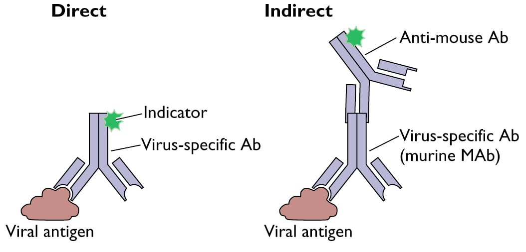 Figure: DFA and IFA for the detection of viral antigen(Image source: virology.ws)
IFA vs DFA in brief
IFA uses two antibodies and DFA uses one. The extra labeled antibody in IFA amplifies the signal, making it more sensitive than DFA, but the two incubation steps make it slower. For the full side-by-side comparison, see Immunofluorescence assay: direct vs indirect
How to Remember
Why two antibodies, and what IFA usually detects
The second antibody is a labeled megaphone. A patient's own antibody is invisible, it carries no dye. The indirect method adds a second antibody that carries the dye and clamps onto the first. Because several of these labeled "megaphones" stack on one patient antibody, the signal gets louder, which is exactly why indirect is more sensitive than direct.
Indirect leans toward antibodies in serum. When the question is "does this patient have antibodies against X" (rabies, syphilis, toxoplasmosis, leishmaniasis, legionellosis), you cannot pre-label the patient's antibody, so you relay through a labeled anti-human secondary. That relay is the indirect method's signature move.
Key exam facts in one table
| Point | What to remember |
|---|---|
| What it detects | Antibodies in patient serum or CSF (and, in sandwich form, antigens in cells) |
| Antibodies used | Two: unlabeled primary + labeled secondary |
| Secondary antibody | Anti-human immunoglobulin, FITC-labeled, binds the patient's antibody |
| Why more sensitive than DFA | Several labeled secondary antibodies stack on one primary, amplifying signal |
| Trade-off vs DFA | Slower (two incubation steps) |
| Fluorophores | FITC emits apple-green; rhodamine emits red |
| Antigen slide | Known antigen is pre-fixed on the slide (commercial slides available) |
| Semi-quantitative | Titer estimated by testing serial serum dilutions |
| Clinical uses | Serodiagnosis of rabies, syphilis, toxoplasmosis, leishmaniasis, legionellosis |
| Syphilis link | Treponemal IFA with an absorption step becomes the FTA-ABS test |
| Reading | Fluorescence microscope; apple-green signal is positive |
Where students get confused
"IFA only detects antibodies." Mostly, but not only. Its classic use is finding antibodies in patient serum, but the same two-antibody logic detects antigens too (unlabeled primary against the antigen, labeled secondary against the primary). The defining feature is two antibodies, not what is being detected.
"The secondary antibody is specific to the disease." No. The secondary antibody is anti-human immunoglobulin, it binds human antibody in general, not the pathogen. That is precisely why one labeled secondary can serve many different IFA tests: the specificity comes from the patient's own antibody (or the unlabeled primary), not the label.
"IFA is better than DFA because it is more sensitive." More sensitive, yes, but not universally better. The two incubation steps make it slower, and the extra antibody adds a route for background. DFA wins when speed and clean background matter; IFA wins when sensitivity and flexibility matter.
"Apple-green and yellow-green are different dyes." They are the same dye. FITC's emission is described both ways. Rhodamine is the red one. Do not read two color words as two fluorophores.
Frequently Asked Questions
What does the indirect fluorescent antibody (IFA) test detect?
Why does IFA use two antibodies?
Why is IFA more sensitive than DFA?
What is the difference between IFA and FTA-ABS?
What colors are seen in an IFA test?
Is IFA quantitative?
References
- Racaniello, V. Detecting viral proteins in infected cells or tissues by immunostaining. Virology blog.
- Tille, P. M. (2022). Bailey & Scott's Diagnostic Microbiology (15th ed.). Elsevier.
- Madigan, M. T., Bender, K. S., Buckley, D. H., Sattley, W. M., & Stahl, D. A. (2018). Brock Biology of Microorganisms (15th ed.). Pearson.
- Procop, G. W., et al. (2017). Koneman's Color Atlas and Textbook of Diagnostic Microbiology (7th ed.). Wolters Kluwer.

Tankeshwar Acharya, MSc (Medical Microbiology)
Tankeshwar Acharya is an Assistant Professor in the Department of Microbiology at Patan Academy of Health Sciences (PAHS), Nepal, where he has been teaching and practicing clinical microbiology for over 14 years. He is the founder of Microbe Online, one of the leading free microbiology education resources on the web, covering bacteriology, mycology, parasitology, immunology, and clinical laboratory diagnostics written from direct experience in both the classroom and the diagnostic laboratory.
Comments
No comments yet. Be the first to share your thoughts.
Leave a comment
All comments are reviewed before they appear.