
Gram-Positive vs Gram-Negative Bacteria: Differences, Cell Wall, and How to Tell Them Apart
Gram-positive and gram-negative bacteria differ in cell wall structure, staining, toxins, and antibiotic response. Compare them side by side and learn to read a Gram stain result.

On this page
The Gram stain is the most important staining procedure in microbiology. It sorts most bacteria into two groups, gram-positive and gram-negative, based on a single structural difference in the cell wall. Gram-positive bacteria stain purple. Gram-negative bacteria stain pink. That color difference predicts how the organism behaves in the body, which antibiotics will work, and what kind of toxin it is likely to produce.
Gram-positive bacteria hold onto the primary stain (crystal violet) even after decolorization, because their thick peptidoglycan wall traps the crystal violet-iodine complex. Gram-negative bacteria have a thin peptidoglycan layer and an outer membrane. The decolorizer dissolves part of that outer membrane and washes the crystal violet out, so the cells appear pink, the color of the counterstain (safranin).
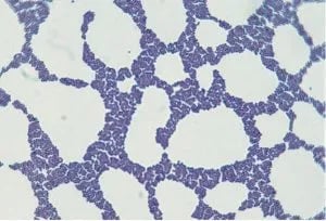 Figure: Staphylococcus in Gram Stain
The major structural difference between the two groups is the thickness of the peptidoglycan layer and the presence or absence of an outer membrane. Gram-positive bacteria have a thick, multilayered peptidoglycan wall and no outer membrane.
Gram-negative bacteria have a thin peptidoglycan layer sandwiched between the inner (cytoplasmic) membrane and an outer membrane. That outer membrane carries lipopolysaccharide (LPS) on its outer face. Every other difference between the two groups, from staining to antibiotic susceptibility to toxin type, follows from this one architectural fact.
The Gram-Positive Cell Wall
The cell wall of gram-positive bacteria is much thicker than of Gram-negative bacteria and consists primarily of a single type of molecule.
Peptidoglycan Layer
The peptidoglycan layer is the outermost covering of the Gram-positive cell wall and makes up the bulk of the Gram-positive cell wall (roughly 50% or more of the wall's dry weight). Gram-positive bacteria have several sheets of peptidoglycan stacked and cross-linked by glycan strands. Many gram-positive bacteria have teichoic acids (polymers of glycerol phosphate or ribitol phosphate) covalently bonded to muramic acid in the wall peptidoglycan or membrane lipids (lipoteichoic acids).
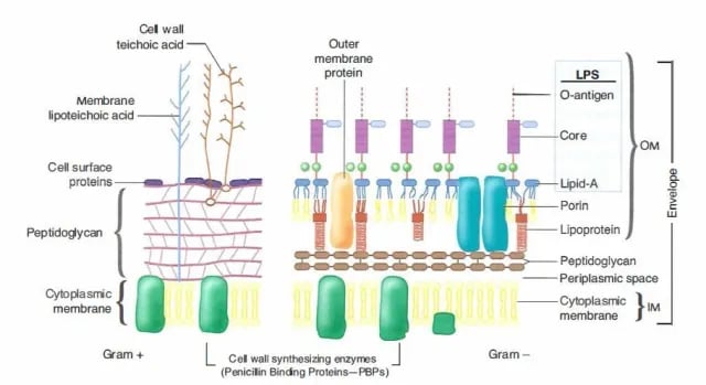 ## The Gram-Negative Cell Wall
The gram-negative cell wall is more chemically complex than the gram-positive and consists of at least two layers.
Lipopolysaccharide Layer
The outer membrane (lipopolysaccharide layer, LPS in short) is the outermost covering of the Gram-negative cell wall.
Peptidoglycan layer
Beneath the outer membrane in Gram-negative bacteria lies a thin sheet of peptidoglycan, which constitutes only 10% of the cell wall of Gram-negative. The outer membrane contains a lipid bilayer bonded with polysaccharides (lipopolysaccharide).
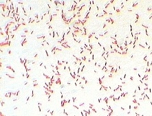 Figure: Gram Stain Reaction of E. coli
Does the periplasmic space exist in gram-positive bacteria?
The periplasm is the compartment between the inner (cytoplasmic) membrane and the outer membrane. In gram-negative bacteria this space is obvious and well defined, because there are clearly two membranes with the thin peptidoglycan layer sitting inside it. The periplasm holds transport proteins (periplasmic binding proteins), enzymes that break down large molecules, and enzymes that inactivate some antibiotics. Beta-lactamases, the enzymes that destroy penicillins, sit in the gram-negative periplasm, which is one reason gram-negative bacteria are often harder to treat.
Gram-positive bacteria are the confusing part, and most textbooks oversimplify it. The old teaching is that gram-positive bacteria have "no periplasm" because they have no outer membrane. That is not quite correct. Gram-positive bacteria do have a distinct compartment between the cytoplasmic membrane and the inner face of the thick peptidoglycan wall. Cryo-electron microscopy studies (Matias and Beveridge) have shown this inner-wall zone directly. It is sometimes called the inner-wall zone or the gram-positive periplasm.
So the accurate answer is this: gram-negative bacteria have a true periplasmic space bounded by two membranes. Gram-positive bacteria have a smaller, less enclosed periplasmic compartment bounded on the outside by the peptidoglycan wall rather than by a membrane. Both groups carry out periplasmic functions such as transport and enzyme activity. Gram-positive bacteria also rely heavily on ABC transporters anchored in the cytoplasmic membrane to move nutrients across the wall.
For an exam, the safe short answer is: the periplasmic space is well developed in gram-negative bacteria and much less developed, but not truly absent, in gram-positive bacteria.
How do you tell gram-positive from gram-negative in the lab?
On a stained slide, you read two things together: the color (gram-positive holds the purple crystal violet, gram-negative takes up the pink safranin) and the cell shape and arrangement (cocci in clusters, cocci in chains, rods). That combination gives a working first guess at the organism before any culture grows.
The Gram stain can mislead, though. An old culture, a smear left too long in the decolorizer, or a smear that is too thick can all make a gram-positive organism read falsely as gram-negative. Because these are technique issues rather than biology, they are covered in full, along with step-by-step method, quality control, and how each organism looks on the slide, in the dedicated guide: Gram staining: procedure, results, and troubleshooting.
Why the cell wall difference matters in the clinic
The wall is not trivia. Three clinical consequences follow directly from it.
Antibiotic choice. The gram-negative outer membrane is a barrier. Many antibiotics that easily reach the target in gram-positive bacteria cannot cross the outer membrane, so they simply do not work against gram-negative organisms. Vancomycin, for example, is a large molecule that cannot cross the outer membrane, which is why it works against gram-positive bacteria but not gram-negative ones.
Type of toxin. Gram-positive bacteria mostly cause disease through exotoxins, proteins they actively secrete. Gram-negative bacteria carry endotoxin, which is the lipid A portion of the LPS in their outer membrane. Endotoxin is not secreted. It is released when the cell is damaged or dies, which is why killing large numbers of gram-negative bacteria can trigger a strong inflammatory response and, in severe cases, septic shock.
Response to lysozyme. Lysozyme, present in tears and saliva, cuts the peptidoglycan backbone. It reaches the exposed thick wall of gram-positive bacteria easily and can strip it, leaving a wall-less protoplast. In gram-negative bacteria the outer membrane shields the thin peptidoglycan, so lysozyme alone is far less effective. Removing the gram-negative wall leaves a spheroplast, which still retains its outer membrane.
Differences between Gram-positive and Gram-negative bacteria
Properties | Gram-Positive Bacteria | Gram-Negative Bacteria |
Thickness of cell wall | Thicker than Gram-negative bacteria, around 20 to 25 nm | The cell wall of Gram-negative bacteria is generally thinner, 11 to 15 nm in diameter |
Gram reaction | Gram-positive bacteria stain violet/purple in the Gram staining technique. | Gram-negative bacteria stain pink to red color in the Gram staining technique. |
Lipopolysaccharide (LPS) layer | The lipopolysaccharide layer, also known as the outer membrane, is absent in Gram-positive bacteria. | LPS is only present in Gram-negative bacteria. |
Peptidoglycan layer | A thick (multilayered) peptidoglycan layer is present in Gram-positive bacteria. It accounts for 50% or more of the dry weight of the wall of some Gram-positive bacteria. | Thin (single-layered). Around 10% weight of the cell wall of Gram-negative bacteria. |
Teichoic acids | Cell wall of gram-positive bacteria contains teichoic acids. | Teichoic acid is absent in Gram-negative bacteria |
Periplasmic space | Smaller, less developed inner-wall periplasmic compartment. Not a true membrane-bounded periplasm, but not truly absent either. | Well-developed true periplasm between the inner (cytoplasmic) membrane and the outer membrane, with the thin peptidoglycan layer inside it. |
Flagellar Structure | Two rings in the basal body | Four rings in the basal body |
Toxins Produced | Primarily exotoxins | Primarily endotoxins, the LPS layer has an endotoxic property. |
Lipid content | Low | High around 11 to 22% of the dry weight of the cell wall (because of the lipid-rich LPS layer). |
Action of Lysozyme | Cell wall of Gram-positive bacteria is easily destroyed by the action of lysozyme. After digestion of the Peptidoglycan layer, Gram-positive bacteria become protoplast. | Gram-negative bacteria are refractory to lysozyme because large protein molecules cannot penetrate the LPS layer. After digestion of the Peptidoglycan layer, Gram-negative bacteria become spheroplasts. |
Examples of gram-positive and gram-negative bacteria
Common gram-positive bacteria:
- Staphylococcus aureus (cocci in clusters)
- Streptococcus pyogenes and Streptococcus pneumoniae (cocci in chains or pairs)
- Enterococcus species
- Bacillus species, including Bacillus anthracis (rods, spore-forming)
- Clostridium species, including Clostridium tetani and Clostridium botulinum (rods, spore-forming, anaerobic)
- Listeria monocytogenes (short rods)
- Corynebacterium diphtheriae (rods)
Common gram-negative bacteria:
- Escherichia coli (rods)
- Klebsiella pneumoniae (rods)
- Pseudomonas aeruginosa (rods)
- Salmonella Typhi and other Salmonella serovars (rods)
- Neisseria gonorrhoeae and Neisseria meningitidis (diplococci)
- Vibrio cholerae (curved rods)
- Haemophilus influenzae (small rods)
A useful memory anchor: most medically important cocci are gram-positive (the main exception is Neisseria), and most medically important rods you meet in a stool or urine sample are gram-negative.
For the gram stain appearance, arrangement, and identifying features of each of these organisms on a slide, see the organism tables in the Gram staining guide.
How to remember gram-positive vs gram-negative
Positive = Purple, thick, Preserves the stain. All three start with P, and all three describe gram-positive: purple color, thick peptidoglycan, and it preserves (keeps) the crystal violet. Everything gram-negative is then the opposite: pink, thin peptidoglycan, loses the stain.
"Negative bacteria have an extra coat." The one thing gram-negative bacteria have that gram-positive bacteria do not is the outer membrane. Picture the gram-negative cell wearing an extra coat (the outer membrane) with LPS studded on the outside. That extra coat is why they resist lysozyme, why they carry endotoxin, and why many antibiotics cannot get in.
Toxin link: exOtoxin from gram-pOsitive, eNdotoxin is iN the gram-Negative membrane. Exotoxin is mostly secreted by gram-positive bacteria. Endotoxin is built into the gram-negative outer membrane and released when the cell breaks.
Key Exam Facts
| Feature | Gram-Positive | Gram-Negative |
|---|---|---|
| Gram stain color | Purple/violet (keeps crystal violet) | Pink/red (takes up safranin) |
| Peptidoglycan | Thick, multilayered (~50% or more of wall dry weight) | Thin, single-layered (~10% of wall) |
| Cell wall thickness | ~20 to 25 nm | ~10 to 15 nm |
| Outer membrane | Absent | Present (carries LPS on outer face) |
| Teichoic acids | Present | Absent |
| Periplasmic space | Small, less developed inner-wall zone (not truly absent) | Well-developed, true periplasm between the two membranes |
| Main toxin type | Exotoxins (secreted proteins) | Endotoxin (lipid A of LPS, released on cell damage) |
| Lipid content of wall | Low | High (lipid-rich LPS) |
| Effect of lysozyme | Wall easily digested, becomes protoplast | Outer membrane protects wall; becomes spheroplast |
| Flagellar basal body rings | 2 rings | 4 rings |
| Vancomycin (large drug) | Effective (can reach target) | Not effective (cannot cross outer membrane) |
| Examples | Staphylococcus, Streptococcus, Bacillus, Clostridium, Listeria | E. coli, Klebsiella, Pseudomonas, Neisseria, Vibrio cholerae |
Where Students Get Confused
"Does gram-positive bacteria have a periplasmic space, yes or no?" The honest answer is "yes, but a smaller and less enclosed one." Gram-negative bacteria have a true periplasm bounded by two membranes. Gram-positive bacteria have an inner-wall periplasmic compartment bounded on the outside by the thick peptidoglycan, not by a membrane. The old textbook claim that gram-positive bacteria have "no periplasm" is an oversimplification. If an exam forces a one-word answer, they usually expect "absent" for gram-positive, but the accurate biology is "much less developed, not absent." Know both so you can answer either version of the question.
Confusing the color with the cause. Students memorize "gram-positive = purple" but forget why. The color is a consequence of the thick peptidoglycan trapping crystal violet. If you understand the wall, you never have to memorize the color, because it falls out of the structure.
Thinking a gram-negative result means "no peptidoglycan." Gram-negative bacteria do have peptidoglycan. It is just thin and hidden under the outer membrane. The difference is thickness and location, not presence or absence.
Endotoxin vs exotoxin direction. Students flip these. Anchor it to structure: endotoxin is a structural part of the gram-negative outer membrane (built in, released on death). Exotoxin is actively secreted, mostly by gram-positive bacteria. The toxin type follows the wall.
Assuming the Gram stain is always right. A gram-positive organism from an old culture can read gram-negative. Over-decolorization is the classic student mistake. When the result does not match the clinical picture or the colony appearance, suspect technique before you suspect a rare organism.
Protoplast vs spheroplast. Both are wall-damaged cells. Protoplast comes from a gram-positive cell that has lost its entire wall. Spheroplast comes from a gram-negative cell that has lost its peptidoglycan but keeps its outer membrane. The gram-negative one still has a "sphere" of outer membrane around it, which is a loose way to remember spheroplast.
Frequently Asked Questions
What is the main difference between gram-positive and gram-negative bacteria?
What is the main difference between gram-positive and gram-negative bacteria?
The main difference is the cell wall. Gram-positive bacteria have a thick peptidoglycan layer and no outer membrane, so they stain purple. Gram-negative bacteria have a thin peptidoglycan layer plus an outer membrane, so they stain pink. Almost every other difference between the two groups follows from this one structural fact.
Why do gram-positive bacteria stain purple and gram-negative bacteria stain pink?
Why do gram-positive bacteria stain purple and gram-negative bacteria stain pink?
Gram-positive bacteria have a thick peptidoglycan wall that traps the crystal violet-iodine complex, so they keep the purple color even after decolorization. Gram-negative bacteria have a thin wall and an outer membrane. The decolorizer washes the crystal violet out, and the pink safranin counterstain shows through.
Do gram-positive bacteria have a periplasmic space?
Do gram-positive bacteria have a periplasmic space?
Yes, but a much smaller and less developed one than gram-negative bacteria. Gram-negative bacteria have a true periplasm bounded by two membranes. Gram-positive bacteria have an inner-wall periplasmic compartment between the cytoplasmic membrane and the thick peptidoglycan wall. The common textbook claim that gram-positive bacteria have "no periplasm" is an oversimplification.
Which bacteria are gram-positive and which are gram-negative?
Which bacteria are gram-positive and which are gram-negative?
Common gram-positive bacteria include Staphylococcus, Streptococcus, Bacillus, Clostridium, and Listeria. Common gram-negative bacteria include Escherichia coli, Klebsiella, Pseudomonas, Neisseria, and Vibrio cholerae. A helpful rule: most medically important cocci are gram-positive, with Neisseria the main exception.
Why are gram-negative bacteria harder to treat with antibiotics?
Why are gram-negative bacteria harder to treat with antibiotics?
Their outer membrane acts as a barrier that many antibiotics cannot cross. Large drugs like vancomycin cannot reach their target inside a gram-negative cell. The gram-negative periplasm also holds enzymes such as beta-lactamases that inactivate some antibiotics before they act.
What is the difference between endotoxin and exotoxin?
What is the difference between endotoxin and exotoxin?
Exotoxins are proteins actively secreted by bacteria, mostly gram-positive. Endotoxin is the lipid A part of the LPS in the gram-negative outer membrane. Endotoxin is not secreted. It is released when the cell is damaged or dies, which is why killing many gram-negative bacteria at once can trigger a strong inflammatory response.
Can a gram-positive bacterium look gram-negative on a slide?
Can a gram-positive bacterium look gram-negative on a slide?
Yes. Old or dying cultures of gram-positive bacteria lose crystal violet and can stain pink. Over-decolorization does the same thing and is the most common student error. Always stain from a fresh culture and decolorize carefully. If a result does not match the clinical picture, repeat the stain before assuming an unusual organism.
References
- Madigan, M. T., Bender, K. S., Buckley, D. H., Sattley, W. M., & Stahl, D. A. (2021). Brock Biology of Microorganisms (16th ed.). Pearson.
- Procop, G. W., Church, D. L., Hall, G. S., Janda, W. M., Koneman, E. W., Schreckenberger, P. C., & Woods, G. L. (2017). Koneman's Color Atlas and Textbook of Diagnostic Microbiology (7th ed.). Wolters Kluwer.
- Tille, P. M. (2022). Bailey & Scott's Diagnostic Microbiology (15th ed.). Elsevier.
- Matias, V. R. F., & Beveridge, T. J. (2005). Cryo-electron microscopy reveals native polymeric cell wall structure in Bacillus subtilis 168 and the existence of a periplasmic space. Molecular Microbiology, 56(1), 240–251.

Tankeshwar Acharya, MSc (Medical Microbiology)
Tankeshwar Acharya is an Assistant Professor in the Department of Microbiology at Patan Academy of Health Sciences (PAHS), Nepal, where he has been teaching and practicing clinical microbiology for over 14 years. He is the founder of Microbe Online, one of the leading free microbiology education resources on the web, covering bacteriology, mycology, parasitology, immunology, and clinical laboratory diagnostics written from direct experience in both the classroom and the diagnostic laboratory.
Comments
No comments yet. Be the first to share your thoughts.
Leave a comment
All comments are reviewed before they appear.