

A wound infection is not responding to antibiotics. The laboratory has grown a gram-positive rod on blood agar; anaerobic, swarming on blood agar, with a characteristic drumstick shape on Gram stain. Endospore staining confirms terminal spores wider than the cell body.
The organism is Clostridium tetani. The patient needs antitoxin, not just antibiotics, because the toxin already produced is what is causing the disease, and no antibiotic can neutralize it.
In a second case, a soil-contaminated wound grows large gram-positive boxcar-shaped rods with subterminal spores on endospore staining. Clostridium perfringens. Surgical debridement is added urgently to antibiotic therapy.
Spore-forming organisms (Bacillus and Clostridium genera) include some of the most toxigenic pathogens known to medicine. The spore position, combined with clinical context, guides identification. And understanding why spores resist ordinary staining is the key to understanding why they resist sterilization and why autoclaving at 121°C for 15 minutes exists.
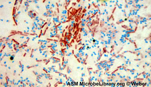 Figure: Bacillus cereus endospore stain
Endospore staining is a differential technique that selectively stains the spores and makes them distinguishable from the vegetative part of the cells. Endospores are produced by a few genera of Gram-positive bacilli, such as Bacillus and Clostridium, in response to adverse environmental conditions. Endospores are highly resistant to environmental conditions such as heat and chemicals (stains and dyes) and therefore require special techniques for staining.
Methods for endospore staining
There are different methods for endospore staining. The most common are
- Schaeffer-Fulton stain technique
- Dorner’s methods
- Modified Ziehl-Neelsen method
- Bartholomew-Mittwer method
- Abbott's method
- Moeller's stain technique
Spores can generally be recognized on Gram’s stains (endospores do not stain and appear as refractile, non-staining bodies). Endospores can also be demonstrated in unstained wet films under a phase-contrast microscope. They appear as large refractile oval or spherical bodies within the mother cell.
Schaeffer-Fulton stain technique
It is the most widely used technique for endospore staining. The technique was first described by Alice B. Schaeffer and MacDonald Fulton in the 1930s. The method utilizes malachite green as the primary stain and safranin as a counterstain.
Principle
When a heat-fixed smear is flooded with aqueous malachite green solution (the primary stain) and steamed, the heat assists the stain in penetrating through the spore. In this technique, heating acts as a mordant.
Once the endospore has absorbed the stain, it is resistant to decolorization, but the vegetative cells are easily decolorized with water (leaving the vegetative cells colorless).
When counter-stained with safranin, the vegetative cells take the color of safranin and appear red or pink, in contrast to the endospores that appear green.
When visualized under microscopy, the cells should have three characteristics:
- the vegetative cells should appear pink/red (i.e., the color of counterstain),
- the vegetative cells containing endospores should stain pink, while the spores should be seen as green ellipses.
- Mature, free endospores should not be associated with vegetative bacteria and should be seen as green ellipses.
Why heat is needed as a mordant: The endospore coat is extraordinarily resistant. It is composed of a protein outer coat, cortex (modified peptidoglycan), and inner membrane that collectively exclude most dyes at room temperature. Heat serves as the mordant by disrupting this resistance, allowing malachite green to penetrate the spore layers.
Once inside, the spore retains the dye so tenaciously that simple water washing (which removes malachite green from vegetative cells) cannot extract it from the spore. This is the same chemical resistance that allows spores to survive boiling, most disinfectants, and 70% ethanol and why autoclaving (pressurized steam at 121°C) is required to destroy them.
Note: Heat fixing requires a Bunsen burner or moist-heat source, which generates aerosols and poses burn risk. For the aerosol-control and fire-safety practices specific to heat-fixing procedures, see microbiology laboratory safety rules, which covers Bunsen burner safety, aerosol generation, and safe cooling practices for hot loops.
Procedure
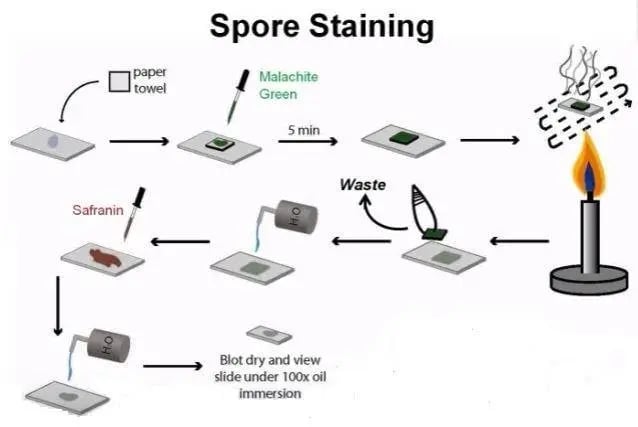 Figure: Spore staining procedure
- Prepare smears of organisms to be tested for the presence of endospores on a clean microscope slide and air dry it.
- Heat fix the smear.
- Place a small piece of blotting paper (absorbent paper) over the smear and place the slide (smear side up) on a wire gauze on a ring stand.
- Saturate the blotting paper with malachite green stain solution and steam for 5 minutes, keeping the paper moist and adding more dye as required. Alternatively, the slide may be steamed over a container of boiling water.
- As the paper begins to dry, add a drop or two of malachite green to keep it moist, but don’t add so much at once that the temperature is appreciably reduced.
- After 5 minutes, remove the slide from the rack using a clothespin.
- Remove the blotting paper and allow the slide to cool to room temperature for 2 minutes.
- Rinse the slide thoroughly with tap water (to wash the malachite green from both sides of the microscope slide).
- Stain the smear with safranin for 2 minutes.
- Rinse both sides of the slide to remove the secondary stain and blot the slide/ air dry.
Result & Interpretation
Observe the bacteria under 1000X (oil immersion) total magnification. When viewed under 1000X of a light microscope, vegetative cells appear pink/red, and spores appear green.
Dorner’s Method
Dorner’s method is an alternative method for staining the endospores published by Dorner in 1922. This method utilizes carbol fuchsin as primary stain, acid alcohol as decolorizer, and nigrosin as counterstain. It employed a lengthy heating step but resulted in differential staining of endospores and vegetative cells in the same sample.
Principle of Dorner’s method for staining endospores
Carbol fuchsin, when applied to a heat-fixed slide and heated, softens the structure of the bacterial spores and the basic fuchsin gets into the spores. When decolorized with acid alcohol, color washes off the vegetative cells, making them colorless.
Since the counterstain nigrosin is negatively charged, bacterial cells don’t easily take up the counterstain. Therefore, vegetative cells appear colorless, endospores stain red, and the background is black.
Procedure for Dorner’s method
- Make a smear on a clean glass slide.
- Allow the slide to dry (air dry) and then heat fix
- Place a blotting paper on the slide (covering the smear) and saturate with carbol fuchsin to steam (for about 5 minutes).This should be repeated while adding drops of carbol fuchsin and avoiding overheating (simply heat to steam)
- Remove the blotting paper and allow the slide to dry for about a minute
- Wash the slide with acid-alcohol for about a minute to decolorize and then rinse with tap water
- Add a drop of nigrosin to the smear to form a thin film.
- Allow the slide to dry.
- Observe under the microscope using oil immersion.
Variation in Dorner’s method
Dorner’s method is further modified by omitting the acid-alcohol decolorizer and using a 7.0% (wt/vol) aqueous solution of the nigrosin. It is performed in a test tube, thus avoiding direct heating procedures.
Procedure of modified Dorner’s method
- Mix an aqueous suspension of bacteria with an equal volume of carbol fuchsin in a test tube.
- Immerse the tube in a boiling water bath for 10 minutes. Allow cooling for some time.
- Mix a loopful of 7% nigrosin on a glass slide with one loopful of the boiled carbol fuchsin-organism suspension and make a thin smear and allow air to dry.
- Examine the slide under the oil immersion lens for the presence of endospores.
Results
Vegetative cells are colorless, endospores are red, and the background is black.
Other techniques of endospore staining
Although the principle of endospore staining is the same, variations exist in the choice of primary stain, counterstain, and whether or not decolorizer is used. Some of them are summarized below.
| Method | Primary Stain | Decolorizer | Counterstain | Interpretation |
|---|---|---|---|---|
| Modified Ziehl-Neelsen method | carbol fuchsin | 0.25-0.5% sulphuric acid | Loeffler's methylene blue | Spores appear red, bacteria are blue |
| Dorner method | carbol fuchsin | Acid-alcohol | Nigrosin | Spores red Bacteria colorless Background Black |
| Schaeffer-Fulton Stain | Malachite Green | Water | Safranin | Spores appear green vegetative cells appear pink/red |
| Bartholomew-Mittwer method | Malachite Green | Water | Safranin | Spores appear green vegetative cells appear pink/red |
| Abbott’s method | Methylene Blue | Acid alcohol | Aniline fuchsin | Spores appear blue bacteria are red |
| Moeller’s stain | carbol fuchsin | Acidified ethanol | Methylene blue | Spores appear red bacteria are Blue |
| Modified Moeller's stain | Kinyoun’s Carbol fuchsin | 2%sulphuric acid and 80% ethanol | Loeffler's methylene blue | Spores appear red bacteria are Blue |
Troubleshooting Endospore Staining
| Problem | Likely Cause | Action |
|---|---|---|
| No green spores visible (all cells pink) | Insufficient steaming time; malachite green not penetrating spore | Steam for full 5 minutes; keep blotting paper moist throughout; ensure visible steam rising but not boiling |
| All cells staining green | Insufficient washing after malachite green; water wash too brief | Wash more thoroughly with tap water before applying safranin |
| Spores and vegetative cells both appear pink | Over decolorization during water wash; malachite green washed from spores | Reduce water wash time; check malachite green concentration |
| No organisms visible | Smear too thin; poor heat fixation | Use a slightly heavier inoculum; ensure proper heat fixation before staining |
| Spores present but difficult to distinguish | Organism produces very few spores | Use older cultures (24–48 hours); subculture to a nutritionally deficient medium to stimulate sporulation |
| False-positive (artefacts appearing green) | Stain precipitate; dirty slides | Use grease-free slides; filter malachite green before use |
Key practical note: Not all cultures of spore-forming organisms will show spores. Sporulation is triggered by nutrient depletion and adverse conditions. Young, actively growing cultures in rich media may show no spores at all.
If spores are clinically suspected but not seen, use older cultures (48–72 hours) or subculture to a nutrient-limited medium.
Spore Position: A Critical Identification Feature
The position of the endospore within the vegetative cell and its size relative to the cell body are important identification features, particularly for differentiating Clostridium species.
| Spore Position | Description | Appearance | Key Organisms |
|---|---|---|---|
| Terminal | Spore at the end of the cell | Drumstick or lollipop shape (spore wider than cell) | Clostridium tetani |
| Subterminal | Spore near but not at the end | Club shape (less pronounced bulge) | Clostridium botulinum, C. perfringens (rarely seen) |
| Central | Spore in the middle of the cell | Spindle or fusiform shape | Clostridium bifermentans, some Bacillus spp |
| Oval, non-distending | Spore fits within the cell width | No cell distortion | Bacillus anthracis, B. subtilis |
| Oval, distending | Spore wider than the vegetative cell | Cell bulges at spore location | Many Clostridium spp |
Note: spore position (central, subterminal, terminal) and distension (whether the spore widens the cell) are two separate features. Bacillus spores are typically central or subterminal and non-distending (the cell keeps its shape); the distending drumstick of Clostridium tetani is what makes it distinctive.
Reading position off the slide:
Two features are read together, the position (terminal, subterminal, or central) and whether the spore distends the cell. The single most useful combination to recognize is a terminal, spherical spore that distends the cell into a drumstick.
Also remember that many clostridia sporulate poorly on routine media, so absent spores do not exclude a spore-former, use an older or nutrient-limited culture if spores are suspected but not seen.
What each spore position means clinically, and the memory device for matching position to organism, is covered in our article on bacterial spores (endospores)..
Clinical Significance of Endospore Detection
Which organisms form clinically relevant endospores?
Two genera matter clinically: Bacillus (aerobic) and Clostridium/Clostridioides (anaerobic). The specific pathogens, their diseases, and how spore features help identify them are covered in bacterial spores (endospores) article.
Why heat is needed to stain what heat cannot kill
There is a neat logic worth noticing here. The spore resists the malachite green dye at room temperature for the same reason it resists disinfectants and boiling: the dehydrated, multilayered spore is nearly impermeable. Heat is what forces the dye in during staining, and heat (as pressurized steam at 121°C) is also what finally destroys the spore during sterilization.
The stain difficulty and the sterilization difficulty are two faces of the same structure. Why the spore is built this way, and what it means for autoclaving, alcohol hand gels, and Clostridioides difficile control, is covered in our article on bacterial spores (endospores).
How to Remember: Endospore Staining
Schaeffer-Fulton: "Green spores on a Pink background": Malachite Green stains spores; Safranin stains vegetative cells pink. G for spores, S for cells: Green Spores.
Why heat is the mordant, the "waxy coat" analogy: Endospores are like wax-coated seeds, impermeable at room temperature. Heat melts the wax (softens the spore coat), allowing the dye to enter. When it cools, the wax reseals and traps the dye inside. Water can wash dye from the vegetative cells (no wax coat) but not from the spores.
Spore position memory: "Tetani Tips, others vary": C. tetani = Terminal (drumstick). All others are subterminal or central and require additional tests for species identification.
Key Exam Facts
| Feature | Detail |
|---|---|
| Spore-forming genera (clinical) | Bacillus (aerobic) and Clostridium (anaerobic) |
| Schaeffer-Fulton primary stain | Malachite green (heat-driven into spore) |
| Schaeffer-Fulton decolorizer | Water (removes malachite green from vegetative cells only) |
| Schaeffer-Fulton counterstain | Safranin |
| Spore color (Schaeffer-Fulton) | Green |
| Vegetative cell color (Schaeffer-Fulton) | Pink/red |
| Dorner's method: spore color | Red |
| Dorner's method: vegetative cell | Colorless |
| Dorner's method: background | Black (nigrosin) |
| C. tetani spore position | Terminal, drumstick/lollipop shape |
| C. perfringens spore visibility | Rarely seen in clinical specimens |
| B. anthracis spore position | Central, non-distending |
| Autoclave effectiveness | 121°C, 15 min kills all spores |
| Clinical relevance of C. difficile spores | Persists on surfaces for months; sporicidal agents (bleach) required |
References and further reading
- Procop, G. W., et al. (2017). Koneman's Color Atlas and Textbook of Diagnostic Microbiology (7th ed.). Wolters Kluwer.
- Hussey, M. A., & Zayaitz, A. (2007, September 29). Endospore Stain Protocol. https://www.asmscience.org/content/education/protocol/protocol.3112
- Murray PR, Rosenthal KS, Pfaller MA. Medical Microbiology. 9th ed. Elsevier; 2020.
- Tille, P. M. (2022). Bailey & Scott's Diagnostic Microbiology (15th ed.). Elsevier.
- Setlow P. Spores of Bacillus subtilis: their resistance to and killing by radiation, heat and chemicals. J Appl Microbiol. 2006;101(3):514–525. https://doi.org/10.1111/j.1365-2672.2005.02736.x

Comments
No comments yet. Be the first to share your thoughts.
Leave a comment
All comments are reviewed before they appear.