
E-Test (Epsilometer): Why It Beats a Plain Disc Test, Procedure & Reading Rules
E-test gives an exact MIC value, not just a Susceptible/Resistant call. Full procedure, strip placement, and the reading rules that trip up most students plus how E-test strips are used to screen for ESBL.

On this page
Epsilometer test (E- test ) is an ‘exponential gradient’ method of determination of antimicrobial resistance. The E-test has been developed to provide a direct quantification of antimicrobial susceptibility of microorganisms. This is a quantitative method that applies both the dilution of antibiotics and diffusion of antibiotics into the medium.
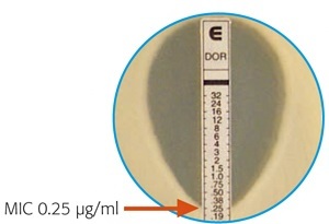 Figure: E-Test showing MIC
The device consists of a predefined, continuous, and exponential gradient of antibiotic concentrations immobilized along a rectangular plastic test strip. After 16 to 24 hours of incubation (longer for slow-growing organisms), a drop-shaped inhibition zone intersects the graded test strip at the inhibitory concentration (IC) of the antibiotic.
Why Use E-Test Instead of a Plain Disc Test?
Standard disc diffusion gives a category (Susceptible, Intermediate, or Resistant) and that's enough for most infections. E-test exists for the cases where a category isn't precise enough and you need the actual number.
Borderline calls matter most where the margin for error is smallest. A vancomycin MIC of 2 μg/mL versus 0.5 μg/mL can both fall in the "Susceptible" range by disc diffusion, yet the higher end of that range is associated with worse outcomes in MRSA bacteremia; a difference a categorical S/I/R report alone won't show you. This is exactly why E-test gets reached for in serious, borderline, or treatment-failure cases rather than run on every isolate by default: it is quantitative precision where it actually changes a decision, not a routine replacement for disc diffusion.
E-test strips are also used in a way disc diffusion can't replicate: dual-ended combination strips carry an antibiotic alone on one end and the same antibiotic plus a β-lactamase inhibitor (like clavulanic acid) on the other. Comparing the two MICs on a single strip is a direct, quantitative way to screen for ESBL production. See the ESBL Detection article for further information.
How It Works
- A predefined stable antimicrobial gradient is present on a thin inert non-porous plastic carrier strip 5mm wide, 60 mm long known as E-test strip.
- When the E-test strip is applied onto an inoculated agar plate, the antibiotic begins to diffuse from the strip into the agar, establishing a stable, continuous concentration gradient in the medium beneath and around the strip.
- After overnight incubation, the tests are read by viewing the strips from the top of the plate, a symmetrical inhibition ellipse is produced.
- The intersection of the lower part of the ellipse-shaped growth inhibition area with the test strip indicates the MIC value.
Requirements
MEDIA: Mueller Hinton agar (MHA) plates (uniform depth of 4 mm)
Others
- E-test strips (of desired antibiotic)
- McFarland standard 0.5
- Forceps
- Sterile cotton swabs
- Sterile normal saline, 4 ml volumes in tubes
Bacterial strains
- Non-fastidious cultures plated out for single colonies.
- Strain for quality control: Escherichia coli ATCC 25922
Procedure
- Inoculum Preparation
- Remove the sealed E-test package from the freezer (at or below -20°C) and let it equilibrate to room temperature before opening, at least 30 minutes. Opening a cold package lets moisture condense on the strip, which can disturb the gradient.
- Emulsify 3 or 4 individual test strain colonies and transfer to a tube of saline.
- Compare turbidity to that in the 0.5 McFarland standards. Adjust turbidity of inoculum to match that standard.
- Inoculation in Mueller Hinton Agar
- Dip a sterile cotton swab into the inoculum, and pulling out slightly, rotate the swab several times against the inside of the tube above the fluid level to remove excess liquid.
- Streak the swab over the entire surface of the agar plate by rotating the plate approximately 60°. Complete inoculation by running the swab around the rim of the agar.
- Leave the lid of the plate ajar for 5 minutes (no more than 15 minutes) to allow any excess moisture to be absorbed before applying strips.
NOTE: Swab plate within 15 minutes of preparing the adjusted inoculum.
- Application of E-test strips:
- Open E-test package by cutting package along the broken line. Apply strips to agar surface using forceps (or E-test applicator if available).
- Place the strip with the ‘E end’ at the edge of the plate and with the scale visible (i.e. facing upwards).
- If strips stick together, they may be pulled apart by handling the section marked E. Do not touch any other area of the strip.
- Use templates to position 4 to 6 strips onto a 150 mm plate or one (seldom two) strips onto a 90 mm plate. Do not remove or replace a strip once it has touched the agar.
- Repeat the entire procedure also for Quality control Strain (E. coli ATCC 25922)
- Incubate plates at 35°C (35 ± 2°C) for 16 to 24 hours; slow-growing organisms may need longer.
How to read the E-test MIC
Reading the E-test is where most errors happen, so read it in a fixed order.
1. Find where the ellipse meets the strip. After incubation, view the plate from above. Growth is inhibited in a symmetrical ellipse (teardrop) around the strip. The MIC is the value on the strip scale at the point where the lower edge of the inhibition ellipse intersects the strip.

2. Read at complete inhibition. Read the intersection at the point of complete inhibition of growth. A faint haze, or a few isolated colonies inside the ellipse, is not counted as growth for the reading (with specific exceptions your breakpoint guidance may note, such as trimethoprim-sulfamethoxazole, where an 80% inhibition endpoint is used).
3. If the value falls between two markings, round up. If the ellipse intersects between two twofold dilution marks, read the higher (next-up) value. This keeps E-test MICs on the same twofold scale as broth and agar dilution, so results from different methods stay comparable.
4. If the two sides disagree, take the higher. If the ellipse intersects the strip at a different point on each side, read the greater (higher) value.
5. Handle off-scale results. If growth runs the full length of the strip with no inhibition ellipse, the organism is resistant across the tested range: report the MIC as greater than or equal to the highest concentration on the strip. If the ellipse clears the strip entirely without touching it, report the MIC as less than or equal to the lowest concentration on the strip.
6. Convert the MIC to a category. Compare the MIC to the current CLSI (or EUCAST, per your laboratory) breakpoint for that organism-drug pair to report susceptible, intermediate, or resistant. The number is the measurement; the breakpoint turns it into a report.
A short worked example makes the rounding rule concrete: if the ellipse crosses between 1 and 2 μg/mL, the MIC is read as 2 μg/mL, not 1.5. There is no 1.5 on a twofold scale.
E-Test vs. Disc Diffusion vs. Broth/Agar Dilution
| Disc Diffusion (Kirby-Bauer) | E-Test | Broth/Agar Dilution | |
|---|---|---|---|
| Result type | Qualitative, S/I/R only | Quantitative MIC + S/I/R | Quantitative MIC + S/I/R |
| Relative cost | Low | Higher (commercial strips) | Low reagent cost, labor-intensive |
| Throughput | High, many drugs per plate | Lower, 4 to 6 strips per large plate | Low — batch setup required |
| Typical role | Routine, first-line screening | Confirming borderline or critical MICs; ESBL screening with combination strips | Reference method; required for fastidious organisms and some drug classes |
| Turnaround | 16–24h | 18–24h (longer for slow growers) | 16–24h |
NOTE: Disc diffusion tells you yes or no; E-test tells you by how much, and "by how much" is exactly what matters when the margin between working and failing is thin.
How to Remember
- The teardrop points to the answer. The MIC is where the pointed lower end of the inhibition ellipse meets the strip. Read from the top of the plate.
- When in doubt, read up. Between two marks, take the higher value. Two sides disagree, take the higher value. Rounding up keeps the E-test comparable to dilution methods.
- The number is not the report. The MIC is a measurement; the breakpoint turns it into S, I, or R. E-test gives you both, but the category still depends on current breakpoints.
- Disc says whether; E-test says how much. Reach for the E-test when "how much" changes the decision: borderline MICs, serious infections, treatment failure, and ESBL screening with combination strips.
Key Exam facts
| Question | Answer |
|---|---|
| E-test strip dimensions? | 5 mm wide, 60 mm long |
| Type of antibiotic gradient on the strip? | Continuous, exponential |
| Media used? | Mueller-Hinton agar, 4 mm uniform depth |
| Standard incubation? | 35°C (35 ± 2°C), 16 to 24 hours; longer for slow-growing organisms |
| QC strain? | E. coli ATCC 25922 |
| If the MIC falls between two twofold dilutions, which way do you round? | Up |
| If the ellipse intersects at different points on either side of the strip? | Read the greater (higher) value |
| How does E-test detect ESBL production? | Dual-ended strip: antibiotic alone versus antibiotic plus a β-lactamase inhibitor; a large MIC drop with the inhibitor present is positive |
Where students get confused
"The MIC is where the growth starts." It is where the inhibition ellipse intersects the strip, read at complete inhibition of growth. Faint haze inside the ellipse is not counted (with breakpoint-specific exceptions).
"Round to the nearest value." Always round up to the next twofold value, not to the nearest. This is what keeps E-test MICs comparable to broth and agar dilution.
"E-test does not replace disc diffusion." Disc diffusion remains the routine screen; the E-test is reached for when a quantitative MIC changes the decision (borderline, critical, or treatment-failure cases, and ESBL screening). Running it on every isolate wastes its value and its cost.
"A susceptible MIC is always fine." Not at the high end of the susceptible range for critical infections. The point of the vancomycin MIC 2 versus 0.5 example is that both read susceptible, yet the higher value is associated with worse outcomes in MRSA bacteremia. The number carries information the category hides.
"The E-test and the automated MIC are different measurements." Both report the minimum inhibitory concentration; they differ in how the gradient is created and read. The MIC concept is the same across E-test, broth dilution, and automated systems.
Frequently Asked Questions
What's the difference between E-test and disc diffusion?
Disc diffusion gives only a category (Susceptible, Intermediate, or Resistant). E-test gives an exact MIC value in µg/mL as well as the S/I/R category, which matters most for borderline or treatment-failure cases where the precise number changes the decision.
Why does E-test use an exponential antibiotic gradient instead of a fixed concentration?
If the inhibition ellipse intersects at different points on either side of the strip, which value do you read?
Always read the greater (higher) value. This is a deliberately conservative rule, plates are never perfectly uniform, and erring toward the higher MIC avoids under-calling resistance.
How is E-test used to screen for ESBL production?
Why is Mueller-Hinton agar used for E-test, and why does the depth matter?
Is E-test used routinely on every isolate?
No. Most labs reserve it for borderline, treatment-failure, or critical cases (such as confirming a borderline vancomycin MIC in MRSA) rather than running it as a routine first-line test, since disc diffusion is faster and cheaper for routine screening.
References
- Clinical and Laboratory Standards Institute (CLSI). M100 — Performance Standards for Antimicrobial Susceptibility Testing. Current annual edition. Wayne, PA: CLSI.
- Tille PM. Bailey & Scott's Diagnostic Microbiology. 15th ed. St. Louis: Elsevier; 2022.
- Procop GW, et al. Koneman's Color Atlas and Textbook of Diagnostic Microbiology. 7th ed. Philadelphia: Wolters Kluwer; 2017.
- Leber AL, editor. Clinical Microbiology Procedures Handbook. 4th ed. Washington, DC: ASM Press; 2016. doi:10.1128/9781683670438.CMPH
- Brown DFJ, Brown L. Evaluation of the E test, a novel method of quantifying antimicrobial activity. J Antimicrob Chemother. 1991;27(2):185-190. doi:10.1093/jac/27.2.185

Tankeshwar Acharya, MSc (Medical Microbiology)
Tankeshwar Acharya is an Assistant Professor in the Department of Microbiology at Patan Academy of Health Sciences (PAHS), Nepal, where he has been teaching and practicing clinical microbiology for over 14 years. He is the founder of Microbe Online, one of the leading free microbiology education resources on the web, covering bacteriology, mycology, parasitology, immunology, and clinical laboratory diagnostics written from direct experience in both the classroom and the diagnostic laboratory.
Comments
No comments yet. Be the first to share your thoughts.
Leave a comment
All comments are reviewed before they appear.