
DNase Test: Three Ways to Read It, and Why It Confirms Staph aureus When Coagulase Won't
A DNase-positive result can look like a fading green, a bright pink, or a clear halo, depending on which agar you use. All three answer one question: did the organism destroy the DNA in the plate? DNase confirms Staphylococcus aureus when coagulase is equivocal, separates Moraxella catarrhalis from Neisseria, and flags the virulence enzyme that helps staph spread. Here is how to read all three methods.
DNA hydrolysis test or Deoxyribonuclease (DNase) test is used to determine the ability of an organism to hydrolyze DNA and utilize it as a source of carbon and energy for growth.
Why It Matters
A blood culture grows Gram-positive cocci in clusters. Catalase positive, so it is a Staphylococcus. The question that decides everything next is whether it is Staphylococcus aureus, the aggressive pathogen, or one of the coagulase-negative staphylococci that are usually contaminants.
The standard answer is the coagulase test. But the slide coagulase came back equivocal, a faint clumping that could be real or could be autoagglutination, and the tube coagulase is still hours from being readable. The clinician needs a call.
The DNase test gives a second, independent line of evidence. S. aureus produces a DNase; most coagulase-negative staphylococci do not. A DNase-positive result on a catalase-positive Gram-positive coccus in clusters supports S. aureus even when coagulase is unclear, and it can be read from a plate the same day.
There is a deeper reason DNase belongs in the S. aureus workup, and it is about virulence, not just identification. When neutrophils try to trap S. aureus, they cast out webs of their own DNA, neutrophil extracellular traps, to snare and hold the bacteria. S. aureus fights back by secreting DNase, which cuts those webs apart and frees the organism to spread. The enzyme this test detects is the same enzyme the organism uses to escape being trapped. So a positive DNase is not only an identification clue; it is a readout of one of the tools that makes S. aureus invasive.
DNase does two more jobs at the bench. It separates Moraxella catarrhalis (DNase positive) from the pathogenic Neisseria (negative), and it helps flag Stenotrophomonas maltophilia (positive) apart from Burkholderia cepacia (negative). But its headline use is the one above: a same-day, independent confirmation of S. aureus when coagulase leaves you guessing.
Principle: why an organism digests DNA
DNA is a very large, negatively charged polymer. It cannot cross the bacterial cell membrane, so an organism cannot import intact DNA and use it. To feed on DNA, the organism must break it down outside the cell first.
That is what DNase (deoxyribonuclease) does. It is an exoenzyme, secreted into the surrounding medium, that hydrolyzes the phosphodiester bonds holding the DNA backbone together, cutting the long polymer into short oligonucleotides and nucleotides small enough to be absorbed and used as a source of carbon, nitrogen, and phosphate.
The test puts DNA into the agar and asks whether the organism can destroy it. Every detection method below is a different way of making that destruction visible: intact DNA looks one way, digested DNA looks another. The organism that clears the DNA around its colony is DNase positive.
The reason this matters clinically is that the same enzyme has a second role in S. aureus. Host neutrophils release webs of DNA (neutrophil extracellular traps) to trap bacteria; S. aureus DNase cuts those webs and releases the organism. So DNase is both a nutritional enzyme and a virulence factor, which is why it appears in the S. aureus identification workup.
Three ways to detect DNase
Three methods exist to detect this activity, distinguished by what's mixed into the DNase agar: methyl green, Toluidine Blue O (TBO), or no indicator at all, which instead requires flooding the plate with HCl after incubation. All three are answering the same question, did the organism break down the DNA in the agar, but they differ in how a positive result looks and in sensitivity for weak producers. This article covers all three, in order of how commonly they're taught.
All three methods answer the same question, did the organism destroy the DNA in the agar, but a positive result looks different in each. This is the single most confusing thing about the test, so anchor on the question, not the color.
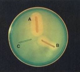
Method 1: Methyl green agar. The agar is pale green because methyl green, a cationic dye, binds intact negatively charged DNA. When a DNase-positive organism digests the DNA, the fragments no longer bind the dye, the green fades, and a colorless zone appears around the colony. Positive = clearing. Negative = the medium stays green.
Method 2: Toluidine blue O (TBO) agar. The agar is blue. Toluidine blue bound to intact DNA is blue; when DNA is hydrolyzed, the dye shifts to a metachromatic form and turns bright pink. Positive = a pink zone around the colony. Negative = the medium stays blue. Note this is the reverse visual logic of methyl green: here the positive is a color appearing (pink), not fading.
Method 3: Plain DNase agar, then flood with HCl. No dye. After incubation, flood the plate with 1N hydrochloric acid. The acid precipitates intact DNA, making the agar turn cloudy/opaque. Where the organism digested the DNA, there is nothing to precipitate, so a clear halo stands out against the opaque background. Positive = a clear zone against cloudiness. This is the original method (Jeffries, 1957) and is read within 5 minutes, because the acid is bactericidal and the plate cannot be re-incubated.
| Method | Uncut DNA looks | DNase-positive zone looks | Reagent needed after incubation |
|---|---|---|---|
| Methyl green | Green | Colorless clearing | None |
| Toluidine blue O | Blue | Bright pink | None |
| Plain agar + HCl | (clear until acid) | Clear halo against opacity | 1N HCl |
Whichever plate is in front of you, ask "did the DNA survive intact?" A positive is wherever the DNA is gone: the green faded, the blue turned pink, or the halo stayed clear while the rest went cloudy.
The heat-stable nuclease shortcut
S. aureus produces a thermostable DNase (thermonuclease) that survives boiling, unlike the DNases of most other organisms. This gives a fast, specific test: boil the specimen or broth to destroy heat-labile enzymes and kill the organism, then apply the boiled material to a DNase (usually toluidine blue) plate. Surviving DNase activity, seen as a pink zone, is strong evidence of S. aureus.
Because it works on a boiled, non-viable sample, the thermonuclease test is used to detect S. aureus even in situations where the organism itself cannot be cultured, including in food samples implicated in staphylococcal food poisoning. It is a good example of an enzyme outlasting the cell that made it.
 Requirements
- Media: DNase Agar with Methyl Green, DNase Agar with Toluidine Blue O, or plain DNase Agar (no indicator), choose one based on the method you're running.
- Reagent: Hydrochloric acid (1N), needed only for the no-indicator method.
- Others: Inoculating loop, Bunsen burner
Procedure
- Dry the surface of agar plates before use. Each plate may be divided into sections by drawing lines on the bottom of the plate.
- Inoculate the test agar:
- Spot inoculation: touch a colony with a loop and inoculate a small area (5-10mm diameter).
- Band/line streak inoculation: use a heavy inoculum and draw a line 3-4cm from the rim to the center of the plate.
- Incubate at 37°C for 18-24 hours.
- Reading the result depends on which agar was used:
- Methyl green or Toluidine Blue agar: examine directly, no additional reagent needed.
- Plain DNase agar (no indicator): flood with 1N HCl, let stand a few minutes to absorb, decant excess acid, and examine within 5 minutes against a dark background.
Expected Results
| Method | Positive result | Negative result |
|---|---|---|
| Methyl green | Colorless zone around growth | Medium remains green |
| Toluidine Blue O | Pink/rose zone around growth | Medium remains blue |
| No indicator + HCl | Clear zone around growth | Cloudy/opaque medium |
Figure -1: DNA Hydrolysis test A. Positive; Staphylococcus aureus B. Positive; Serratia marcescens C. Negative: Staphylococcus epidermidis on methyl green agar
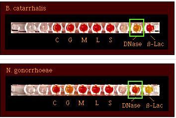 Fig 2: DNase test separating Moraxella catarrhalis (positive) from Neisseria gonorrhoeae (negative). The positive organism has hydrolyzed the DNA in the agar, producing a zone of change around its growth; the negative organism leaves the DNA intact.
Test results
- DNase-positive organisms: Serratia marcescens, Staphylococcus aureus, Campylobacter jejuni (some strains), Moraxella catarrhalis
- DNase-negative organisms: Staphylococcus epidermidis, Neisseria gonorrhoeae
Quality control
- Positive control: Staphylococcus aureus ATCC 25923 — DNase positive
- Negative control: Staphylococcus epidermidis ATCC 12228 — DNase negative
Run controls with each new lot of DNase agar. For the toluidine blue method, a heavy inoculum improves sensitivity, because the test detects preformed enzyme and some organisms grow poorly on the dye-containing medium.
Uses of DNase Test
- Differentiates S. aureus (DNase positive) from other staphylococci. Particularly useful when plasma isn't available for a coagulase test, or when coagulase results are difficult to interpret.
- Distinguishes M. catarrhalis from other Gram-negative diplococci of human origin (e.g., N. gonorrhoeae, N. meningitidis).
Limitation of DNase Test
- Some MRSA strains do not give positive DNase test result and some coagulase-negative staphylococci such as Staphylococcus capitis may give weak reactions.
- Serratia and Moraxella species also produce deoxyribonuclease.
- 1N HCl is bactericidal for Staphylococci. Once the HCl has been applied, the test must be read within 5 minutes and cannot be continued by re-incubation.
How to remember
One question beats three colors. The three methods look contradictory: green fades to clear, blue turns pink, or a halo stays clear against cloudiness. Do not memorize three color rules. Ask one question: did the DNA in this agar survive intact? The positive is always wherever the DNA is gone. Green faded, blue went pink, halo stayed clear: all the same answer.
DNase backs up coagulase for S. aureus. When the coagulase test is equivocal, DNase is the independent second opinion. S. aureus is DNase positive; most coagulase-negative staphylococci are not. Same-day, from a plate.
The enzyme that cuts the net. Picture neutrophils throwing a net of their own DNA to trap S. aureus, and the organism cutting the net with DNase and walking free. That image is why DNase is a virulence factor, not just an ID marker, and it makes the test memorable: you are detecting the organism's escape tool.
Thermonuclease survives the boil. S. aureus DNase is heat stable. Boil the sample to kill everything and destroy other enzymes, and if DNase activity remains, it is S. aureus. An enzyme that outlives its own cell.
Key exam facts in one table
| Question | Answer | The reason behind it |
|---|---|---|
| What does the test detect? | DNase (deoxyribonuclease), an exoenzyme | It hydrolyzes DNA outside the cell |
| Why does an organism make DNase? | To use DNA as a carbon, nitrogen, phosphate source | DNA is too large to import intact |
| What bond does DNase cut? | Phosphodiester bonds of the DNA backbone | Produces absorbable oligo- and nucleotides |
| Methyl green positive | Green fades to a colorless zone | Digested DNA no longer binds the cationic dye |
| Toluidine blue positive | Blue turns bright pink | Metachromatic shift on DNA hydrolysis |
| HCl method positive | Clear halo against a cloudy background | Acid precipitates intact DNA; digested zones stay clear |
| The unifying question | Did the DNA survive intact? | Positive is wherever the DNA is gone |
| Primary clinical use | Confirm S. aureus when coagulase is equivocal | S. aureus DNase +, most CoNS − |
| Second use | Separate M. catarrhalis (+) from Neisseria (−) | Among Gram-negative diplococci |
| Third use | Separate S. maltophilia (+) from B. cepacia (−) | Among the non-fermenters |
| Virulence role | DNase degrades neutrophil extracellular traps (NETs) | Lets S. aureus escape being trapped and spread |
| Thermonuclease | Heat-stable DNase of S. aureus | Survives boiling; a rapid, specific S. aureus marker, used even in food |
| Why read HCl plates in 5 min? | 1N HCl is bactericidal; no re-incubation | The reaction is fixed once acid is added |
| Why heavy inoculum on dye plates? | Detects preformed enzyme; some organisms grow poorly on dye media | Avoids false negatives |
| Positive QC | S. aureus ATCC 25923 | DNase positive |
| Negative QC | S. epidermidis ATCC 12228 | DNase negative |
| False negatives to know | Some MRSA; weak CoNS like S. capitis | DNase-negative S. aureus strains exist |
| Other DNase producers | Serratia marcescens, Moraxella | Not all DNase positives are S. aureus |
Where students get confused
Memorizing three color rules instead of one question. The three methods genuinely look contradictory: a positive is fading green, appearing pink, or a clear halo, depending on the agar. Trying to hold all three color rules separately is where students get lost. Ask instead: did the DNA survive intact? The positive is always where the DNA is gone. That one question converts three rules into one.
Assuming DNase positive means S. aureus. It does not, on its own. Serratia marcescens, Moraxella catarrhalis, and some Campylobacter are DNase positive. DNase supports S. aureus only in the right context: a catalase-positive Gram-positive coccus in clusters. Read it as a confirmation alongside coagulase and Gram stain, not as a standalone S. aureus test.
Re-incubating an HCl-flooded plate. Once you flood a plain DNase plate with 1N HCl, the plate is finished. The acid is bactericidal and the result is fixed. Read within 5 minutes and do not put it back in the incubator hoping a weak reaction will strengthen.
Using too light an inoculum on dye plates. The toluidine blue and methyl green media can inhibit growth, and the test reads preformed enzyme, so a light inoculum can give a false negative, especially for Moraxella and Gram-positive cocci that grow poorly on these media. Use a heavy inoculum.
Forgetting DNase-negative S. aureus exists. Some MRSA strains and occasional other S. aureus are DNase negative, and some coagulase-negative staphylococci like S. capitis give weak positives. This is exactly why DNase is a supporting test, not a sole confirmation. Pair it with coagulase.
Confusing the pink of toluidine blue with a pH indicator. The color changes here are about dye binding to DNA, not about pH. DNase agar has no acid/alkaline pH indicator. The pink of the toluidine blue method is a metachromatic dye shift when DNA is cut, not an acid color change.
Missing the virulence meaning. The DNase you detect in the tube is the same enzyme S. aureus uses to cut neutrophil DNA traps and spread. It is a virulence factor, not just an identification marker.
References
- Jeffries CD, Holtman DF, Guse DG. Rapid method for determining the activity of microorganisms on nucleic acids. J Bacteriol. 1957;73(4):590-591. doi:10.1128/jb.73.4.590-591.1957
- Procop GW, Church DL, Hall GS, Janda WM, Koneman EW, Schreckenberger PC, Woods GL. Koneman's Color Atlas and Textbook of Diagnostic Microbiology. 7th ed. Philadelphia: Wolters Kluwer; 2017.
- Pimenta FP, Souza MC, Pereira GA, Hirata R Jr, Camello TC, Mattos-Guaraldi AL. DNase test as a novel approach for the routine screening of Corynebacterium diphtheriae. Lett Appl Microbiol. 2008;46(3):307-311. doi:10.1111/j.1472-765X.2007.02310.x
- Kateete DP, Kimani CN, Katabazi FA, Okeng A, Okee MS, Nanteza A, Joloba ML, Najjuka FC. Identification of Staphylococcus aureus: DNase and mannitol salt agar improve the efficiency of the tube coagulase test. Ann Clin Microbiol Antimicrob. 2010;9:23. doi:10.1186/1476-0711-9-23
- Tille PM. Bailey and Scott's Diagnostic Microbiology. 15th ed. St. Louis: Elsevier; 2022.
Frequently Asked Questions
Why would a lab use toluidine blue instead of methyl green for the DNase test?
Is a positive DNase test enough to identify an isolate as Staphylococcus aureus?
Why does the HCl-flooded DNase test need to be read within 5 minutes?
Why does a positive DNase test look different on different agars?
Why is DNase considered a virulence factor in S. aureus?
What is thermonuclease and how is it used?

Tankeshwar Acharya, MSc (Medical Microbiology)
Tankeshwar Acharya is an Assistant Professor in the Department of Microbiology at Patan Academy of Health Sciences (PAHS), Nepal, where he has been teaching and practicing clinical microbiology for over 14 years. He is the founder of Microbe Online, one of the leading free microbiology education resources on the web, covering bacteriology, mycology, parasitology, immunology, and clinical laboratory diagnostics written from direct experience in both the classroom and the diagnostic laboratory.