
Lipase (Lipid Hydrolysis) Test: The Clear Zone That Reveals a Fat-Digesting Enzyme
Lipids are too large for a bacterium to import, so lipase-producing organisms digest them outside the cell, clearing the opaque tributyrin agar into a transparent halo. That clearing identifies lipase producers like Staphylococcus aureus and separates them from non-lipolytic bacteria. Here is the extracellular-enzyme mechanism, why lipase is a virulence factor, and how the test differs from the lecithinase (Nagler) test.
Why It Matters
Lipase is not just a metabolic convenience for a bacterium. For some pathogens it is a weapon.
Human skin is coated in sebum, an oily, lipid-rich secretion that is part of the body's barrier. Staphylococcus aureus and Cutibacterium acnes (formerly Propionibacterium acnes) both produce lipases that break sebum down, and this matters clinically: it helps these organisms colonize and persist on skin, contributes to the tissue damage of wound and skin infections, and, in the case of C. acnes, generates the free fatty acids that drive the inflammation of acne. So when the lipid hydrolysis test shows a clear zone around a staphylococcal colony, it is detecting the same enzyme that helps that organism live on and damage skin.
The test has a second, older use in the dairy and food world. Lipase-producing bacteria spoil high-fat products, butter, cream, cheese, by breaking down their fats into free fatty acids that turn the food rancid. The lipid hydrolysis test was developed partly to identify these lipolytic spoilage organisms.
Mechanically, the test asks one question: can this organism secrete an enzyme that digests fat? The answer appears as a clear halo in an otherwise cloudy, fat-containing agar. This article covers how that halo forms, why lipase matters as a virulence factor, and how this test differs from the lecithinase (Nagler) test that it is easily confused with.
Lipid hydrolysis or lipase test helps determine the ability of bacteria to produce lipase and hydrolyze lipids. Lipids are fatty, waxy, and oily bio-compounds. These include fats and triglycerides.
Some bacteria catabolizes lipid, especially in high-fat dairy product, to generate energy (ATP). These bacteria may also use lipids for synthesizing various biomolecules by entering different pathways.
Since lipids are complex fatty acids, they require further degradation before bacteria metabolize them. The extracellular enzyme lipase produced by bacteria helps in the hydrolysis of lipids.
The bacteria that can produce lipase belong to Enterobacteriaceae, Clostridium, Staphylococcus, and Neisseria. Many fungal species also demonstrate lipolytic ability.
Principle of Lipid Hydrolysis Test
Lipids are large, water-insoluble molecules. A bacterium cannot import an intact triglyceride across its membrane, so to use fat as a food source it must break the fat down outside the cell first. That is the job of lipase, an extracellular enzyme the organism secretes into its surroundings. Lipase cleaves the ester bonds of the triglyceride, releasing glycerol and free fatty acids small enough to be absorbed.
This is why the test works visually. Tributyrin agar is opaque because it is an emulsion of tributyrin fat suspended in the medium, like the cloudiness of milk. Where a lipase-positive organism grows, its secreted lipase digests the surrounding tributyrin, and the emulsion clears to a transparent halo. The clear zone is a map of where the fat has been destroyed. No lipase, no clearing, the medium stays cloudy right up to the colony.
Although many oils can be used for lipid hydrolysis tests, tributyrin oil is the most commonly used lipid for determining the lipolytic effects of the bacteria. The oil is simple fat or triglycerides or triacylglycerols. Triglycerides comprise glycerol and three long-chain fatty acids. Although tributyrin is simple fat, it is large and cannot enter the cell. Some cells can secrete the enzyme lipase to break down tributyrin before cellular uptake.
The breaking down of tributyrin releases glycerol converted into dihydroxyacetone phosphate, an intermediate product of glycolysis. Beta-oxidation catabolizes the fatty acids; two carbons combine with Coenzyme A to form acetyl Coenzyme A.
The acetyl CoA enters into the Krebs cycle to produce energy. Glycerol and fatty acids are useful in both anabolic and catabolic pathways alternatively. Tributyrin agar is opaque, and when the plate is inoculated with lipase-positive organisms, clear zones appear around the growth. The absence of the clear indicates the organism does not produce lipase.
The testing lipid hydrolysis can also occur in spirit blue agar. The composition of the agar is an emulsion with tributyrin oil and spirit blue dye as a color indicator. The media is opaque and light blue. Lipase-positive bacteria produce clear halos around the growth. However, the lightening of the medium is not considered positive growth.
Materials Required for Lipid Hydrolysis Test
A lipid hydrolysis test requires some laboratory equipment and materials. The equipment and materials needed for the test are as follows:
- Tributyrin agar base: It has peptone, yeast extract, and agar. The pH of the base is 7.5 ± 0.2 at 25℃.
- Tributyrin: It is also known as glycerol tributyrate or propane-1,2,3-triyl tributyrate. Its molecular formula is C15H26O6.
- Petri plates: The use of Petri plates is for preparing culture media or tributyrin agar.
- Test tubes: The test tubes are used for preparing bacterial inoculum.
- Autoclave: This equipment is used for sterilizing tributyrin agar during preparation.
- Incubator: Tributyrin plate inoculated with bacterial isolates is incubated during the test.
- Beaker: The glassware is used to transfer liquid during the tributyrin agar preparation.
- Inoculating loop: The inoculating loop of standard size is used for streaking bacterial inoculum in the tributyrin agar.
- Bunsen burner: It is used to sterilize inoculating loops and preparing tributyrin agar.
- Analytical balance: It is used for weighing powder for preparing tributyrin agar.
- Conical flask: It is used for preparing tributyrin agar.
Composition of Tributyrin Agar Base
| Composition | Amount |
|---|---|
| Peptone | 5.00 g |
| Yeast Extract | 3.00 g |
| Agar | 15.00 g |
| Final pH | 7.5 ± 0.2 at 25°C |
Procedure of Lipid Hydrolysis Test
The lipid hydrolysis test involves streaking bacterial inoculum in tributyrin agar. The clear zone around the growth indicates a positive for lipase production.
Preparation of Tributyrin Agar
Before conducting a lipid hydrolysis test, preparing tributyrin agar is an important step. The steps for preparing tributyrin agar depend on the manufacturing company. The steps for the preparation of tributyrin agar manufactured by Himedia are as follows:
- In 990 ml of distilled water, mix 23 grams of tributyrin agar base.
- After that, add 10 ml of tributyrin to the mixture above.
- Then, boil the solution to mix the base and tributyrin thoroughly.
- Sterilize the solution by autoclaving at 121℃ or 15 lbs pressure for 15 minutes.
- Cool the solution to 40-45℃.
- After that, pour the solution into sterile Petri plates.
Steps of Lipid Hydrolysis Test
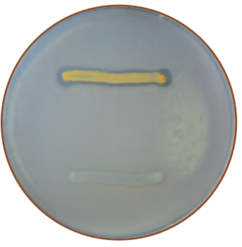 Figure: Above: Positive for lipase test (presence of clear zone around the growth).Below: Negative lipase test (absence of clear zone around the growth).
Following are the steps for performing a lipid hydrolysis test:
- In a tributyrin agar, inoculate a loopful of freshly isolated bacterial samples in a straight streak line or circle the size of a dime using a sterile inoculation loop.
- Incubate the streaked plate at 37℃ for 24-48 hours for aerobic bacteria and 72 hours for anaerobic bacteria.
- After incubation, observe the plate for a clear zone and interpret the result.
Precautions
There are some precautionary steps to follow during performing lipid hydrolysis tests; they are as follows:
- While sterilizing the inoculation loop, heating should be done until the loop turns red hot.
- Cooling down to the right temperature is necessary before picking up the bacterial colony.
- Maintaining proper incubation time is required. At least incubate for four days for anaerobic bacteria.
Result Interpretation
The lipid hydrolysis test’s result interpretation is based on the presence or absence of the clear zone around the bacterial growth.
- Positive result: There is a clear zone around the inoculation area. Staphylococcus aureus, S saprophyticus, Clostridium botulinum, Bacillus subtilis, Pseudomonas aeruginosa, etc., are bacteria that show positive results.
- Negative result: There is no clear zone around the inoculation area. Clostridium perfringens, C difficile, Escherichia coli, Klebsiella pneumoniae, K oxytoca, etc, are bacteria that shows negative result.
Quality Control
The quality control bacteria used for this test are as follows:
- Positive control: Staphylococcus aureus ATCC 12600 is used as the positive control.
- Negative control: Clostridium perfringens ATCC 1292 is used as the negative control.
Uses of Lipid Hydrolysis Test
The lipid hydrolysis test is a biochemical test to determine the lipolytic properties of some bacteria. It helps identify the bacteria. Its use is as follows:
- Differentiating lipolytic bacteria from non-lipolytic bacteria, especially in dairy industries.
- Identification of bacteria of the genus Clostridium, Corynebacterium, Bacillus, and Moraxella.
Limitation of Lipid Hydrolysis Test
The lipid hydrolysis test is a practical test for identifying lipolytic bacteria. However, it also has some limitations, which are as follows:
- The time for incubation is longer compared to another biochemical test.
- The lipid hydrolysis test is not confirmatory. Further biochemical testing is required to confirm the bacteria.
- The growth of fastidious bacteria is not possible.
Lipase vs lecithinase: two different lipid tests
The lipid hydrolysis (lipase) test is easily confused with the lecithinase test (the Nagler reaction on egg yolk agar). Both involve lipids and both produce a zone around the growth, but they detect different enzymes acting on different substrates and mean different things:
| Lipase test (this test) | Lecithinase test (Nagler) | |
|---|---|---|
| Enzyme detected | Lipase | Lecithinase (phospholipase C) |
| Substrate | Triglycerides (tributyrin) | Lecithin (a phospholipid, in egg yolk) |
| Medium | Tributyrin agar (opaque) | Egg yolk agar |
| Positive result | Clear (transparent) zone | Opaque/white (precipitate) zone |
| Key organism | S. aureus (positive) | Clostridium perfringens (positive) |
The visual logic is opposite: lipase clears the medium (digesting the fat emulsion into transparency), while lecithinase clouds it (splitting lecithin into an insoluble white precipitate). Clostridium perfringens is the classic split: it is lecithinase-positive (the basis of the Nagler test for its identification) but lipase-negative on tributyrin. So the two tests, done on the same organism, can give opposite-looking results while both being correct. See the lecithinase (Nagler) test article for that test's full method.
A related test uses Tween 80 (a synthetic fatty-acid ester) instead of tributyrin; there, a lipase-positive organism produces an opaque precipitate of calcium salts of the released fatty acids rather than a clear zone. The tributyrin method (clear zone) is the more common teaching version.
How to remember
Clear means clean: the fat is gone. Tributyrin agar is cloudy because it is full of suspended fat. A lipase-positive organism digests that fat and leaves a clear window. Clear zone = lipase present = the fat was destroyed. If the medium stays cloudy up to the colony, no lipase.
Lipase digests outside, then the cell eats. Fat is too big to import, so the enzyme has to work outside the cell first. The clear zone is literally the reach of the secreted enzyme. This is the same "digest outside, absorb inside" logic behind the other extracellular-enzyme tests (starch, gelatin, DNase).
Lipase is a skin weapon. Remember lipase clinically through S. aureus and C. acnes: both use it to break down skin sebum, which is why the enzyme this test detects is also a virulence factor in skin and wound infection and in acne.
Lipase clears, lecithinase clouds. The two lipid tests are visual opposites. Lipase (tributyrin) makes a clear zone; lecithinase (egg yolk, Nagler) makes a white opaque zone. C. perfringens is lecithinase-positive but lipase-negative, the split that keeps them straight.
Key exam facts in one table
| Question | Answer | The reason behind it |
|---|---|---|
| What does the test detect? | Lipase, an extracellular enzyme | Hydrolyzes lipids (triglycerides) |
| Why must lipase be extracellular? | Lipids are too large to import | The cell digests fat outside, then absorbs the products |
| Substrate | Tributyrin (glycerol tributyrate), a triglyceride | Simple fat used in the standard medium |
| What bond is cleaved? | Ester bonds of the triglyceride | Releases glycerol + free fatty acids |
| Medium appearance | Opaque (fat emulsion) | Clears where lipase acts |
| Positive result | Clear/transparent zone (halo) around growth | Fat digested to transparency |
| Negative result | Medium stays cloudy up to the colony | No lipase |
| Positive organisms | S. aureus, S. saprophyticus, C. botulinum, B. subtilis, P. aeruginosa | Lipolytic |
| Negative organisms | C. perfringens, C. difficile, E. coli, Klebsiella | Non-lipolytic |
| Metabolic fate of products | Glycerol → DHAP → glycolysis; fatty acids → beta-oxidation → acetyl-CoA → Krebs | Fat as an energy source |
| Clinical significance | Lipase is a virulence factor | S. aureus/C. acnes digest skin sebum |
| Dairy/food significance | Lipolytic spoilage of high-fat foods | Rancidity from fatty-acid release |
| Incubation | 37°C, 24-48h aerobes, up to 72h+ anaerobes | Slow reaction |
| pH of medium | 7.5 ± 0.2 | (Not ±2, a common typo) |
| Positive QC | S. aureus ATCC 12600 | Clear zone |
| Negative QC | C. perfringens | No clear zone |
| vs lecithinase (Nagler) | Lipase clears; lecithinase clouds | Different enzyme, substrate, and visual result |
Where students get confused
Confusing lipase with lecithinase (Nagler). The biggest one. Both are lipid tests, but lipase (on tributyrin) produces a clear zone, while lecithinase (on egg yolk agar, the Nagler reaction) produces a white opaque zone. They detect different enzymes on different substrates. C. perfringens is lecithinase-positive but lipase-negative, so the same organism gives opposite-looking results on the two tests, both correct.
Reading the clearing backwards. Because most agar tests show a positive as something appearing (color, precipitate), students sometimes expect lipase-positive to add something. It is the opposite: lipase-positive removes the cloudiness, leaving a clear window. The clear zone is the positive.
Mistaking the spirit blue variant's color for the result. On spirit blue agar, the medium is light blue, and lipase activity produces clear halos. A general lightening of the whole medium is not the positive result; the discrete clear halo around growth is. Do not read diffuse lightening as positivity.
Reading too early. Lipase reactions are slow. Aerobes need 24-48 hours and anaerobes up to 72 hours or more; some organisms take up to a week for a good zone. A plate read at 24 hours may look negative on a slow lipase producer. Hold before calling negative.
Expecting all staphylococci or all clostridia to behave alike. Lipolysis varies within a genus. S. aureus is lipase-positive but not every staphylococcus is strongly so; C. botulinum is positive while C. perfringens and C. difficile are negative. The test contributes to identification within a genus, it does not label a whole genus.
Treating a positive as a species identification. Lipase positivity is a characteristic, not an identification. Many genera produce lipase (Staphylococcus, Clostridium, Pseudomonas, Corynebacterium, Moraxella, members of Enterobacteriaceae). Read it alongside other tests, not as a standalone ID.
References
- Leboffe MJ, Pierce BE. A Photographic Atlas for the Microbiology Laboratory. 4th ed. Englewood, CO: Morton Publishing; 2011.
- Madigan MT, Bender KS, Buckley DH, Sattley WM, Stahl DA. Brock Biology of Microorganisms. 15th ed. Boston: Pearson; 2018.
- Tille PM. Bailey and Scott's Diagnostic Microbiology. 15th ed. St. Louis: Elsevier; 2022.
- Procop GW, Church DL, Hall GS, Janda WM, Koneman EW, Schreckenberger PC, Woods GL. Koneman's Color Atlas and Textbook of Diagnostic Microbiology. 7th ed. Philadelphia: Wolters Kluwer; 2017.
- Tributyrin agar base. HiMedia Laboratories technical data sheet M157.
Frequently Asked Questions
What does a positive lipid hydrolysis (lipase) test look like?
Why is lipase considered a virulence factor?
What is the difference between the lipase test and the lecithinase (Nagler) test?
Why does lipase have to act outside the bacterial cell?
Why does the lipid hydrolysis test need long incubation?

Tankeshwar Acharya, MSc (Medical Microbiology)
Tankeshwar Acharya is an Assistant Professor in the Department of Microbiology at Patan Academy of Health Sciences (PAHS), Nepal, where he has been teaching and practicing clinical microbiology for over 14 years. He is the founder of Microbe Online, one of the leading free microbiology education resources on the web, covering bacteriology, mycology, parasitology, immunology, and clinical laboratory diagnostics written from direct experience in both the classroom and the diagnostic laboratory.