
Cellulose Acetate Electrophoresis: Principle and Application
Cellulose acetate electrophoresis separates serum proteins and hemoglobin variants by net charge at alkaline pH. Learn the principle, why Hb S co-migrates with Hb D and Hb G, and why a sickle cell result must be confirmed at acid pH.
A three-year-old is brought to the outpatient department with painful, swollen hands and feet. She has been pale since infancy. Her mother says an older cousin had similar episodes. Her hemoglobin is 7.2 g/dL and the blood film shows target cells and a few elongated, curved red cells.
Her blood goes for hemoglobin electrophoresis. A drop of hemolysate is applied to a cellulose acetate strip soaked in an alkaline buffer at pH 8.6. Four samples run side by side: a normal adult control, a sickle cell trait control, a sickle cell disease control, and the child.
Twenty minutes later, the strip is stained. The normal control shows one dense band. The trait control shows two. The child's lane shows no band where Hb A should be, one dense band further back, and a faint band between them.
Her band sits exactly where the sickle cell control's band sits. It would be easy, and it would be wrong, to stop here and call it sickle cell disease.
Because at pH 8.6, the strip does not know what a hemoglobin is. It only knows how much net negative charge it carries. And three different hemoglobins, S, D, and G, happen to carry almost exactly the same charge at that pH. They all land in the same place. Hb D-Punjab in particular is clinically silent on its own, but a child who inherits Hb S from one parent and Hb D from the other has severe disease. The strip cannot tell you which situation you are looking at.
So the laboratory runs the sample again, this time on citrate agar at pH 6.2. At acid pH the charges shift, and Hb S pulls away from Hb D and Hb G. The child's band moves. It is Hb S.
The diagnosis is homozygous sickle cell disease. It took two pH values to get there.
That is the whole logic of this technique. A cellulose acetate strip reports one number: net charge at one pH. Everything below, the alkaline buffer, the strip, the wicks, the stain, exists to measure that number accurately. What it cannot do is tell you the identity of a molecule from a single measurement. Understand that, and you will never over-read a band.
What is cellulose acetate electrophoresis?
Cellulose acetate electrophoresis is a form of zone electrophoresis in which proteins are separated on a thin cellulose acetate membrane soaked in an alkaline buffer, usually around pH 8.4 to 8.6.
Its pores are large compared with a protein molecule, so the membrane does almost no sieving. Separation is by net charge, not by size. At alkaline pH the buffer is above the isoelectric point of every serum protein, so every protein carries a net negative charge and migrates toward the anode. The greater the net negative charge, the further it travels. Albumin, with the lowest isoelectric point, travels furthest; the gamma globulins barely move at all.
When analyzing a biochemical sample, separating its components is the essential first step. Electrophoresis is one such method, in which the components of a mixture are separated according to how they move under an applied electric field. Electrophoretic techniques are distinguished by the supporting medium used: polyacrylamide gel, agarose gel, capillary, and cellulose acetate.
Cellulose acetate electrophoresis was introduced by Joachim Kohn in 1957 as the successor to paper electrophoresis, and it was among the first electrophoretic methods adopted for routine clinical diagnosis.
Principle of Cellulose Acetate Electrophoresis
The principle follows the general principle of electrophoresis. A support matrix, here a cellulose acetate strip, holds a buffer, and sample components placed on it migrate under an applied electric field.
Cellulose acetate is obtained by acetylating cellulose paper (filter paper) with acetic anhydride. Its pores are large compared with a protein molecule, much larger than the pores of an agarose or polyacrylamide gel. Because the matrix therefore does almost no sieving, separation does not depend on molecular size. This is not a weakness. It is precisely what allows the technique to resolve molecules of near-identical size, such as hemoglobin variants that differ by a single amino acid.
Separation instead depends on net charge. The buffer is alkaline, typically pH 8.4 to 8.6, which is above the isoelectric point of every serum protein. Each protein has therefore lost protons, carries a net negative charge, and migrates toward the anode. The isoelectric point still governs the outcome, but indirectly: albumin has the lowest isoelectric point (about 4.7), so at pH 8.6 it sits furthest above its pI, carries the greatest net negative charge, and travels furthest. The gamma globulins have the highest isoelectric points, carry the least net negative charge at this pH, and travel least.
A protein sitting exactly at its isoelectric point carries no net charge and would not migrate at all. Separating molecules at their isoelectric points is a different technique, called isoelectric focusing.
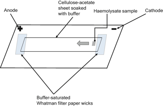 Figure: Diagram of cellulose acetate electrophoresis
The cellulose acetate strip is first wetted in the electrophoretic buffer. Then the sample is loaded in the strip (approximately ⅓ area of the strip). The end of the strip is in contact with the buffer with the help of a filter paper wick. A field strength of approximately 6 to 8 volts per centimetre is applied across the strip. Across a strip of usable length this corresponds to a few hundred volts in total, which is why the power supply must deliver up to 400 V.
The components are separated in the form of a band based on their charge. (1) The positively charged substance moves to the cathode, and the negatively charged substance moves to the anode.
Once the run is complete, the separated bands must be made visible. The standard sequence for serum proteins and hemoglobin is:
Stain the strip with a protein dye. Ponceau S and Amido Black (Naphthol Blue Black) are the classical stains; Coomassie brilliant blue is also used.
Destain in dilute acetic acid to remove background dye, leaving only the protein bands colored.
Clear the strip in a methanol or acetic acid mixture. Cellulose acetate becomes optically transparent, which is the distinctive advantage of this support.
Scan the cleared strip in a densitometer. Because the strip is now transparent, band densities can be read directly and converted into a quantitative tracing giving the percentage of each fraction. The cleared strip can also be dried and stored as a permanent record.
Where the components of interest are enzymes rather than structural proteins, a zymogram may be used instead. The strip is laid on filter paper soaked in buffer and the appropriate substrate and incubated, so that bands are revealed by their enzymatic activity rather than by a protein stain. This is how isoenzymes are visualized, and it is not the routine method for serum protein or hemoglobin analysis.
Materials Required for Cellulose Acetate Electrophoresis
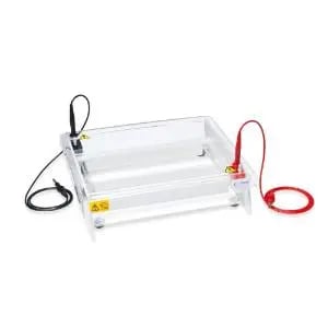 Figure: Horizontal cellulose acetate electrophoresis unit
- Cellulose acetate strip or membrane: cellulose paper acetylated by treatment with acetic anhydride. It serves as the support matrix. The strip itself is not an electrode. Its two ends are placed in contact with the anodal and cathodal buffer compartments of the tank by means of filter paper wicks.
- Electrophoresis buffer: an alkaline buffer. Tris-EDTA-borate (TEB) at pH 8.4 to 8.6 is standard for hemoglobin analysis. Barbital (veronal) buffer at pH 8.6 is classical for serum protein electrophoresis.
- Electrophoresis tank: a horizontal tank with an adjustable bridge, containing two buffer compartments and an electrode in each.
- Power supply: capable of delivering a constant current or constant voltage, typically up to 400 volts.
- Filter paper wicks: 3 mm Whatman filter paper, cut to the width of the tank, connecting each end of the strip to the buffer.
Application of Cellulose Acetate Electrophoresis
The most common application of cellulose acetate electrophoresis is in the diagnostic area for determining abnormalities in human hemoglobin.
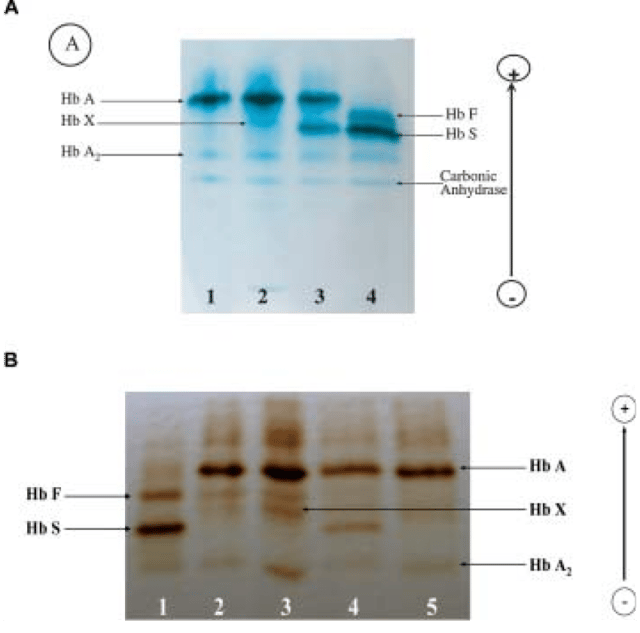 Figure: Electrophoretic study. A) Cellulose acetate electrophoresis at alkaline pH. Lane 1: normal adult control; lane 2: proband; lane 3: sickle cell trait control; lane 4: homozygous sickle cell. B) Isoelectric focusing on polyacrylamide gel. Lane 1: homozygous sickle cell; lane 2: β-thal trait and Hb X lane 3: proband; lane 4: sickle cell trait control; lane 5: normal adult control. (5)
So, it is sometimes referred to as hemoglobin electrophoresis. The applications of cellulose acetate electrophoresis are:
- Blood analysis: The cellulose acetate electrophoresis is applicable to determine hemoglobin abnormalities, especially in people with undiagnosed sickle cell anemia. Likewise, serum proteins like glycoproteins and albumin are also analyzed using this technique. It is used in antenatal diagnosing of the beta thalassemia. (4)
- Analysis of proteins: Different amino acids present in the proteins are separated on the basis of charge under the influence of electric current while performing cellulose acetate electrophoresis. This analysis is helpful in forensic, molecular, and clinical laboratories. Cellulose acetate membrane electrophoresis (CAME) is a classic approach to analyzing protein panels. (3)
- Analysis of polypeptides, dyes, and polysaccharides: Kohn's original work applied the technique to each of these, and it remains useful for separating small charged molecules and dye mixtures.
- Analysis of nucleic acids: cellulose acetate can be used to separate nucleic acids, although agarose and polyacrylamide gels are far more common for this purpose because they sieve by size.
Hemoglobin electrophoresis at alkaline pH
Hemoglobin variants differ from one another by one or two amino acids. They are essentially identical in size, so no sieving matrix could ever separate them. What differs is charge, and that is exactly what a cellulose acetate strip at pH 8.6 measures.
Order of migration toward the anode (fastest first): Hb A, Hb F, Hb S, Hb C.
In sickle hemoglobin, glutamate at position 6 of the beta chain is replaced by valine. Glutamate is negatively charged; valine is neutral. Hb S therefore carries less net negative charge than Hb A and lags behind it. In Hb C, the same glutamate is replaced by lysine, which is positively charged, so Hb C lags further still.
The trap. Charge is not identity. Several clinically distinct hemoglobins carry nearly identical charge at alkaline pH and land on top of one another:
| Band position at pH 8.6 | Hemoglobins that co-migrate there |
|---|---|
| Hb S position | Hb S, Hb D, Hb G, Hb Lepore |
| Hb C position | Hb C, Hb E, Hb O-Arab, Hb A2 |
This is not a technical nuisance. Hb D-Punjab is clinically silent in the heterozygous state, but a compound heterozygote for Hb S and Hb D has severe sickling disease. Reporting "Hb S present" on the basis of an alkaline strip alone can be seriously misleading.
Confirmation is mandatory. A band at the S position must be confirmed by electrophoresis on citrate agar at acid pH (about 6.0 to 6.2), where Hb S separates cleanly from Hb D and Hb G, and Hb C separates from Hb E, Hb O-Arab, and Hb A2. A sickle solubility test provides supporting evidence that the variant is a sickling hemoglobin. Alkaline and acid electrophoresis are complementary, not alternatives.
Reading the common patterns:
| Pattern | Bands seen | Interpretation |
|---|---|---|
| AA | One dense band at A | Normal adult |
| AS | Two bands; A denser than S (roughly 60:40) | Sickle cell trait |
| SS | No A band; dense S band; Hb F often raised | Sickle cell disease |
| SC | Bands at S and C, no A | Hb SC disease |
Two cautions. A recently transfused patient will show a donor-derived Hb A band regardless of genotype. And in the newborn, Hb F predominates, so newborn screening results are reported in order of abundance: "FA" is normal, "FAS" is sickle cell trait, and "FS" is sickle cell disease. The letter order carries the diagnosis.
Advantages
Cellulose acetate electrophoresis is an old technique that remains in routine use, because it is simple, fast, inexpensive, and produces sharp, quantifiable zones. Its advantages are described below.
- Easy setup: The instrument setup in the case of cellulose acetate electrophoresis is very simple. It does not require complicated instruments like setting gels in case of gel electrophoresis or properly maintaining capillaries for capillary electrophoresis.
- Simple detection and permanent records: the strip is stained, destained, and then cleared until it becomes optically transparent, so it can be scanned directly in a densitometer for quantification and stored indefinitely as a permanent record. No gel offers this. Where the analytes are enzymes, a zymogram may be used instead.
- Fast: the entire procedure, including staining and clearing, is generally completed within two hours, and the separation itself takes only 20 to 30 minutes. This is considerably quicker than gel electrophoresis, where casting and running the gel take longer. capillary electrophoresis is faster still and fully automated, but it requires an expensive instrument, which is why cellulose acetate remains the practical choice in many laboratories.
- No tailing effect: While separating the proteins, other methods can leave a tailing effect after separation. This electrophoresis separation method does not leave such effects after separation.
- Better zone development: The zone formed after separation in cellulose acetate electrophoresis is more apparent than in paper electrophoresis. The bands are better in resolution and sharpness.
Limitations
The cellulose acetate electrophoresis is highly advantageous, but it has some limitations, which are discussed below:
- Run at alkaline pH, with consequences. The technique is conventionally performed at pH 8.4 to 8.6, where all serum proteins are negatively charged and migrate toward the anode. This single pH cannot resolve hemoglobins that happen to carry similar net charge at that pH: Hb S co-migrates with Hb D and Hb G, and Hb C co-migrates with Hb E, Hb O-Arab, and Hb A2. Complementary electrophoresis on citrate agar at acid pH (about 6.0 to 6.2) is required to separate them.
- Electroendosmosis. Fixed negative charges on the support attract cations from the buffer, and the resulting bulk flow of buffer moves toward the cathode, opposing the anodal migration of the proteins. Albumin has enough net charge to be largely unaffected. The gamma globulins do not, and are dragged back to, or slightly behind, the point of application. This is why gamma globulins appear on the cathodal side of the origin on a serum protein strip, which surprises students who expect them to sit between albumin and the origin.
- Semi-quantitative by nature. Densitometry of a stained strip gives the percentage of each fraction rather than an absolute concentration, and dye binding is not perfectly proportional to protein mass for every fraction. Where precise quantification of hemoglobin variants is needed, HPLC or capillary electrophoresis is preferred.
In well-resourced laboratories, high-performance liquid chromatography (HPLC) and capillary electrophoresis have largely replaced cellulose acetate for hemoglobin variant analysis, because they are automated, quantitative, and resolve variants that co-migrate at alkaline pH. Cellulose acetate remains widely used where those instruments are unavailable, which is often where the burden of hemoglobinopathy is greatest. It is a current technique, not a historical one
References
- Kohn J. A cellulose acetate supporting medium for zone electrophoresis. Clinica Chimica Acta. 1957;2(4):297-303. [VERIFY volume, issue, page range]
- Wild BJ, Bain BJ. Detection and quantitation of normal and variant haemoglobins: an analytical review. Annals of Clinical Biochemistry. 2004;41(5):355-369.
- Bain BJ. Haemoglobinopathy Diagnosis. 3rd ed. Chichester: Wiley-Blackwell; 2020.
- Wilson K, Walker J. Principles and Techniques of Biochemistry and Molecular Biology. 8th ed. Cambridge: Cambridge University Press; 2018. Chapter: Electrophoretic Techniques.
- Boccacci M, Massa A, Tentori L. Application of cellulose acetate electrophoresis to globin chain separation for antenatal diagnosis of beta thalassemia. Clinica Chimica Acta. 1981;116(2):137-142.
- Nakayama A, Kubota R, Sakatsume M, Suzuki H, Katayama A, Kanamori K, et al. Cellulose acetate membrane electrophoresis based urinary proteomics for the identification of characteristic proteins. Journal of Clinical Laboratory Analysis. 2016;30(5):359-367.
Frequently Asked Questions
What is the principle of cellulose acetate electrophoresis?
Does cellulose acetate electrophoresis separate proteins by their isoelectric point?
Why is cellulose acetate used for hemoglobin rather than a gel?
In what order do hemoglobins migrate on alkaline cellulose acetate?
Can sickle cell disease be diagnosed from an alkaline cellulose acetate strip alone?
How do you distinguish sickle cell trait from sickle cell disease on the strip?
Why are newborn screening results written as FS or FAS?
Why do gamma globulins appear behind the point of application?
How are the bands visualized on a cellulose acetate strip?
Is cellulose acetate electrophoresis still used?

Tankeshwar Acharya, MSc (Medical Microbiology)
Tankeshwar Acharya is an Assistant Professor in the Department of Microbiology at Patan Academy of Health Sciences (PAHS), Nepal, where he has been teaching and practicing clinical microbiology for over 14 years. He is the founder of Microbe Online, one of the leading free microbiology education resources on the web, covering bacteriology, mycology, parasitology, immunology, and clinical laboratory diagnostics written from direct experience in both the classroom and the diagnostic laboratory.