

The CAMP test is used for the presumptive identification of Group B beta-hemolytic streptococci; Streptococcus agalactiae. The test is effective for the “prompt and reliable” identification of Streptococcus agalactiae in the clinical lab. The result appears in as little as 18 hours and requires few manipulations.
A naming caution before anything else: CAMP has nothing to do with cyclic AMP. If you have done any biochemistry, that is the association your brain will reach for, and it is wrong here. CAMP is an eponym: Christie, Atkins, and Munch-Petersen, the three researchers who described this synergistic hemolysis in 1944.
Principle of CAMP Test
The basis of the CAMP test is the enhanced hemolytic activity of beta-hemolysin-producing strains of Staphylococcus aureus by an extracellular protein ( CAMP factor) produced by group B streptococci. Synergistic hemolysis in the blood agar plate results from the interaction of the beta-hemolysin with the factor. Both hemolytic and non-hemolytic strains of group B streptococci show this phenomenon.
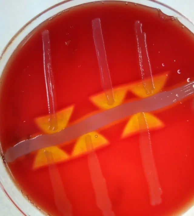 Figure: CAMP Test Positive (arrowhead increased zone of hemolysis)
Quality control
Test each lot of beta-lysin reagent or disks with a positive and negative control by streaking them in a line parallel to the test organism.
Organisms
- CAMP test positive: S. agalactiae ATCC 13813
- CAMP test negative: Streptococcus pyogenes ATCC 19615
- Periodically use an in-house laboratory strain of Arcanobacterium haemolyticumto demonstrate the reverse CAMP test for training purposes.
Procedure for CAMP test
Standard Method
- Firstly, make a single straight line streak of beta-hemolysin producing Staphylococcus aureus down the center of a blood agar plate,
- Then, inoculate a streak of the test organism (beta-hemolytic streptococci to be identified) perpendicular to the staphylococcal streak. Take care not to intersect the staphylococcal streak.
Note: Make these streaks in such a way that, after incubation, the growth of the two organisms will not be touching.
- The streptococcal streak should be 3 to 4 cm long.
- Similarly, inoculate known group A and B streptococcal strains on the same plate as negative and positive controls.
- Ensure labeling the location of each streak on the back of the plate.
- Finally, incubate the plate at 35°C in ambient air for 18-24 hours.
Disk method
- Firstly, place disks containing beta-lysin of S. aureus on a warmed blood agar plate.
- Then, streak microorganisms 2 to 3 mm from the edge of the disk.
- Finally, incubate the plate overnight at 35°C in ambient air. (Avoid CO₂, candle jar, or anaerobic incubation.)
Spot rapid method
- At first, place one drop or a 10 μl loopful of CAMP liquid reagent next to a presumptive S. agalactiae colony growing on a blood agar plate.
Note: Do not worry if the liquid touches or engulfs the colony.
- Then, incubate the plate right side up to prevent the spot CAMP reagent from running over the plate’s surface for 20 min at 35°C.
- After that, examine the plate with transmitted light for a zone of enhanced hemolysis next to the colony.
- Again, re-incubate for up to 30 min if the reaction is initially negative. Use a hand lens if necessary for examining the plate.
- Refrigeration may enhance reaction after incubation.
Results and Interpretations
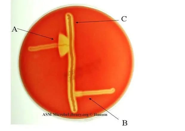 Figure: CAMP test : A. Streptococcus agalactiae (positive) B. Streptococcus pyogenes (Negative) Image Source: ASM
- CAMP Test: A positive test is the formation of a distinct arrowhead of hemolysis at the intersection of the staphylococcus and test organism streaks. If any gram-positive, catalase-negative, beta-hemolytic cocci, bacitracin-resistant, trimethoprim-sulfamethoxazole-resistant shows such arrowhead hemolysis, it can be reported as group B. streptococci (Streptococcus agalactiae).
- A positive reverse CAMP test or phospholipase D is indicated by a distinct arrow of no hemolysis at the intersection of the two hemolytic organisms. C. perfringens is reverse CAMP positive.
- In the disk test, a positive result is indicated by a distinct crescent- or arc-shaped zone of complete hemolysis at the intersection of the disk of beta-lysin and the isolate.
- In the rapid spot test, the presence of clear enhanced hemolysis only where the diffused hemolysis overlaps is a positive result.
- A negative test is a lack of enhanced hemolysis near the colony being tested.
Other organisms that give a standard (positive) CAMP reaction
Besides S. agalactiae, these give an enhanced-hemolysis CAMP reaction (not to be confused with the reverse CAMP organisms below):
- Listeria monocytogenes
- Rhodococcus equi
- Certain Corynebacterium species (e.g., C. glucuronolyticum, C. striatum group)
Limitation of CAMP test
- Some group A streptococci will be CAMP test positive if the test plate is incubated in a candle jar, in a CO₂ atmosphere, or under anaerobic conditions. Therefore, ambient air incubation should be used.
- S. pyogenes can give a reaction that may be interpreted as positive. When there is confusion, check for the pyrrolidonyl-β-naphthylamide (PYR) test. S. pyogenes is PYR positive, whereas S. agalactiae is PYR negative.
- It has a 98% sensitivity for detecting S. agalactiae, so isolates with a negative CAMP test could still be S. agalactiae and may require further testing.
- The reaction may be very weak if the agar is too thin or hemolyzed.
Reverse CAMP Test
The name "reverse CAMP" covers two related but distinct bench observations. Keeping them apart is what prevents misreading a plate.
1. Inhibition of staphylococcal beta-hemolysin. Some organisms produce a phospholipase that blocks the S. aureus beta-hemolysin instead of enhancing it. Where the test organism approaches the staph streak, hemolysis is suppressed, giving a triangular zone (arrowhead) of inhibited hemolysis. The organisms that behave this way include Arcanobacterium haemolyticum (phospholipase D), Corynebacterium pseudotuberculosis, and C. ulcerans.
2. Two-toxin synergy to identify Clostridium perfringens. In the reverse CAMP test used for C. perfringens, S. agalactiae is streaked as the source of CAMP factor and C. perfringens is the test organism. The alpha-toxin of C. perfringens (a phospholipase C) and the GBS CAMP factor act together, producing the classic bow-tie zone of enhanced hemolysis at the junction. Note the naming trap: here S. agalactiae is the reagent, not the reverse-CAMP-positive organism, and the result is synergy (extra hemolysis), which is the opposite of the inhibition pattern above.
So a single "reverse CAMP" label spans an arrow of no hemolysis (inhibition) and a bow-tie of extra hemolysis (C. perfringens synergy). Always confirm which version the protocol in front of you is describing.
Where students actually get confused
- The arrowhead is not made by GBS alone. A common misread is treating the arrowhead as something S. agalactiae produces by itself. It does not. The visible clearing appears only where the GBS CAMP factor and the S. aureus beta-hemolysin diffuse into the same zone. No staph streak, no arrowhead, even for a strong CAMP-factor producer.
- A bow-tie is not a strong forward CAMP. Students who have seen a forward arrowhead sometimes read any junction clearing as "very positive." The bow-tie belongs to the C. perfringens reverse test, where GBS is only the reagent. Reporting it as a forward CAMP positive misidentifies the organism.
- "Listeria is CAMP positive" is a forward result, not reverse. Listeria monocytogenes enhances hemolysis toward the S. aureus streak, exactly like GBS. It is easy to slot it mentally next to C. perfringens under "reverse," which is wrong.
Because Listeria and GBS give the same arrowhead, they are separated by catalase (Listeria positive, GBS negative) and Gram morphology (rod vs. coccus), not by CAMP shape.
Key exam facts
| Feature | Detail | Memory hook |
|---|---|---|
| What CAMP stands for | Christie, Atkins, Munch-Petersen | Not cyclic AMP |
| Identifies | Streptococcus agalactiae (GBS) | |
| Mechanism | Synergy: CAMP factor + S. aureus beta-hemolysin | Neither organism alone produces it |
| Positive result | Arrowhead zone of complete hemolysis | At the streak junction |
| Sensitivity | ~98% | Negative ≠ automatic exclusion |
| False-positive risk | Some GAS strains under CO2/anaerobic incubation | Use ambient air |
| Resolves ambiguity | PYR test (GAS positive, GBS negative) | |
| Reverse CAMP positive organisms | C. perfringens, Arcanobacterium haemolyticum | Arrow of no hemolysis |
How to Remember
The arrow is a collaboration, not a solo. Neither organism makes the arrowhead alone. S. aureus beta-hemolysin partially damages the red cells; GBS CAMP factor on its own does nothing visible. Where the two diffusing substances overlap, the partial damage is finished off completely, and that overlap zone is the arrowhead, pointing toward the Staph streak because that is where the synergy is strongest.
Standard arrow = enhanced hemolysis; reverse arrow = no hemolysis. A positive standard CAMP is an arrow of extra clearing (synergy). A positive reverse CAMP is an arrow of absent clearing (inhibition). Same arrow shape, opposite biology. Always know which version you are reading.
PYR breaks the GAS tie. If a beta-hemolytic strep gives a borderline CAMP under the wrong incubation, PYR settles it: S. pyogenes (GAS) is PYR-positive, S. agalactiae (GBS) is PYR-negative.
References
- Tille PM. Bailey and Scott's Diagnostic Microbiology. 15th ed. St. Louis: Elsevier; 2022.
- Procop GW, Church DL, Hall GS, Janda WM, Koneman EW, Schreckenberger PC, Woods GL. Koneman's Color Atlas and Textbook of Diagnostic Microbiology. 7th ed. Philadelphia: Wolters Kluwer; 2017.
- Leber AL, editor. Clinical Microbiology Procedures Handbook. 4th ed. Washington, DC: ASM Press; 2016. doi:10.1128/9781555818814
- Christie R, Atkins NE, Munch-Petersen E. A note on a lytic phenomenon shown by group B streptococci. Aust J Exp Biol Med Sci. 1944;22:197-200.

Comments
No comments yet. Be the first to share your thoughts.
Leave a comment
All comments are reviewed before they appear.