
Acridine Orange Staining: Principle, Procedure, Results, and Clinical Applications
Acridine orange is a fluorescent dye that detects bacteria in blood cultures, mycobacteria, fungi, and malaria parasites. Learn the principle, procedure, results interpretation, and when to use it over Gram staining.
A blood culture bottle flags positive at 2 AM. The automated system has detected bacterial growth, but the Gram stain shows nothing — the organisms are too few, too small, or morphologically ambiguous. The laboratory scientist reaches for acridine orange.
Under the fluorescence microscope, orange-glowing bacteria appear instantly against a green background of human cells and debris. Where the Gram stain failed, acridine orange delivers. The result guides the physician's antibiotic choice hours before the subculture grows.
Acridine orange (AO) is a fluorescent nucleic acid dye with a unique property: it changes emission colour depending on what it binds. This single property gives it clinical utility across bacteriology, mycology, parasitology, and mycobacteriology — wherever conventional staining fails or is too slow.
Acridine orange is a fluorescent dye that intercalates or binds with the nucleic acid (either DNA or RNA) present in organisms and fluoresces to emit various colors that help differentiate cellular organelles. This binding results from the electrostatic interactions of acridine molecules between the nucleic acid-base pairs. Due to its metachromatic properties, acridine orange (AO) is commonly used in fluorescence microscopy and flow cytometry analysis of cellular physiology and cell cycle status, including the fluorescent microscopic examination of microorganisms.
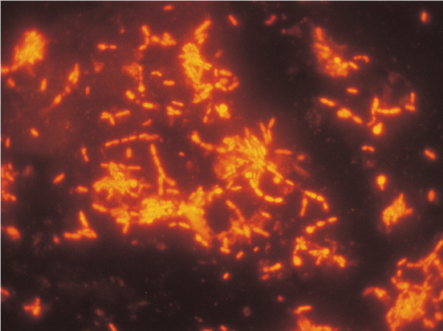 Figure: Fluorescent acridine orange stain coryneforms bacteria (Imagesource)
Principle
Acridine orange is a cell-permeable, nucleic acid selective fluorescent dye that emits green fluorescence when bound to dsDNA (at 520 ) and red fluorescence when bound to ssDNA or RNA (at 650 nm). Since it is a cationic dye, it also enters acidic compartments such as lysosomes which, in low pH conditions, will emit orange light.
Acridine orange is a carcinogen when absorbed through the skin. Wear gloves when working with this stain.⚠
Why colour shift matters clinically:
The differential fluorescence is pH-dependent and concentration-dependent:
- At physiological pH, DNA-bound AO fluoresces green (emission ~520 nm)
- RNA-bound AO (in ribosomes and cytoplasm) fluoresces orange-red (emission ~650 nm)
- At low pH (acidic compartments such as lysosomes), AO emits orange
In practice for clinical microbiology:
- Bacteria — orange-red fluorescence (high RNA content relative to DNA, active metabolism)
- Human cells and debris — green to yellow-green background
- Fungi — orange-red (RNA-rich cytoplasm) with green nuclear fluorescence
- Malaria parasites — yellow-green to orange within red blood cells
This differential makes bacteria and parasites stand out vividly against host cell background — the key advantage over conventional staining in paucibacillary or difficult specimens.
⚠ Safety: Acridine Orange is a Carcinogen
Acridine orange is classified as a potential carcinogen and mutagen. Safe handling requires:
- Wear nitrile gloves at all times when handling powder or solution
- Work in a well-ventilated area; avoid generating aerosols
- Avoid skin and eye contact — wash immediately with water if contact occurs
- Store in brown/amber bottles at 4°C away from light (AO degrades on light exposure)
- Dispose of waste solutions according to institutional hazardous waste protocols — do not pour down the drain
- The stock solution (50 mg in 10 mL water) is significantly more concentrated and hazardous than the working solution — handle with extra careClinical Applications of Acridine Orange Staining
Staining procedure
Staining procedures vary according to their use
- For staining clinical specimens with acridine orange at low pH (Acridine orange acid stain)
- Requirements: acridine orange, glacial acetic acid, distilled water
- Preparation of reagent: 50 mg acridine orange is dissolved in 10 ml of distilled water to prepare a stock solution and stored in the refrigerator.1 ml of acridine orange stock solution and 0.5 ml of glacial acetic acid is added to 50 ml of distilled water to prepare a working solution.
- Staining procedure:
- Prepare a smear in a clean grease-free slide and allow it to air dry.
- The slide is then fixed with methanol and dried again.
- It is then put in a trough with an acridine orange staining working solution (i.e 0.01 percent).
- After 2 minutes of staining, the slides are washed gently with water, dried, and examined in a fluorescent microscope.
Troubleshooting Acridine Orange Staining
| Problem | Likely Cause | Action |
|---|---|---|
| Background too bright (green haze) | Working solution too concentrated; overstaining | Reduce staining time to 1 minute; dilute working solution |
| Bacteria not visible / faint fluorescence | AO degraded (light exposure); filter mismatch | Replace stain; verify fluorescence microscope has correct excitation filter (490 nm excitation, 520 nm emission for green; 490 nm excitation, 630 nm for orange) |
| Cannot distinguish bacteria from debris | pH wrong; overstaining | Ensure pH of working solution is correct (acidic); reduce staining time |
| False positives (non-bacterial fluorescent particles) | Stain precipitate; environmental contamination | Filter working solution before use; use fresh slides |
| Smear washing off slide | Inadequate fixation | Ensure complete air-drying and proper heat or methanol fixation |
Observance:
Bacteria stain orange against a green to a yellow background of human cells and debris.
- For staining cells for analysis by flow cytometry.
- Requirements: 0.1M Citric Acid (dissolve 1.921g per 100ml distilled water), 0.2M Dibasic Sodium Phosphate (dissolve 2.839g per 100ml distilled water) ,Triton X-100 (Baker), 0.5M EDTA, Sodium chloride(NaCl), Acridine Orange (Powder) and Sucrose.
- Preparation of reagents:
- Stock Buffer I:20mM Citrate-Phosphate, pH 3.0, 0.1mM EDTA, 0.2M Sucrose, 0.1% Triton X-100(To 125ml distilled water add 40µl 0.5M EDTA, 26.48ml 0.1M Citric Acid, 6.85ml 0.2M Dibasic Sodium Phosphate, 13.69g Sucrose, 0.2ml Triton X-100. QS to 200ml and 0.2µ filter. Store at 4°C)
- Stock Buffer II:10mM Citrate-Phosphate, pH 3.8, 0.1M NaCl (To 150 ml distilled water, add 9.92ml 0.1M Citric Acid, 5.46ml 0.2M Dibasic Sodium Phosphate, 1.7g NaCl. QS to 200ml and 0.2m filter. Store at 4°C)
Staining Procedure
- Make a 2mg/ml solution of acridine orange in distilled water and dilute to 1:100 in Buffer II
- Aliquot cells: 105- 106 in 100µl PBS or media.
- Add Buffer I (0.5ml) at room temp, agitate to suspend.
- Add Buffer II + AO (0.5ml) at room temp, agitate to suspend.
- Run on flow cytometer. Excitation 488 nm; dot plot of green fluorescence at 530nm versus red fluorescence >600 nm).
Observation
- Green fluorescence when bound to dsDNA and
- Red fluorescence when bound to ssDNA or RNA.
Limitations
- Acridine orange stain is non-specific. It stains all nucleic acid-containing cells, so it does not discriminate between gram-negative and gram-positive bacteria.
Quality Control
- Examine the acridine orange staining solution for color and clarity. The solution should be clear, orange, and without evidence of precipitate.
- Each use time, stain a prepared slide of known bacteria, such as Escherichia coli mixed with staphylococci, and examine for the desired results. Record results and refer out-of-control results to the supervisor. Gram-negative rods and Gram-positive cocci are fluorescent (orange).The background is nonfluorescent (green-yellow).
Clinical Applications of Acridine Orange Staining
1. Bacteria in Blood Cultures — The Primary Clinical Application
Acridine orange staining is most valuable when a blood culture bottle signals positive but the Gram stain is negative or equivocal. This occurs when:
- Bacterial density is below the Gram stain detection threshold
- Organisms are morphologically distorted (due to antibiotic treatment)
- Gram reaction is variable or ambiguous (e.g., Acinetobacter, gram-variable organisms)
Appearance: Bacteria fluoresce orange against a green-yellow background of human cells and debris. Even single bacteria are detectable because the fluorescence signal amplifies visibility dramatically compared to a Gram stain at 1,000x.
Research evidence: Studies consistently show AO is more sensitive than Gram stain for detecting bacteria in blood culture broths early during incubation — particularly for gram-negative organisms and for specimens from patients on antibiotics where bacterial morphology is distorted.
Practical workflow:
- When blood culture bottle signals positive → Gram stain first (standard)
- If Gram stain negative or equivocal → Acridine orange staining immediately
- Report AO result; subculture proceeds simultaneously
- Never delay subculture waiting for AO result — both are done in parallel
2. Mycobacteria — Direct Specimen Examination
Acridine orange stains mycobacteria directly in sputum and other specimens, producing bright orange-red fluorescence against a pale green background. This application is sometimes used as an alternative or supplement to auramine-rhodamine fluorochrome staining.
Comparison with auramine-rhodamine:
- Both are fluorochrome methods for mycobacteria
- Auramine-rhodamine is more commonly used and WHO-recommended for TB
- AO can be used when auramine-rhodamine reagents are unavailable
- Both require a fluorescence microscope
For the dedicated fluorochrome procedure for mycobacteria, see: Auramine-Rhodamine Fluorochrome Staining
3. Malaria Parasites — Quantitative Buffy Coat (QBC) Method
The Quantitative Buffy Coat (QBC) technique uses acridine orange pre-coated capillary tubes to stain malaria parasites directly in whole blood. Infected red blood cells are concentrated by centrifugation in the buffy coat layer, where parasites fluoresce yellow-green under UV light.
Advantages over conventional Giemsa smear:
- Faster — result in 8–10 minutes
- More sensitive for low-parasitaemia cases
- Less technically demanding for initial screening
Limitations:
- Cannot reliably speciate Plasmodium (species identification requires Giemsa smear confirmation)
- Requires fluorescence microscope and specific QBC equipment
- More expensive than conventional smear
Read details about QBC procedure
4. Fungal Detection in Specimens
Acridine orange stains fungal elements in tissue sections and smears. Fungal cytoplasm (RNA-rich) fluoresces orange-red while nuclei fluoresce green, providing useful contrast for identifying:
- Yeast cells in blood culture or CSF
- Hyphal elements in tissue sections
- Pneumocystis jirovecii (less commonly — calcofluor white is preferred)
When AO is used for fungi: Primarily as a rapid screen when calcofluor white is unavailable or when a single stain needs to serve multiple purposes (simultaneous bacterial and fungal detection in a BAL specimen, for example).
For dedicated fungal staining methods, see: Fungal Staining Methods and Uses
5. Detection of Mycoplasma and Cell Wall-Deficient Bacteria
Mycoplasma species lack a cell wall and stain poorly or not at all by Gram stain. Acridine orange detects them reliably because it targets nucleic acid directly, bypassing cell wall-dependent staining mechanisms.
Clinical relevance: Mycoplasma pneumoniae causes atypical ("walking") pneumonia — a patient who appears well enough to continue daily activities but has persistent dry cough and bilateral infiltrates on CXR. Mycoplasma genitalium causes urethritis and cervicitis. Neither is detectable on routine Gram stain. Serology and PCR are the primary diagnostic tools, but AO can provide rapid microscopic evidence in direct specimens.
When to Choose Acridine Orange Over Gram Stain
Acridine orange does not replace Gram staining — it complements it in specific situations:
| Situation | Recommended Approach |
|---|---|
| Routine clinical specimen (wound, urine, sputum, CSF) | Gram stain first |
| Blood culture positive, Gram stain negative | AO immediately |
| Patient on antibiotics, distorted morphology on Gram | AO as supplement |
| Suspected mycoplasma (no cell wall) | AO or serology/PCR |
| Rapid malaria screen needed | QBC-AO method |
| Fungal detection in BAL, calcofluor unavailable | AO acceptable |
| Mycobacteria, fluorescence microscope available | Auramine-rhodamine preferred; AO acceptable |
How to Remember: Acridine Orange
The colour rule — "DNA is green, RNA is red-orange":
- DNA (double-stranded, stable) → Green fluorescence (D and G — both "solid/stable")
- RNA (single-stranded, active) → Red-orange fluorescence (R and R — both "reactive/active")
Bacteria are metabolically active and RNA-rich → they fluoresce orange and stand out against the green host cell background. This is how one bacteria in a sea of human cells becomes visible.
The "BMMF" applications:
- Blood culture (primary use — Gram stain negative but clinically suspect)
- Mycobacteria (fluorochrome alternative to auramine-rhodamine)
- Malaria (QBC method — rapid parasitaemia screen)
- Fungi (tissue sections and BAL when calcofluor unavailable)
Key Exam Facts in One Table
| Feature | Detail |
|---|---|
| Dye type | Fluorescent nucleic acid dye; cationic; cell-permeable |
| dsDNA binding | Green fluorescence (~520 nm) |
| ssDNA/RNA binding | Orange-red fluorescence (~650 nm) |
| Microscope required | Fluorescence microscope |
| Primary clinical use | Bacteria detection in blood cultures when Gram stain negative |
| Bacteria appearance | Orange against green-yellow background |
| Human cells/debris | Green to yellow-green |
| Advantage over Gram stain | Higher sensitivity in paucibacillary specimens; detects mycoplasma |
| QBC malaria method | AO-precoated capillary tubes; parasites fluoresce yellow-green |
| Mycobacteria | Detects AFB; auramine-rhodamine preferred for TB |
| Safety | Carcinogen and mutagen — gloves mandatory; store in dark |
| Key limitation | Requires fluorescence microscope; not universally available |
References
- Bailey & Scott’s Diagnostic Microbiology, Forbes, 11th edition
- Review of Medical Microbiology and Immunology, Lange Medical Books, 13th edition
- Murray PR, Rosenthal KS, Pfaller MA. Medical Microbiology. 9th ed. Elsevier; 2020.
- Welch DF. Applications of cellular fatty acid analysis. Clin Microbiol Rev. 1991;4(4):422–438.
- Gosselin M, Giguère A. Acridine orange staining for rapid detection of microorganisms in blood culture systems. J Clin Microbiol. 1986;23(5):964–966.
- Kawamoto F. Rapid diagnosis of malaria by fluorescence microscopy with light microscope and interference filter. Lancet. 1991;337(8735):200–202.
Frequently Asked Questions
What colours do different structures appear when stained with acridine orange?
When should acridine orange staining be used instead of Gram staining?

Tankeshwar Acharya, MSc (Medical Microbiology)
Tankeshwar Acharya is an Assistant Professor in the Department of Microbiology at Patan Academy of Health Sciences (PAHS), Nepal, where he has been teaching and practicing clinical microbiology for over 14 years. He is the founder of Microbe Online, one of the leading free microbiology education resources on the web, covering bacteriology, mycology, parasitology, immunology, and clinical laboratory diagnostics written from direct experience in both the classroom and the diagnostic laboratory.