
Stokes Disc Diffusion Method: Why the Control Shares the Plate

On this page
A rural laboratory receives a shipment of antibiotic discs that has spent three days in transit through the summer heat, with no guarantee the cold chain held. The discs may have lost potency, and there is no way to be sure. Running a standard Kirby-Bauer technique would compare each zone against a fixed millimeter chart that assumes the disc delivers its full stated potency. If the discs are weak, every isolate could read as falsely resistant, and the lab would never know.
The Stokes method solves exactly this problem. Instead of trusting the disc and comparing to a chart, it runs a known, fully susceptible control organism on the same plate as the patient's isolate, under the identical disc, medium, and incubation. If the disc has lost potency, the control's zone shrinks too, and the comparison stays valid because both zones moved together.
The test does not ask "how big is the zone against a standard," it asks "how does this isolate compare to a known susceptible strain that faced the same conditions." That single design choice is why Stokes remains in wide use wherever disc quality, media lots, or storage conditions cannot be tightly guaranteed.
Why This Matters
Kirby-Bauer is the more standardized method and the one most guidelines are written around, but it depends on every variable being tightly controlled: disc potency, agar depth, inoculum density, and incubation temperature all have to be within narrow limits, because the zone is read against a fixed breakpoint chart. When any of those cannot be guaranteed, the chart's assumptions break and the result becomes unreliable.
Stokes builds the control into the plate itself. Because the reference strain and the test strain sit on the same plate and face identical conditions, any drift in disc potency, media quality, incubation temperature, or atmosphere affects both equally. The comparison between the two zones stays meaningful even when the absolute zone sizes would not. This is why comparative disc diffusion based on the Stokes method has remained in wide use in the United Kingdom and in many resource-limited laboratories, where reliably storing and standardizing discs is not always possible. The trade-off is throughput: a Stokes plate tests one or two antibiotics against one isolate plus its control, where a Kirby-Bauer plate can carry many discs for a single isolate.
Principle
The Stokes method is a comparative disc diffusion technique. A known control organism of the same or a similar species as the test isolate is inoculated on the same plate as the test organism, and both are exposed to the same antimicrobial disc under identical technical conditions of medium, inoculum, incubation time, atmosphere, and temperature.
Because the control and test organisms grow adjacent to each other on one plate, the difference between their zone sizes can be measured directly. The control strain is a fully susceptible reference, so its zone represents what a susceptible organism looks like under that day's exact conditions. The test isolate is then classified by how its zone compares to the control's, rather than against a fixed millimeter value from a chart. Any variable that would distort a Kirby-Bauer reading, a weak disc, a thin plate, a warm incubator, distorts both zones equally and therefore cancels out of the comparison.
Conventional vs Modified Stokes: Which Strain Goes in the Middle
There are two layouts, and the difference is simply which organism occupies the center of the plate. This is a common point of confusion.
| Conventional Stokes | Modified Stokes | |
|---|---|---|
| Center of plate | Test organism | Control organism |
| Outer thirds | Control organism | Test organism |
| Reading | Compare the central test zone to the outer control zones | Compare the outer test zones to the central control zone |
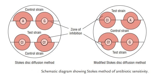 Both give the same kind of comparison; only the physical arrangement differs. The layout described in the procedure below is the modified Stokes method (control in the center), which is the more commonly used version.
Procedure
Preparing the inoculum
- Select at least three to five well-isolated colonies of the same morphological type from both the test and the control cultures. Touch the top of each colony with a loop and transfer the growth into a tube containing 4 to 5 mL of a suitable broth, such as tryptic soy broth.
- Incubate the broth cultures at 37°C until the turbidity reaches or exceeds that of a 0.5 McFarland standard (usually 2 to 6 hours).
- Adjust the turbidity of each actively growing broth culture with sterile saline or broth so it is optically comparable to a 0.5 McFarland standard. This corresponds to approximately 1 to 2 x 10⁸ CFU/mL for Escherichia coli ATCC 25922. Use a photometric device, or visually compare each tube against the 0.5 McFarland standard using a card with a white background and contrasting black lines under adequate light.
- Within 15 minutes of adjusting the turbidity, dip a sterile cotton swab into each suspension, rotate it several times, and press it firmly against the inside wall of the tube above the fluid level to remove excess inoculum.
Inoculating and applying discs
- Divide a dried Mueller-Hinton agar plate into three sections in your mind: two outer thirds and a central third.
- Inoculate the control strain evenly across the central third of the plate (modified Stokes layout). Inoculate the test strain across the upper and lower thirds, leaving a gap of not more than 5 mm between the test and control areas on each side.
- Allow the inocula to dry for a few minutes with the lid on.
- Place each antimicrobial disc in the gap between the test and control areas, using sterile forceps, and press gently to ensure full contact with the agar. The disc sits at the boundary so that its zone extends into both the test and the control lawns.
- Within 30 minutes of applying the discs, incubate the plates aerobically at 35°C for 18 to 24 hours.
Note: extremes in inoculum density must be avoided. Never use undiluted overnight broth cultures or other unstandardized inocula for streaking plates, as an inoculum that is too heavy or too light will distort the zones.
Interpretation of Results
Measure the radius of each zone of inhibition, from the edge of the disc to the edge of the zone, for both the test and the control. Then compare the test radius to the control radius.
| Category | Criterion |
|---|---|
| Sensitive (S) | Test zone radius is equal to, wider than, or not more than 3 mm smaller than the control zone radius |
| Intermediate (I) | Test zone radius is more than 2 mm, but smaller than the control by more than 3 mm |
| Resistant (R) | No zone of inhibition, or test zone radius is 2 mm or less |
The logic is that a truly susceptible isolate should produce a zone close to the fully susceptible control's zone. A meaningfully smaller zone signals reduced susceptibility, and little or no zone signals resistance. Because the comparison is against the control on the same plate, the reading holds even if that day's discs or media were not perfectly standard.
Advantages of the Stokes Method
The strengths of Stokes all follow from having the control on the same plate:
- The control and test strains are read on the same plate, under identical conditions, so the comparison is direct.
- It is more reliable when disc quality cannot be guaranteed, because a weak disc shrinks the control zone too and is immediately obvious.
- Variation in environmental conditions such as temperature, incubation time, or atmosphere affects the control and test simultaneously, minimizing error.
- Errors from an inoculum that is too heavy or too light tend to show up in the control as well, flagging the problem rather than silently producing a wrong result.
How to Remember
The whole method collapses to one idea: the control shares the plate, so every error is shared too. If the disc is weak, the media is off, or the incubator drifts, the control zone changes right alongside the test zone, and the comparison survives. That is the entire reason Stokes exists.
For the two layouts: in conventional Stokes the test organism takes the center; in modified Stokes the control takes the center. A way to hold it: "modified moves the control to the middle."
One sentence that captures it: Kirby-Bauer trusts the disc and reads against a chart; Stokes trusts nothing and reads against a control that faced the same day, which is why it holds up where standardization cannot be guaranteed.
Key exam facts in one table
| Feature | Detail |
|---|---|
| Method type | Comparative disc diffusion; control and test on the same plate |
| Key difference from Kirby-Bauer | Test zone compared to an on-plate control strain, not to a fixed breakpoint chart |
| Main use case | Labs where disc potency, media, or storage cannot be tightly standardized (widely used in the UK) |
| Conventional layout | Test organism in the center, control on the outer thirds |
| Modified layout | Control organism in the center, test on the outer thirds |
| What is measured | Zone radius (disc edge to zone edge), for both test and control |
| Sensitive (S) | Test radius equal to, wider than, or not more than 3 mm smaller than the control |
| Intermediate (I) | Test radius more than 2 mm, but smaller than the control by more than 3 mm |
| Resistant (R) | No zone, or test radius 2 mm or less |
| Inoculum | 0.5 McFarland (about 1 to 2 x 10⁸ CFU/mL for E. coli ATCC 25922) |
| Incubation | 35°C, aerobic, 18 to 24 hours |
| Medium | Mueller-Hinton agar |
| Main trade-off | Lower throughput; one or two antibiotics per plate versus many on a Kirby-Bauer plate |
Where Students Get Confused
Stokes measures the radius, Kirby-Bauer measures the diameter. This is the single most missed difference. In Kirby-Bauer you measure the full zone diameter (including the disc) and compare it to a chart. In Stokes you measure the radius, from the disc edge to the zone edge, on both the test and the control, and compare the two. Mixing up radius and diameter will make every Stokes reading wrong.
Conventional and modified Stokes are not different tests, only different layouts. The confusion is over which organism sits in the middle. Conventional puts the test organism in the center with controls outside; modified puts the control in the center with the test outside. The comparison and the interpretation are identical; only the physical arrangement changes.
The control is a fully susceptible strain, not a resistant one. The control exists to show what a susceptible zone looks like under that day's exact conditions. It is not there to represent resistance. If the control's own zone is unexpectedly small, that is a warning that the discs or media are faulty, not a normal result to read past.
A big test zone is not automatically "sensitive" in isolation. The whole point is the comparison. You read the test zone against the control zone, not against your memory of a normal zone size. An isolate is sensitive because its zone is close to the control's, not because the zone looks large on its own.
Stokes does not give an MIC. Like Kirby-Bauer, it produces a categorical result (S, I, or R). If an exact minimum inhibitory concentration is needed, that requires a broth or agar dilution method or an E-test.
References and further reading
- Gosden PE, Andrews JM, Bowker KE, Holt HA, MacGowan AP, Reeves DS, Sunderland J, Wise R. Comparison of the modified Stokes' method of susceptibility testing with results obtained using MIC methods and British Society of Antimicrobial Chemotherapy breakpoints. J Antimicrob Chemother. 1998;42(2):161-169. https://doi.org/10.1093/jac/42.2.161
- Tille PM. Bailey and Scott's Diagnostic Microbiology. 15th ed. St. Louis: Elsevier; 2022.
- CLSI. M02, Performance Standards for Antimicrobial Disk Susceptibility Tests. Clinical and Laboratory Standards Institute; current edition.

Tankeshwar Acharya, MSc (Medical Microbiology)
Tankeshwar Acharya is an Assistant Professor in the Department of Microbiology at Patan Academy of Health Sciences (PAHS), Nepal, where he has been teaching and practicing clinical microbiology for over 14 years. He is the founder of Microbe Online, one of the leading free microbiology education resources on the web, covering bacteriology, mycology, parasitology, immunology, and clinical laboratory diagnostics written from direct experience in both the classroom and the diagnostic laboratory.
Comments
No comments yet. Be the first to share your thoughts.
Leave a comment
All comments are reviewed before they appear.