
Löwenstein-Jensen (LJ) Medium: Principle, Preparation, Uses, and Colony Characteristics
Löwenstein-Jensen (LJ) medium is the standard solid culture medium for Mycobacterium tuberculosis. Learn its principle, preparation by inspissation, colony characteristics of M. tuberculosis and NTM, and how LJ compares to MGIT liquid culture.
A 34-year-old man presents with three weeks of productive cough, night sweats, and weight loss. His sputum smear is negative for acid-fast bacilli on Ziehl-Neelsen staining — but his clinician's index of suspicion remains high. In a laboratory with access to GeneXpert, the molecular result comes back within two hours: MTB detected, rifampicin resistance not detected. But the treating physician also wants to know which other drugs the organism is susceptible to. That requires growing the organism in culture, testing it against a panel of anti-TB drugs, and waiting.
The culture medium that will grow M. tuberculosis from that smear-negative sputum, and sustain it long enough for drug susceptibility testing, is Löwenstein-Jensen medium. It may take three to six weeks to produce a visible result — but in the absence of liquid culture systems, it is irreplaceable.
Principle
Mycobacterium tuberculosis is an obligate aerobe with a generation time of 16–24 hours — approximately 10 to 20 times slower than common bacteria like E. coli. It has a lipid-rich cell wall that makes it hydrophobic, resistant to many conventional media components, and unusually dependent on specific nutrients for primary isolation. Standard bacteriological media (nutrient agar, blood agar, TSA) will not support its growth.
Löwenstein-Jensen medium was developed to meet these specific requirements. Each component serves a defined purpose:
| Component | Role |
|---|---|
| Whole eggs (inspissated) | Primary nutrient source — provide albumin, lipids, carbohydrates, vitamins, and minerals. Coagulation by inspissation (heating at 85°C) solidifies the egg protein into a firm slope without agar |
| Asparagine | Preferred nitrogen source for M. tuberculosis; stimulates growth and enhances colony formation |
| Glycerol | Carbon and energy source; specifically stimulates growth of M. tuberculosis; M. bovis does NOT utilise glycerol and grows poorly on glycerol-containing LJ |
| Pyruvate (in M. bovis variant) | Replaces glycerol; required for M. bovis growth; LJ with pyruvate should be used in regions where bovine TB or M. bovis infection is endemic |
| Malachite green | Selective agent — bacteriostatic dye that inhibits the growth of most contaminating bacteria from sputum and other specimens, while Mycobacterium species are resistant to its inhibitory effect due to their lipid-rich cell wall |
| Mineral salts (KH₂PO₄, MgSO₄, magnesium citrate) | Provide essential ions for bacterial metabolism |
Key physical property: LJ medium contains no agar. It solidifies entirely through the thermal coagulation of egg albumin during inspissation at 85°C. This is why the temperature and duration of inspissation are critical — undercoagulation produces a liquid medium, overcoagulation degrades nutrients and produces a brittle, cracked surface.
Selective but not highly selective: Malachite green suppresses most contaminating bacteria but does not eliminate all of them. Respiratory specimens (sputum, BAL) require decontamination before culture — typically using N-acetyl-L-cysteine and sodium hydroxide (NALC-NaOH method) — to reduce contaminating bacterial load before inoculation onto LJ slopes.
Uses of Löwenstein-Jensen Medium
1. Primary isolation of Mycobacterium tuberculosis from clinical specimens LJ medium is inoculated with processed (decontaminated and concentrated) specimens from suspected TB cases:
- Sputum (the most common specimen; three morning samples recommended)
- Bronchoalveolar lavage (BAL) for smear-negative cases or suspected pulmonary TB
- Gastric lavage or induced sputum in children (who cannot produce sputum)
- CSF, urine, tissue biopsies, and pleural fluid in extrapulmonary TB
2. Drug susceptibility testing (DST) — indirect method After primary isolation, the M. tuberculosis culture is used to perform drug susceptibility testing. The proportion method on LJ medium remains the reference standard in many resource-limited settings: the organism is inoculated onto LJ slopes containing individual anti-TB drugs at critical concentrations, and the proportion of colonies growing in the presence of drug versus drug-free control determines susceptibility or resistance.
3. Species identification within the Mycobacterium genus Colony characteristics on LJ medium, combined with growth rate, pigmentation patterns (Runyon classification), and biochemical tests (niacin, nitrate reduction, catalase), allow presumptive identification of M. tuberculosis complex and differentiation from non-tuberculous mycobacteria (NTM). See the colony characteristics table below.
4. M. bovis differentiation By inoculating the same specimen onto both glycerol-LJ and pyruvate-LJ slopes, growth on pyruvate-LJ only (or markedly better growth on pyruvate-LJ) suggests M. bovis — relevant in regions where bovine TB is endemic and zoonotic transmission occurs.
LJ Medium vs. MGIT Liquid Culture
Modern TB diagnostic guidelines increasingly recommend liquid culture systems — particularly the Mycobacterium Growth Indicator Tube (MGIT, Becton Dickinson) — as the primary culture method for M. tuberculosis because:
- Time to detection is 9–14 days on average (vs. 3–6 weeks on LJ)
- Sensitivity is higher (detects smaller inocula)
- Automated systems (BACTEC MGIT 960) can process large numbers of specimens
However, LJ medium retains a critical role in many settings:
| Feature | LJ Solid Medium | MGIT Liquid Culture |
|---|---|---|
| Time to detection | 3–6 weeks | 9–14 days (average) |
| Sensitivity | Moderate | Higher |
| Equipment required | Inspissator, incubator | Automated BACTEC system |
| Cost | Low | Higher |
| Colony morphology visible | Yes — important for identification | No |
| Drug susceptibility testing | Proportion method (reference standard) | MGIT DST (WHO-endorsed) |
| M. bovis differentiation | Yes (glycerol vs. pyruvate slopes) | Requires additional testing |
| Resource-limited settings | Suitable | Often not feasible |
In Nepal and similar settings: LJ medium remains the primary culture method in most district and provincial laboratories. GeneXpert/Xpert MTB-RIF has reduced dependence on culture for initial M. tuberculosis detection, but culture on LJ remains essential for drug susceptibility testing beyond rifampicin and for extrapulmonary specimens where GeneXpert sensitivity is lower.
Preparation of Löwenstein–Jensen Media
Ingredients
A. Mineral salt solution
- Potassium dihydrogen phosphate anhydrous (KH2PO4): 2.4 g
- Magnesium sulphate anhydrous: (MgSo4.7H20): 0.24 g
- Magnesium citrate: 0.6 g
- Asparagine: 3.6 g
- Glycerol (reagent grade): 12 ml
- Distilled water: 600 ml
Dissolve the ingredientsin orderin the distilled water by heating. Autoclave at 121°C for 30 minutes to sterilize. Cool to room temperature. This solution keeps indefinitely and may be stored in suitable amounts in the refrigerator.
B. Malachite green solution 2%
- Malachite green dye: 2.0 g
- Distilled water: 100 mlDissolve the dye in the distilled water completely. Filter and store in the refrigerator.
C. Homogenized whole eggs
- Get a fresh (those are not more than seven days old), hen’s eggs
- Clean the eggs by scrubbing thoroughly with a hand brush in water and soap.
- Let the eggs soak for 30 minutes in a soap solution.
- Rinse eggs thoroughly in running water and soak them in 70% ethanol for 15 minutes.
- Before handling the clean dry eggs scrub and wash the hands with a disinfectant.
- Crack the eggs with the edge of the beaker into a sterile flask and beat them in a sterile blender for 30 seconds to one minute.
Preparation of complete medium
Aseptically pool the following reagents in a large, sterile flask and mixed well:
- Mineral salt solution: 600ml
- Malachite green: 20 ml
- Homogenised eggs (25-30 eggs, depending on size): 1000ml
The complete egg medium is distributed in 6-8ml volumes in sterile universal containers or culture bottles (14 ml or 28 ml) and the caps tightly closed and inspissated without delay to prevent sedimentation of heavier ingredients.
Cultures are usually made in bottles rather than in Petri dishes because of the long incubation time required. Use of bottle limits both chances of contamination and drying of the culture media (if the caps are tightly closed).
Coagulation of medium
- Heat the inspissator to 85°C to quicken the build-up of the temperature before loading.
- Place the bottles in a slanted position in the inspissator and coagulate the medium for 50 minutes at 85°C (since the medium has been prepared with sterile precautions this heating is to solidify the medium, not to sterilize it).
The quality of egg media deteriorates when coagulation is done at too high a temperature or for too long. Discoloration of the coagulated medium may be due to excessive temperature. The appearance of little holes or bubbles on the surface of the medium also indicates faulty coagulation procedures.
Sterility check
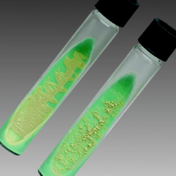 Figure: Lowenstein Jensen Medium
- After inspissation, the whole media batch of the media bottles should be incubated at 35°C-37°C for 24 hours as a check for bacterial sterility.
- After 24 hours 5% of the slopes should be picked up randomly and continued for incubation for 14 days to check for fungal sterility.
- In both cases the contamination rate should not be > 10 %.
Storage
The Löwenstein–Jensen medium should be dated and stored with the batch number in the refrigerator and can keep for up to 4 weeks if the caps are tightly closed to prevent drying of the medium.
Inoculation and Incubation
Two slopes of LJ medium should be inoculated per specimen (an additional one slope with pyruvate in M. bovis endemic areas).
- Remove the condensed moisture before inoculation.
- Inoculate each slope with 0.2-0.4 ml (2-4 drops or 2-4 loopful) of the centrifuged sediment, distributed over the surface.
- Incubate the cultures at 35-37°C until growth is observed or discarded as negative after eight weeks.
Examination Schedule
All cultures should be examined 72 hours after inoculation to check that liquid has completely evaporated, to tighten the caps in order to prevent drying out of media and to detect contaminants. Thereafter, cultures are examined weekly, or if this is not operationally feasible, on at least three occasions, viz
- after one week to detect rapidly growing mycobacteria which may be mistaken for M. tuberculosis
- after 3-4 weeks to detect positive cultures of M. tuberculosis as well as other slow-growing mycobacteria which may be either harmless saprophytes or potential pathogens
- after 8 weeks to detect very slow-growing mycobacteria, including M. tuberculosis, before judging the culture to be negative
Results
Visible colonies are usually produced 2-3 weeks after incubation (M. tuberculosis is a SLOW GROWERS, do not grow in primary culture in less than one week and may take 3-4 weeks to give visible growth), but cultures should be incubated for up to 8 weeks before being discarded.
When cultured on Lowenstein Jensen (LJ) medium at 35-37°C, M.tuberculosis produces rough (having the appearance of bread crumbs or cauliflower), raised, dry, non-pigmented (cream/buff colored) colonies.
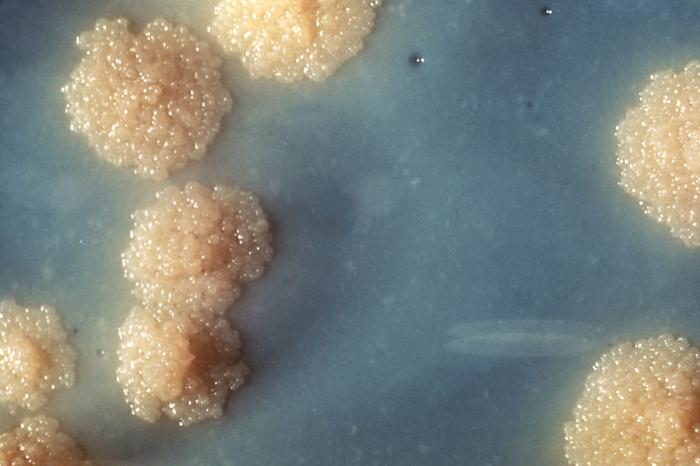 Figure: Close view of Mycobacterium tuberculosis colonies(Image source: CDC/ Dr. George Kubica)
With doubtful cultures or when less experienced staff read cultures, the acid fastness of the isolate should be confirmed by Ziehl-Neelsen (ZN) staining.
Colony Characteristics of Mycobacteria on LJ Medium
The Runyon classification groups non-tuberculous mycobacteria by growth rate and pigmentation — both directly observable on LJ medium.
| Organism | Growth rate | Pigmentation | Colony morphology | Clinical significance |
|---|---|---|---|---|
| M. tuberculosis | Slow (3–6 weeks) | Non-pigmented (cream/buff) | Rough, dry, raised, "breadcrumb" or "cauliflower" surface; not easily emulsified | Pulmonary TB; extrapulmonary TB |
| M. bovis | Slow (5–8 weeks) | Non-pigmented | Smooth, flat, dysgonic; grows better on pyruvate-LJ | Zoonotic TB; intestinal TB via unpasteurised milk |
| M. kansasii (Runyon I — photochromogen) | Slow | Non-pigmented in dark; yellow-orange in light | Smooth | Pulmonary disease in COPD patients |
| M. scrofulaceum (Runyon II — scotochromogen) | Slow | Yellow-orange in dark AND light | Smooth | Cervical lymphadenitis in children |
| M. gordonae (Runyon II) | Slow | Yellow-orange in dark | Smooth; very common tap water contaminant | Rarely pathogenic; common culture contaminant |
| M. avium complex (Runyon III — non-chromogen) | Slow | Non-pigmented | Smooth, flat, translucent | Disseminated disease in HIV; pulmonary disease |
| M. fortuitum (Runyon IV — rapid grower) | Rapid (3–7 days) | Variable | Rough or smooth; grows within 1 week | Wound infections, catheter infections, post-surgical |
| M. chelonae (Runyon IV) | Rapid (3–7 days) | Non-pigmented | Smooth, small | Skin and soft tissue infections; keratitis |
Key exam point — Runyon classification:
- Group I (Photochromogens): Pigment only in light — "photo" = light
- Group II (Scotochromogens): Pigment in dark and light — "scoto" = dark
- Group III (Non-chromogens): No pigment in either condition
- Group IV (Rapid growers): Colonies visible within 7 days
Rapid growers are the key differentiator. If colonies appear on LJ medium within the first week of incubation, the organism is a rapid grower and cannot be M. tuberculosis. Record the day of first visible growth on every LJ slope — this single observation rules in or rules out M. tuberculosis complex.
How to Remember
LJ medium has three defining features — each one critical:
- Eggs, not agar — LJ solidifies by inspissation of egg albumin at 85°C, not by agar. This is why temperature control during preparation is so important: too hot and the egg protein degrades; too cool and the slopes stay liquid.
- Malachite green selects for mycobacteria — the lipid-rich mycobacterial cell wall resists the bacteriostatic effect of malachite green that kills most contaminating bacteria. Think of it as the mycobacterium's armour.
- Glycerol for MTB, pyruvate for M. bovis — always inoculate both slope types when M. bovis is a clinical possibility (cattle farmers, veterinarians, patients with unexplained intestinal TB).
Growth timeline — the three checkpoints:
- 72 hours: Check liquid has evaporated; tighten caps; look for contaminants
- 1 week: Rule out rapid growers (Runyon IV); if growth already visible, not M. tuberculosis
- 3–4 weeks: First positive M. tuberculosis cultures typically appear here
- 8 weeks: Final read; discard as negative only after 8 full weeks
Inspissation problems and what they mean:
- Discoloured medium (brown/dark) = temperature too high during inspissation
- Holes or bubbles on slope surface = faulty coagulation technique
- Liquid medium that did not solidify = temperature too low or duration too short
Where LJ fits in the diagnostic pyramid:
Smear microscopy (minutes) → GeneXpert (2 hours) → LJ culture (weeks) → DST on LJ (weeks more) Each step answers a different question. LJ answers: "Is the organism alive and culturable? What drugs will kill it?"
Identification
Mycobacterium tuberculosis can be identified presumptively on the basis of its colony characteristics. Though there is not a completely reliable single test that will differentiate M. tuberculosis from other mycobacteria.
The following tests when used in combination help to identify the M.tuberculosis strains.
- Niacin Test
- Nitrate Reduction Test
- Catalase Test
References and Further Reading
- World Health Organization. (1998). Laboratory Services in Tuberculosis Control, Part III: Culture. WHO/TB/98.258. Geneva: WHO. Available at: https://apps.who.int/iris/bitstream/10665/65942/3/WHO_TB_98.258\_(part3).pdf
- Cheesbrough, M. (2006). District Laboratory Practice in Tropical Countries, Part 2 (2nd ed.). Cambridge University Press.
- World Health Organization. (2014). Companion Handbook to the WHO Guidelines for the Programmatic Management of Drug-Resistant Tuberculosis. Geneva: WHO.
- Tille, P. M. (2017). Bailey and Scott's Diagnostic Microbiology (14th ed.). Elsevier.
- Camus, J. C., Pryor, M. J., Médigue, C., & Cole, S. T. (2002). Re-annotation of the genome sequence of Mycobacterium tuberculosis H37Rv. Microbiology, 148(10), 2967–2973.

Tankeshwar Acharya, MSc (Medical Microbiology)
Tankeshwar Acharya is an Assistant Professor in the Department of Microbiology at Patan Academy of Health Sciences (PAHS), Nepal, where he has been teaching and practicing clinical microbiology for over 14 years. He is the founder of Microbe Online, one of the leading free microbiology education resources on the web, covering bacteriology, mycology, parasitology, immunology, and clinical laboratory diagnostics written from direct experience in both the classroom and the diagnostic laboratory.