
Trichomonas vaginalis: Clinical Features, Lab Diagnosis, and Differentiation from Bacterial Vaginosis
Trichomonas vaginalis vs Gardnerella vaginalis: how to tell trichomoniasis apart from bacterial vaginosis using pH, wet mount, whiff test, and clue cells. Complete lab diagnosis: microscopy, culture, NAAT.

On this page
A 28-year-old woman presents with vaginal discharge and an unpleasant odor. Two organisms are on the differential, and getting the diagnosis right matters: one is a sexually transmitted parasitic infection requiring partner treatment and STI counselling; the other is a non-sexually-transmitted disturbance of the normal vaginal microbiome. Both can produce a malodorous discharge. Both can give a positive whiff test. Both can raise the vaginal pH above 4.5.
This is precisely the kind of clinical overlap that trips up students moving from textbook definitions to an actual patient with a swab in hand. Trichomonas vaginalis and bacterial vaginosis (commonly, if imprecisely, called "Gardnerella vaginitis") are frequently confused, not because the organisms are similar, but because the clinical presentation and several screening tests overlap heavily. The diagnosis hinges on one thing neither organism's name tells you directly: what you actually see under the microscope.
What Is Trichomonas vaginalis?
Trichomonas vaginalis is a flagellated protozoan parasite and the causative agent of trichomoniasis, one of the most common non-viral sexually transmitted infections worldwide. It is one of the three common causes of vaginitis; for the differential with candidiasis and bacterial vaginosis, see the list of sexually transmitted infections and their causative agents.
Its primary habitat in the human body is the vagina and the male urethra/prostate. Unlike most protozoan parasites, which have both a trophozoite and a cyst stage, T. vaginalis exists in only one form, the trophozoite.
There is no cyst stage, which has a direct diagnostic consequence: the organism does not survive well outside the body and specimens must be examined promptly while the trophozoites are still motile.
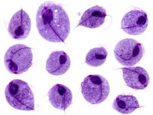 Clinical presentation:
- Infected women: Approximately 25% are symptomatic, presenting with vaginal itching, burning, and a profuse, foul-smelling, yellow-green discharge. The remaining majority are asymptomatic carriers.
- Infected men: Generally asymptomatic; approximately 10% develop urethritis. Men play a critical role in transmission despite rarely having symptoms themselves. T. vaginalis is not an exclusively female infection, and male partners of infected women are frequently also infected without knowing it.
Critical point: Symptoms alone cannot reliably diagnose trichomoniasis. The discharge, itching, and odor overlap substantially with bacterial vaginosis and other causes of vaginitis. Laboratory confirmation is required.
Why This Matters: Trichomonas vs Bacterial Vaginosis
Bacterial vaginosis (BV) is not an infection caused by a single pathogen, it is a dysbiosis. The normal vaginal flora, dominated by hydrogen-peroxide-producing Lactobacillus species, shifts toward overgrowth of Gardnerella vaginalis and other anaerobic organisms. Gardnerella vaginalis is present in small numbers in many healthy women. Its mere presence on a culture is not diagnostic of BV, which is why culturing for Gardnerella is not recommended as a diagnostic strategy. BV is not classified as a sexually transmitted infection, and male partners do not require treatment.
Trichomoniasis is a true sexually transmitted parasitic infection caused by a single, identifiable organism that can be directly visualized. It does require partner treatment and STI counselling.
Why the overlap happens
Both conditions can produce:
- Vaginal pH above 4.5 (trichomoniasis: often >5.4; BV: typically >5)
- A positive whiff test (amine odor with KOH). Classically associated with BV, but the whiff test can also be positive in trichomoniasis
- Vaginal discharge and odor as the presenting complaint
Because these overlapping features are part of the same bedside screening tools (the Amsel criteria for BV: pH >4.5, positive whiff test, homogeneous discharge, and clue cells, at least 3 of 4 required for diagnosis), relying on pH or whiff test alone cannot distinguish the two conditions.
What actually differentiates them
| Feature | Trichomoniasis | Bacterial Vaginosis |
|---|---|---|
| Causative agent | Trichomonas vaginalis (protozoan parasite) | Dysbiosis: Lactobacillus depletion + Gardnerella vaginalis/anaerobic overgrowth |
| Classification | Sexually transmitted infection | NOT classified as an STI |
| Partner treatment required? | Yes | No |
| Diagnostic hallmark on microscopy | Motile trophozoites (pear-shaped, jerky motility, 4 anterior flagella) | Clue cells (epithelial cells with borders obscured by adherent coccobacilli) |
| WBCs on wet mount | Typically increased (>10/hpf) | Typically normal |
| Vaginal pH | Often >5.4 | Typically >4.5–5 |
| Whiff test | Can be positive | Classically positive |
| Discharge character | Frothy, yellow-green | Thin, grey, homogeneous |
| Gram stain utility | Not primary diagnostic tool | Nugent score (Gram stain–based scoring system) is a standard diagnostic method |
| Culture as diagnostic tool | Yes, more sensitive than wet mount (roughly 44 to 75%), useful when smear is negative and NAAT is unavailable | NOT recommended (low specificity, Gardnerella is part of normal flora in low numbers) |
The one-sentence version for exam purposes: Trichomoniasis is diagnosed by finding the motile organism itself; bacterial vaginosis is diagnosed by finding evidence of a disturbed bacterial ecosystem (clue cells, Nugent score), you are looking for a parasite in one case and a pattern in the other.
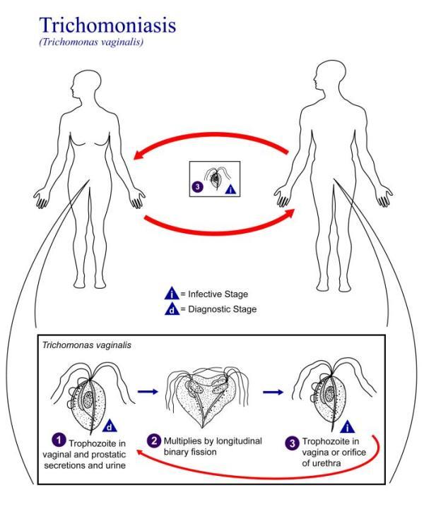 Figure: Life cycle of Trichomonas vaginalis
Morphological Features of Trichomonas vaginalis Trophozoites
- Pear-shaped organism with a central nucleus
- Four anterior flagella
- An undulating membrane extending approximately two-thirds the length of the body
- Characteristic jerky, twitching motility on a wet mount. This motility is the single most important diagnostic feature, since the organism is roughly the same size as a white blood cell and can otherwise be overlooked or mistaken for one
There is no cyst form. The trophozoite is the only morphological stage of T. vaginalis, which is why prompt specimen processing matters so much (see Laboratory Diagnosis below).
Laboratory Diagnosis
Specimen Collection
Specimens are collected using urogenital swabs:
- Female: Urine sediment, vaginal secretions
- Male: Urethral discharge, prostatic secretions, or the first few drops of voided urine
For culture, specimens should be placed immediately into a tube containing 0.5 mL of sterile saline, or into a commercially available transport/culture pouch (e.g., InPouch TV, BIOMED) for direct examination and culture.
Why timing matters: Because Trichomonas vaginalis exists only as a trophozoite without a resistant cyst stage and loses motility relatively quickly outside the body, particularly with temperature changes, slides should be prepared and examined as soon as possible after collection. A delayed specimen risks a false-negative result simply because the organisms have stopped moving and become difficult to distinguish from white blood cells.
1. Microscopy and Staining
Direct microscopic examination of a wet preparation of vaginal discharge is the simplest and most rapid diagnostic test. The diagnostic finding is motile, pear-shaped trophozoites with characteristic jerky motility.
Direct Fluorescent Antibody (DFA) staining can also be used to visualize the parasite when available.
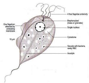 Figure: Trophozoites of T. vaginalis
Wet mount microscopy is only moderately sensitive. Its reported sensitivity ranges from roughly 40–90% depending on the population studied (lower in some series, particularly when organism load is low or specimens are not examined promptly).
This is precisely why a negative wet mount does not rule out trichomoniasis, and why culture or molecular methods are recommended as the next step when clinical suspicion remains.
2. Culture
Culture was historically the reference standard and is more sensitive than wet mount, though modern estimates place its sensitivity at roughly 44 to 75%, below that of NAATs. It remains useful when a direct smear is negative and molecular testing is unavailable, and for antimicrobial resistance testing in suspected treatment failure.
Johnson's and Trussell's medium supports good growth of T. vaginalis and consists of proteose peptone, NaCl, sodium thioglycolate, and human serum. Optimal growth occurs at 35–37°C under anaerobic conditions (less well aerobically), at an optimal pH of 5.5 to 6.0.
T. vaginalis can also be cultured using commercially available Diamond's medium or plastic transport/culture pouches.
3. Serology
No reliable serologic test for T. vaginalis is currently available for routine clinical use.
4. Molecular Methods
PCR and nucleic acid hybridization methods can detect T. vaginalis directly in clinical specimens with high sensitivity. The Aptima Trichomonas vaginalis Assay is a nucleic acid amplification test (NAAT) that detects ribosomal RNA (rRNA) of T. vaginalis, using Target Capture, Transcription-Mediated Amplification (TMA), and Hybridization Protection Assay (HPA) technologies, run on the Tigris DTS System or Panther System.
Why molecular methods matter clinically: Nucleic acid amplification tests (NAATs) are now recognized as more sensitive than wet mounts or even culture in many settings. Critically, they make it practical to test asymptomatic men, who are a population traditionally under-tested because of the invasive or low-yield nature of older methods, despite being a major source of ongoing transmission.
Treatment
Trichomoniasis is treated with a nitroimidazole, most commonly metronidazole (tinidazole is an alternative). The great majority of cases resolve with treatment. Because trichomoniasis is sexually transmitted, sexual partners must be treated simultaneously, regardless of whether they are symptomatic, to prevent reinfection. This is the single biggest practical difference from bacterial vaginosis management, where partner treatment is not indicated.
How to Remember
The core distinction: "Trich moves, BV doesn't" is a classic diagnostic pearl: on a wet mount, trichomoniasis reveals a living, swimming organism, the trophozoite, characterized by its jerky motility. In contrast, bacterial vaginosis shows a pattern of disturbed ecology, specifically clue cells, where nothing is swimming.
Why Gardnerella culture is a trap: "Present ≠ guilty". Gardnerella vaginalis lives in small numbers in many healthy women. Finding it on culture proves nothing on its own; this is exactly why culturing for it is not a recommended diagnostic strategy. BV is diagnosed by the overall pattern (Amsel criteria or Nugent score), not by isolating one organism.
The four flagella: "Four in front, one behind (sort of)". T. vaginalis has four anterior flagella plus a fifth flagellum incorporated into the undulating membrane running along two-thirds of its body. Visualizing "four flagella at the front, driving it forward in jerky bursts" helps fix both the motility pattern and the morphology together.
Where Students Get Confused
1. "A positive whiff test means bacterial vaginosis." The whiff test (fishy amine odor on adding KOH to discharge) is classically associated with BV and is one of the four Amsel criteria, but it can also be positive in trichomoniasis. A positive whiff test alone does not distinguish the two conditions, as microscopy for either motile trophozoites or clue cells is required for a specific diagnosis.
2. "Gardnerella vaginalis is the cause of bacterial vaginosis the way Trichomonas is the cause of trichomoniasis." This framing is imprecise and is a major source of confusion. Trichomoniasis has one specific causative parasite. Bacterial vaginosis is a polymicrobial dysbiosis, a shift away from normal Lactobacillus-dominant flora toward overgrowth of Gardnerella and various anaerobes (and organisms like Mobiluncus).
Gardnerella is often called the "marker organism" of BV, but it is not solely responsible, and its presence alone (e.g., on a culture) is not diagnostic, since it can be found in low numbers in healthy women.
3. "Trichomonas has a cyst stage like other protozoa." No. T. vaginalis has only the trophozoite form. There is no environmentally resistant cyst stage, unlike Entamoeba or Giardia. This is precisely why the organism is fragile outside the host and why prompt specimen processing is so important for accurate wet mount diagnosis.
4. "A negative wet mount rules out trichomoniasis." Wet mount sensitivity is moderate at best and is highly dependent on organism load and how quickly the specimen is examined after collection. Delayed examination allows the fragile trophozoites to lose motility, making them difficult to distinguish from white blood cells of similar size. A negative wet mount in a symptomatic patient should prompt culture or NAAT testing.
5. "Men don't need to be tested or treated for trichomoniasis since they're usually asymptomatic." This is a dangerous assumption with real public health consequences. Most infected men are asymptomatic, but they remain infectious and are frequently the source of reinfection in treated female partners. Current STI management guidelines require simultaneous partner treatment regardless of symptoms. Failing to treat the male partner is one of the most common causes of treatment failure/recurrence in clinical practice.
Key Exam Facts
| Fact | Detail | Memory hook |
|---|---|---|
| Causative organism | Trichomonas vaginalis (flagellated protozoan) | Not bacterial, a parasite |
| Forms | Trophozoite ONLY, no cyst stage | Fragile outside host |
| Number of flagella | 4 anterior + 1 in undulating membrane | "Four in front, one along the side" |
| Undulating membrane | Extends ~2/3 of body length | Distinctive structural feature |
| % symptomatic women | ~25% | Most infections are silent |
| % symptomatic men | ~10% (urethritis) | Men are usually asymptomatic carriers |
| Classic discharge | Frothy, foul-smelling, yellow-green | Distinct from BV's thin grey discharge |
| Best initial test | Wet mount (motile trophozoites) | Quick, but moderate sensitivity only |
| Traditional reference standard | Culture (roughly 44 to 75% sensitive), now surpassed by NAAT | Johnson's and Trussell's medium |
| Culture conditions | 35–37°C, anaerobic, pH 5.5–6.0 | Use when wet mount is negative |
| Most sensitive overall | NAAT (e.g., Aptima assay) | Detects rRNA; enables male testing |
| Serology | NOT available/reliable | Don't rely on antibody testing |
| BV diagnostic hallmark | Clue cells (Amsel criteria / Nugent score) | NOT a single organism, a pattern |
| Gardnerella culture | NOT recommended | Low specificity, present in healthy women too |
| Trichomoniasis = STI? | Yes. Partner treatment required | Differentiates from BV |
| BV = STI? | No. Partner treatment NOT required | Dysbiosis, not infection transmission |
| Treatment | Metronidazole (nitroimidazole); treat partner simultaneously | Partner treatment prevents reinfection |
Frequently Asked Questions
How is Trichomonas vaginalis different from Gardnerella vaginalis / bacterial vaginosis?
How is Trichomonas vaginalis different from Gardnerella vaginalis / bacterial vaginosis?
Trichomonas vaginalis is a flagellated protozoan parasite causing trichomoniasis, a true sexually transmitted infection diagnosed by finding motile trophozoites on microscopy. Bacterial vaginosis is not caused by a single organism but is a dysbiosis, a shift from normal Lactobacillus-dominant vaginal flora toward overgrowth of Gardnerella vaginalis and anaerobes. BV is diagnosed by finding clue cells or using the Amsel criteria/Nugent score, not by isolating Gardnerella alone, since it is present in low numbers in many healthy women. Trichomoniasis requires partner treatment; bacterial vaginosis does not.
What is the best test to diagnose Trichomonas vaginalis infection?
What is the best test to diagnose Trichomonas vaginalis infection?
Direct wet mount microscopy is the fastest initial test, looking for motile, pear-shaped trophozoites with characteristic jerky movement, but its sensitivity is moderate and depends on prompt specimen examination. Culture in Johnson’s and Trussell’s medium is highly sensitive (95%) and recommended when wet mount is negative. Nucleic acid amplification tests (NAAT), such as the Aptima Trichomonas vaginalis Assay, offer the highest sensitivity and are increasingly used, especially for testing asymptomatic men.
Why does Trichomonas vaginalis need to be examined quickly after specimen collection?
Why does Trichomonas vaginalis need to be examined quickly after specimen collection?
Trichomonas vaginalis exists only as a trophozoite, it has no cyst stage. The trophozoite is fragile outside the host and loses motility relatively quickly, particularly with temperature changes. Since motility (the characteristic jerky movement) is the key diagnostic feature on wet mount, a delayed specimen can lead to a false-negative result simply because the organisms have stopped moving and become hard to distinguish from white blood cells of similar size.
Should male partners of women with trichomoniasis be treated?
References
- Madigan, M. T., Bender, K. S., Buckley, D. H., Sattley, W. M., & Stahl, D. A. (2021). Brock Biology of Microorganisms (16th ed.). Pearson.
- Procop, G. W., et al. (2017). Koneman's Color Atlas and Textbook of Diagnostic Microbiology (7th ed.). Wolters Kluwer.
- Sherrard, J., Wilson, J., Donders, G., Mendling, W., & Jensen, J. S. (2018). 2018 European (IUSTI/WHO) International Union against sexually transmitted infections (IUSTI) World Health Organisation (WHO) guideline on the management of vaginal discharge. International Journal of STD & AIDS, 29(13), 1258–1272. https://doi.org/10.1177/0956462418785451
- Hobbs, M. M., & Sena, A. C. (2013). Modern diagnosis of Trichomonas vaginalis infection. Sexually Transmitted Infections, 89(6), 434–438. https://doi.org/10.1136/sextrans-2013-051057
- Coleman, J. S., & Gaydos, C. A. (2018). Molecular diagnosis of bacterial vaginosis: an update. Journal of Clinical Microbiology, 56(9), e00342-18. https://doi.org/10.1128/JCM.00342-18
- Workowski, K. A., Bachmann, L. H., Chan, P. A., et al. (2021). Sexually transmitted infections treatment guidelines, 2021. MMWR Recommendations and Reports, 70(4), 1–187. https://doi.org/10.15585/mmwr.rr7004a1

Tankeshwar Acharya, MSc (Medical Microbiology)
Tankeshwar Acharya is an Assistant Professor in the Department of Microbiology at Patan Academy of Health Sciences (PAHS), Nepal, where he has been teaching and practicing clinical microbiology for over 14 years. He is the founder of Microbe Online, one of the leading free microbiology education resources on the web, covering bacteriology, mycology, parasitology, immunology, and clinical laboratory diagnostics written from direct experience in both the classroom and the diagnostic laboratory.
Comments
No comments yet. Be the first to share your thoughts.
Leave a comment
All comments are reviewed before they appear.