
Type II Hypersensitivity: How One Antibody Kills a Cell Three Different Ways
Type II hypersensitivity explained by mechanism: the three ways an antibody bound to a cell causes damage, why Graves disease and myasthenia gravis sit at opposite ends of the same mechanism, and the exam discriminations students miss. Transfusion reactions, HDN, Goodpasture, and more.

On this page
A patient receives the wrong blood by mistake. Within minutes, before even a full unit has run in, they spike a fever, their back aches, their blood pressure drops, and their urine turns dark. Nothing was injected that is toxic on its own. The transfused red cells are healthy. What is killing them is the patient's own antibody, which recognized a foreign marker on those donor cells and called in the machinery to destroy them. This is Type II hypersensitivity: the damage is not done by the antigen, but by an antibody that binds a cell and marks it for death.
Type II hypersensitivity is an antibody-mediated reaction. It is not an "immediate" reaction in the way Type I is. The response develops over hours, not minutes, because it depends on antibody binding, complement activation, and cell recruitment rather than the instant degranulation of a pre-armed mast cell. It is also called antibody-mediated cytotoxic hypersensitivity, because in most cases the bound antibody leads to destruction of the target cell. In a smaller group of diseases, the antibody does not destroy the cell but interferes with a receptor, switching it on or blocking it. The target antigens in Type II are not harmless environmental substances. They are markers fixed on the body's own cells or tissues, such as a blood group antigen on a red cell or a receptor on a muscle cell.
A hypersensitivity reaction type II requires a target cell with bound antigen and antibody, which activates mechanisms to damage the target cell. It is mediated by IgG or IgM antibodies against antigens on cells or extracellular space.
The one idea that explains everything below
Every Type II reaction starts the same way: an antibody (IgG or IgM) binds to an antigen that is fixed on a cell surface or in a tissue. The antigen does not float freely. It sits on a red cell, a basement membrane, a thyroid cell, a muscle receptor. That single fact, a fixed target, is what separates Type II from Type III, where the antigen and antibody meet in the blood and form floating complexes.
Once the antibody is bound, the damage happens in one of three ways. Keep these three in mind and every disease in this article sorts itself into one of them.
First, destruction. The bound antibody either activates complement to punch holes in the cell, or it coats the cell so that phagocytes eat it. This is how mismatched blood and Rh disease destroy red cells.
Second, ADCC. When the target is too big to swallow, killer cells (NK cells, macrophages, neutrophils, eosinophils) latch onto the antibody's tail and pour toxic granules onto the target from outside. The cell is killed without being eaten.
Third, dysfunction. Here nothing is destroyed at all. The antibody binds a receptor and either switches it on or jams it. No cell dies, but the tissue misbehaves. This is the mechanism behind Graves disease and myasthenia gravis, and it is the one students find most surprising, because it is a hypersensitivity reaction with no cell killing in it.
So the whole topic is three verbs: destroy, kill from outside, or disrupt. Ask which verb applies and any Type II disease becomes readable.
Components of Hypersensitivity Type II
- Antigen
Antigens involved in type II hypersensitivity reactions are intrinsic and exogenous antigens.
Intrinsic antigen
- Protein on the cell membrane, e.g., Rh ag on RBCs
- An antigen on a space/matrix between cells, like on a basement membrane
- Self-antigen (causing autoimmune diseases)
- Receptors antigens on cells, like hormone receptors
Exogenous antigens
- Microbes, parasites, drugs
- Antigens of blood transfusion reaction
- Antibody
Type II reactions are driven mainly by IgG, and in some cases by IgM. IgM is especially important in ABO-mismatched transfusion reactions, where the naturally occurring anti-A and anti-B antibodies are IgM. The reason the isotype matters is mechanistic: both IgG and IgM fix complement, and complement fixation is central to how these reactions destroy cells.
- The effector cells
The effector cells of hypersensitivity type II reaction are macrophages, neutrophils, eosinophils, and natural killer (NK) cells.
Mechanism
Hypersensitivity type II reactions affect the cells where the target antigens are present. Hence such reactions are not often systemic. But sometimes, target antigens may be present on the surface of mobile cells like erythrocytes and leukocytes, affecting the whole body.
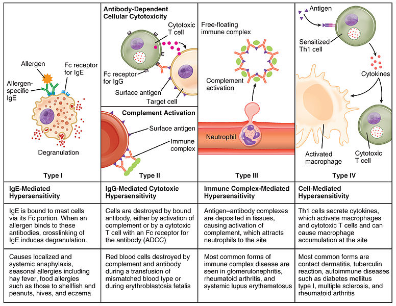 Figure: Mechanism of different types of hypersensitivity
Type II hypersensitivity has three mechanism: A target cell is restricted either by antibody and complement-mediated lysis or by antibody-dependent cell-mediated cytotoxicity (ADCC) or dysfunction of the target cell.
Antibody and complement-mediated destruction
Here, the destruction is mediated either by a complement system or antibodies via opsonization.
The antibody attaches to the antigen on the surface of cells and activates the complement system via the classical pathway. This leads to the formation of membrane attack complex (MAC), which creates pores in foreign cell membranes leading to cell lysis. In opsonization, antibodies coat an antigen and make it a target for phagocytosis.
Examples of this mechanism are erythroblastosis fetalis and mismatched blood transfusion reaction.
ADCC (Antibody-Dependent Cell-Mediated Cytotoxicity)
When the antibody-coated target is a whole cell that is too large to be swallowed by a phagocyte, a different set of cells finishes the job. NK cells, macrophages, neutrophils, and eosinophils carry Fc receptors. They grip the tail (Fc region) of the antibody that is already stuck to the target, then release toxic granules directly onto the target cell surface. The target is killed from the outside, without being engulfed. This is antibody-dependent cell-mediated cytotoxicity, or ADCC. It matters clinically because it is one way antibody-coated cells are destroyed even when complement is not the main driver.
Target cell dysfunction
This is the non-cytotoxic arm of Type II. No cell is destroyed. The antibody binds a receptor and changes what that receptor does. There are two opposite outcomes, and telling them apart is a common exam point.
The antibody can switch the receptor on. In Graves disease, an autoantibody binds the TSH receptor on thyroid cells and acts like TSH itself, driving the gland to overproduce thyroid hormone. The result is hyperthyroidism. Nothing is killed. The thyroid is simply forced to run without its normal off switch.
The antibody can jam the receptor. In myasthenia gravis, an autoantibody binds the acetylcholine receptor at the neuromuscular junction and blocks it (and promotes its internalization), so nerve signals no longer reach the muscle. The result is muscle weakness that worsens with use, classically drooping eyelids by the end of the day.
So the same mechanism, an antibody on a receptor, produces opposite diseases depending on whether it stimulates or blocks. One overworks an organ, the other silences it.
A note for exams: some classifications separate these receptor-mediated diseases into their own category called Type V (stimulatory) hypersensitivity, keeping Type II strictly for cytotoxic reactions. Most textbooks used in South Asia still teach Graves and myasthenia gravis under Type II. Know both framings, and answer according to the classification your course uses.
Clinical Manifestation
Various clinical manifestations are seen as a result of hypersensitivity type II reactions.
Transfusion reactions
ABO or blood group incompatibility is a type of hypersensitivity reaction. The red blood cell surface bears a large number of proteins and glycoproteins. An individual acquiring one allelic form of a blood group antigen can recognize other allelic forms on transfused blood as foreign, stimulating an antibody response.
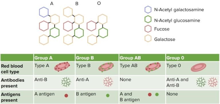 Figure: Image source:https://www.lecturio.com/concepts/type-ii-hypersensitivity-reaction/
The naturally occurring antibodies against A and B antigens are called isohemagglutinins, and they are usually of the IgM class.
Blood group A recipient reacts with a type AB or B donor as it has anti-B antibodies. Group B recipient reacts with a type AB and A donor as it has anti-A antibodies. Blood group O recipient reacts with type AB, A, and B donor as it has anti-A and anti-B antibodies.
Massive intravascular hemolysis of transfused RBCs by antibody and complement takes place if mismatched blood is transferred. Immediately ABO blood-group incompatibilities are seen, which lead to complement-mediated lysis triggered by IgM isohemagglutinins. Within hours, free hemoglobin can be detected in the plasma, filtered through the kidneys, and leads to hemoglobinuria. Some hemoglobin is converted to bilirubin, a high toxic level. Fever, chills, nausea, clotting within blood vessels, urticaria, respiratory distress, and hypotension are seen.
With repeated exposure to incompatible blood, the recipient can mount an IgG response against minor blood-group antigens. This produces a slower, delayed reaction: fever, a falling hemoglobin, rising bilirubin, mild jaundice, and anemia developing over days rather than minutes.
Hemolytic disease of the newborn
This is known as erythroblastosis fetalis, occurring against RBCs and RhD-incompatibility. It is also antibody and complement-mediated. When a Rhesus-negative woman conceives a rhesus-positive fetus, fetal RBCs leak into the mother. It survived long enough to elicit an IgG response. During the second pregnancy, maternal anti-D antibodies cross the placenta and attack fetal RBC of the second rhesus-positive fetus. The symptoms may vary from self-limiting hemolytic anemia to hydrops fetalis.
Goodpasture syndrome
Antibodies attack antigens in the basement membrane of alveoli (causing pulmonary hemorrhage) and kidneys (causing nephritis). Systemic complaints, followed by renal (hematuria) and pulmonary symptoms (dyspnea, hemoptysis, cough), are seen.
Grave disease
It is an autoimmune disease. A follicular cell of the thyroid gland has receptors for thyroid stimulating hormones (TSH). Severe illness occurs when a body mistakenly produces autoantibodies, thyroid stimulating immunoglobulin (TSI). TSI binds to TSH receptors, activate them to cause high T3 and T4, and results in hyperthyroidism.
Myasthenia Gravis
Autoimmune antibodies block or destroy acetylcholine (ACH) receptors, which limits communication between neurons and skeletal muscle. It leads to muscle weakness. E.g., droopy eyelids.
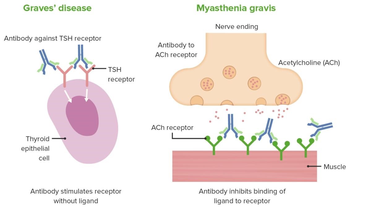 Figure: Image source:https://www.lecturio.com/concepts/type-ii-hypersensitivity-reaction/
Autoimmune hemolytic anemia (against RBCs)
IgG-mediated (warm autoimmune hemolytic anemia) or IgM-mediated (cold autoimmune hemolytic anemia). Common symptoms are weakness, shortness of breath, anemia, jaundice, icterus, and dark urine from hemolysis.
Pernicious anemia (against intrinsic factor)
Pernicious anemia involves two antibody targets. One set of antibodies attacks the parietal cells of the stomach, which are the cells that make intrinsic factor. Another set attacks intrinsic factor itself, blocking the site where it would bind vitamin B12. Either way, vitamin B12 cannot be absorbed in the ileum, and a megaloblastic anemia follows. Symptoms include the general features of anemia, a sore smooth tongue (glossitis), and tingling or numbness (paresthesias) from the effect of B12 deficiency on nerves.
Drug-induced hemolytic anemia, thrombocytopenia, or neutropenia
Antibiotics like penicillin, cephalosporin, and streptomycin can adsorb non-specifically to proteins on membranes of RBC. It forms a complex similar to the hapten-carrier complex. This induces complement-mediated lysis of RBC, and thus, progressive anemia occurs. It disappears in the removal of the drug.
Similarly, thrombocytopenia or neutropenia may also result from medication.
Penicillin is remarkable because it can induce all four types of hypersensitivity with different clinical manifestations.
Autoimmune thrombocytopenic purpura
In this case, phagocytes destroy sensitized platelets in the blood. Thus, there is an increased bleeding risk leading to severe hemorrhage.
Acute rheumatic fever
Antibodies against the cell wall of Streptococcus pyogenes react with the self-antigen on myocardial cells. It results in arthritis, fever, carditis, and subcutaneous nodules.
Lab Diagnosis
Diagnosis is based on the type of disease observed:
- Coombs test is done in transfusion reaction and anemia.
- In pernicious anemia, CBC and vitamin levels are examined.
- To diagnose erythroblastosis fetalis, pregnancy history is required; ultrasound, fetal lab test, and maternal antibody test are performed.
- TSH and TFT help know graves disease.
- In rheumatoid arthritis, CRP, ASO, and ESR are performed.
Control
- Hypersensitivity type II is controlled according to the symptoms and disease.
- A blood transfusion reaction is controlled by rapid cessation of transfusion, and supportive care is required. Urine flow should be maintained with a diuretic; acute tubular necrosis can occur because of hemoglobin accumulation.
- Control of disseminated intravascular coagulation (DIC) and bleeding control should be carried out in thrombocytopenic purpura.
- Glucocorticoids, immunosuppressive drugs, and appendectomy may require in autoimmune hemolytic anemia.
- Antithyroid drugs, Thyroidectomy, or immunotherapy are used for Grave’s disease.
- Antibiotics against Streptococcus are used to control rheumatic fever.
- Erythroblastosis fetalis prevention is conducted by giving anti-RhD at 28 weeks gestation and within 72 hours of birth.
How to remember Type II Hypersensitivity
Three verbs: destroy, kill from outside, disrupt. Every Type II disease is one of three actions. Destroy (complement or phagocytosis eats the cell): transfusion reaction, Rh disease. Kill from outside (ADCC, killer cells pour granules on): antibody-coated cells. Disrupt (receptor switched on or off, nothing dies): Graves, myasthenia. If you can name the verb, you understand the disease.
Fixed target is Type II. Floating target is Type III. The single line that separates the two antibody-mediated types. In Type II the antigen is stuck on a cell or tissue. In Type III the antigen and antibody float in the blood and clump together. Fixed versus floating.
Graves goes up, myasthenia goes down. Same mechanism, opposite direction. Graves stimulates the TSH receptor, so thyroid hormone goes up (hyperthyroidism). Myasthenia blocks the acetylcholine receptor, so muscle signaling goes down (weakness). Up and down, from the same kind of antibody.
Penicillin is the four-type villain. Penicillin can trigger all four hypersensitivity types. If an exam asks which single agent can cause Type I, II, III, and IV, the answer is penicillin.
Key exam facts
| Point | Fact |
|---|---|
| Also called | Antibody-mediated cytotoxic hypersensitivity |
| Antibodies | IgG (mainly), IgM |
| Antigen location | Fixed on a cell surface or in tissue (not floating) |
| Timing | Hours (not immediate like Type I) |
| Mechanism 1 | Complement-mediated lysis or opsonization and phagocytosis |
| Mechanism 2 | ADCC (NK cells, macrophages, neutrophils, eosinophils via Fc receptors) |
| Mechanism 3 | Receptor dysfunction (stimulating or blocking), no cell death |
| Effector cells | Macrophages, neutrophils, eosinophils, NK cells |
| Complement pathway | Classical, forming the membrane attack complex |
| Transfusion reaction antibodies | Isohemagglutinins, usually IgM |
| Rh disease (HDN) antibody | Anti-D IgG, crosses the placenta |
| Graves disease | Antibody stimulates the TSH receptor, hyperthyroidism |
| Myasthenia gravis | Antibody blocks the acetylcholine receptor, muscle weakness |
| Pernicious anemia | Antibodies against parietal cells and against intrinsic factor |
| Goodpasture syndrome | Antibody against basement membrane of lung and kidney |
| Acute rheumatic fever | Cross-reacting antibody to streptococcal cell wall attacks myocardium |
| Key diagnostic test | Direct and indirect Coombs (antiglobulin) test |
| HDN prevention | Anti-D at 28 weeks and within 72 hours of delivery |
| Type V variant | Some classifications place receptor-stimulating diseases (Graves) in a separate Type V |
Where students get confused
"Type II is an immediate reaction." No. Only Type I is the immediate type. Type II develops over hours because it depends on antibody binding, complement, and cell recruitment, not on instant mast cell degranulation.
"The antigen in Type II is a harmless environmental substance." No, that is Type I. In Type II the target is a fixed marker on the body's own cells or tissue, such as a blood group antigen or a receptor. This is why Type II so often shows up as autoimmune disease.
"Type II and Type III are basically the same because both use antibodies." They differ on one decisive point. In Type II the antigen is fixed on a cell or tissue and the antibody comes to it. In Type III the antigen and antibody are both free in the blood, meet there, and form floating immune complexes that later deposit. Fixed target versus floating complex.
"Every Type II reaction destroys cells." Not the receptor-dysfunction diseases. In Graves disease and myasthenia gravis nothing is destroyed. The antibody binds a receptor and either switches it on or blocks it. It is a hypersensitivity reaction with no cell killing.
"Graves and myasthenia are opposite mechanisms." They are the same mechanism (antibody on a receptor) with opposite effects. Graves stimulates, myasthenia blocks. Same category, different direction.
"Pernicious anemia is just poor B12 absorption." The absorption fails because of antibodies: some destroy the parietal cells that make intrinsic factor, and some block intrinsic factor directly. It is an antibody-mediated disease, which is why it belongs in Type II.
References
- Punt J, Stranford SA, Jones PP, Owen JA. Kuby Immunology. 8th ed. New York: W. H. Freeman; 2019.
- Abbas AK, Lichtman AH, Pillai S. Cellular and Molecular Immunology. 10th ed. Philadelphia: Elsevier; 2022.
- Delves PJ, Martin SJ, Burton DR, Roitt IM. Roitt's Essential Immunology. 13th ed. Chichester: Wiley-Blackwell; 2017.
- Bajwa SF, Mohammed RHA. Type II Hypersensitivity Reaction. In: StatPearls. Treasure Island (FL): StatPearls Publishing; 2025.
Frequently Asked Questions
What is the difference between Type II and Type III hypersensitivity?
What is the difference between Type II and Type III hypersensitivity?
Both use IgG or IgM antibodies. The difference is where the antigen is. In Type II the antigen is fixed on a cell surface or in a tissue, and the antibody binds it there. In Type III the antigen and antibody are both free in the blood, form floating immune complexes, and those complexes later deposit in tissues. Fixed target means Type II; floating complex means Type III.
Why is Type II called cytotoxic if some of its diseases do not kill cells?
Why is Type II called cytotoxic if some of its diseases do not kill cells?
Most Type II reactions do destroy the target cell, which is why the name fits the majority. The receptor-dysfunction diseases such as Graves disease and myasthenia gravis are the exception: the antibody changes receptor function without killing the cell. Some classifications separate these into a distinct Type V for exactly this reason.
How can Graves disease and myasthenia gravis share a mechanism but cause opposite diseases?
How can Graves disease and myasthenia gravis share a mechanism but cause opposite diseases?
Both are caused by an antibody binding a receptor. In Graves disease the antibody stimulates the TSH receptor, so the thyroid overproduces hormone. In myasthenia gravis the antibody blocks the acetylcholine receptor, so muscle signaling fails. The same kind of antibody produces opposite outcomes depending on whether it switches the receptor on or off.
Which test is central to diagnosing Type II reactions?
Which test is central to diagnosing Type II reactions?
The Coombs test, also called the antiglobulin test. The direct Coombs test detects antibody already bound to a patient's red cells, and the indirect Coombs test detects antibody free in the serum. It is the key test in transfusion reactions and autoimmune hemolytic anemia.
How is hemolytic disease of the newborn prevented?
How is hemolytic disease of the newborn prevented?
An Rh-negative mother carrying an Rh-positive fetus is given anti-D immunoglobulin, typically at 28 weeks of pregnancy and again within 72 hours of delivery. The anti-D clears fetal Rh-positive cells from the mother's circulation before she can mount her own lasting antibody response, protecting future pregnancies.
Why can penicillin cause a Type II reaction?
Why can penicillin cause a Type II reaction?
Penicillin can attach to proteins on the red cell surface, acting as a hapten. Antibodies then form against the drug-cell combination and trigger destruction of the red cell, causing a drug-induced hemolytic anemia. Notably, penicillin can trigger all four types of hypersensitivity.

Tankeshwar Acharya, MSc (Medical Microbiology)
Tankeshwar Acharya is an Assistant Professor in the Department of Microbiology at Patan Academy of Health Sciences (PAHS), Nepal, where he has been teaching and practicing clinical microbiology for over 14 years. He is the founder of Microbe Online, one of the leading free microbiology education resources on the web, covering bacteriology, mycology, parasitology, immunology, and clinical laboratory diagnostics written from direct experience in both the classroom and the diagnostic laboratory.
Comments
No comments yet. Be the first to share your thoughts.
Leave a comment
All comments are reviewed before they appear.