
Gram-Negative Cocci and Coccobacilli of Medical Significance: List, Diseases, and Lab Identification
The medically important Gram-negative cocci include Neisseria gonorrhoeae (gonorrhoea, ophthalmia neonatorum), N. meningitidis (meningitis), and Moraxella catarrhalis (otitis media, COPD). This article covers all GN cocci and coccobacilli with diseases, key properties, and lab identification links.

On this page
A university student presents with a sudden-onset fever, headache, and a non-blanching petechial rash spreading across her trunk. She deteriorates within hours and the culprit is, Neisseria meningitidis, serogroup B, causing meningococcal septicemia.
A 24-year-old man attends a sexual health clinic with a four-day history of purulent urethral discharge and dysuria. Gram stain of the discharge shows Gram-negative diplococci inside neutrophils, and the etiology is Neisseria gonorrhoeae, the second most common STI globally.
An 18-month-old presents to the emergency department with ear pain, fever, and tugging at his ear. Middle ear fluid grows Moraxella catarrhalis, the third most common cause of otitis media in children after S. pneumoniae and non-typeable Haemophilus influenzae.
All three organisms are Gram-negative cocci or coccobacilli, a small group with disproportionate clinical importance. The most common gram-negative cocci of medical importance belong to the family Neisseriaceae which includes the genera Neisseria, Moraxella, Kingella, Acinetobacter, etc.
This article is about the overview of these organisms and you can find the links, if you wish to read in detail about individual organisms.
All GN Cocci and Coccobacilli of Medical Importance
Gram-Negative Cocci (true cocci):
| Organism | Key disease(s) | Unique feature | Detailed article |
|---|---|---|---|
| N. meningitidis | Bacterial meningitis, meningococcemia | Polysaccharide capsule; 6 major serogroups; vaccine available | N. meningitidis |
| N. gonorrhoeae | Gonorrhea, PID, ophthalmia neonatorum | No capsule; ferments glucose only (not maltose) | N. gonorrhoeae |
| Moraxella catarrhalis | Otitis media, COPD exacerbations, sinusitis | β-lactamase producer; oxidase positive; hockey-puck colonies | Moraxella catarrhalis |
| Veillonella spp. | Rarely pathogenic; oral and gut flora | Obligate anaerobe; only GN anaerobic diplococcus |
Gram-Negative Coccobacilli:
| Organism | Key disease(s) | Unique feature | Detailed article |
|---|---|---|---|
| Haemophilus influenzae | Meningitis (type b), pneumonia, otitis media, epiglottitis | Requires X factor (hemin) and V factor (NAD); type b polysaccharide vaccine | H. influenzae lab diagnosis |
| Haemophilus ducreyi | Chancroid (painful genital ulcers) | STI; "school of fish" arrangement on Gram stain | |
| Acinetobacter baumannii | HAP, VAP, wound infections; ICU outbreaks | Multi-drug resistant nosocomial pathogen; survives on surfaces >1 month | Acineobacter |
| Bordetella pertussis | Whooping cough (pertussis) | Tiny GN coccobacillus; Bordet-Gengou agar; DFA for rapid diagnosis | |
| Brucella spp. | Brucellosis (undulant fever); zoonosis | Tiny, poorly staining GN coccobacillus; BSL-3; cattle/goat/sheep reservoir | Brucella |
| Francisella tularensis | Tularemia | BSL-3; tick/rabbit transmission; bioterrorism agent | Francisella tularensis |
| Pasteurella multocida | Animal bite wound infections | Cat/dog bite; GN coccobacillus; satellite growth around S. aureus | |
| Kingella kingae | Osteomyelitis, septic arthritis in children <5 years | Increasingly recognized pediatric pathogen; HACEK group |
Gram Negative Cocci
Neisseria Species
Both N. meningitidis and N. gonorrhoeae are Gram-negative diplococci that share morphology, cultural requirements (chocolate agar, Thayer-Martin medium, 5% CO₂), and oxidase-positive biochemistry, yet they cause completely different diseases in completely different body sites. The single most important laboratory test distinguishing them is maltose fermentation: meningococci ferment maltose; gonococci do not.
For a full systematic comparison of their properties, diseases, virulence factors, and lab identification tests, see the dedicated article: Differences between N. gonorrhoeae and N. meningitidis
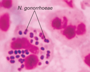 Figure: Neisseria gonorrhoeae (Gram-negative diplococci)
N. meningitidis
N. meningitidis (meningococcus) is a Gram-negative diplococcus with a prominent polysaccharide capsule, its most important virulence determinant. It is a commensal of the nasopharynx in approximately 10% of the population but in susceptible individuals breaches the mucosal barrier to cause invasive meningococcal disease (IMD): meningitis (75–80% of IMD) and meningococcemia with septicemia (15–20%). Meningococcemia classically presents with a petechial or purpuric non-blanching rash reflecting Disseminated Intravascular Coagulation (DIC), a medical emergency.
Six serogroups (A, B, C, W, X, Y) cause the vast majority of global disease. Serogroup A predominates in the African meningitis belt; B and C predominate in Europe and the Americas; W has caused recent outbreaks in Sub-Saharan Africa and among Hajj pilgrims.
Detailed article: Neisseria meningitidis: Properties, Pathogenesis, Lab Diagnosis
N. gonorrhoeae
N. gonorrhoeae (gonococcus) is a Gram-negative diplococcus without a capsule. It is an exclusive human pathogen transmitted sexually or perinatally. It causes gonorrhoea, the second most common bacterial STI globally. Gonorrhea manifest as urethritis in men and cervicitis in women, with complications including pelvic inflammatory disease (PID), ectopic pregnancy, and infertility. Neonates exposed during delivery develop ophthalmia neonatorum, which can cause blindness without prophylaxis.
Antimicrobial resistance in N. gonorrhoeae is an urgent global health concern: sequential resistance to penicillin, tetracycline, fluoroquinolones, and now reduced susceptibility to cephalosporins has left very limited treatment options.
Detailed article: Neisseria gonorrhoeae: Disease, Pathogenesis, Lab Diagnosis
Moraxella catarrhalis
Moraxella catarrhalis is a Gram-negative diplococcus, morphologically similar to Neisseria on Gram stain but clinically and biochemically distinct. It is an exclusively human pathogen that colonizes the upper respiratory tract and causes opportunistic infections when host defences are impaired. It is the third most common cause of otitis media in young children after S. pneumoniae and non-typeable H. influenzae, and is a significant cause of acute exacerbations of COPD in adults with underlying chronic lung disease.
Lab identification: oxidase-positive, DNase-positive, "hockey puck" colonies that can be pushed across agar surface intact, positive butyrate esterase.
Read more about Moraxella catarrhalis
Veillonella species
Veillonella are obligate anaerobic Gram-negative diplococci, the only GN anaerobic cocci of any clinical significance. They form part of the normal oral, gastrointestinal, and vaginal flora. They are rarely primary pathogens but are occasionally isolated in polymicrobial infections including dental abscesses, aspiration pneumonia, and bacteremia in immunocompromised patients.
Their significance in a clinical specimen is usually as a commensal contaminant rather than a causative organism. They are resistant to vancomycin (a useful distinguishing feature from Gram-positive cocci).
Gram Negative Coccobacilli
A coccobacillus is a type of bacterium with a shape intermediate between cocci and bacilli, i.e., they are very short rods that may be mistaken for cocci. These tiny, pleomorphic gram-negative bacteria range in shape from round (cocci) to short, thin rods (bacilli); hence the bacteria are called “coccobacilli.”
Haemophilus influenzae
Haemophilus influenzae is a small Gram-negative coccobacillus that is a strict human commensal and pathogen. It requires two growth factors provided by red blood cells: X factor (hemin) and V factor (NAD): this dependence on both factors is the key identifying characteristic used in satellite growth tests and factor requirement testing.
Two epidemiologically distinct populations cause disease:
- Encapsulated type b (Hib): Before the introduction of the Hib conjugate vaccine, this was the leading cause of bacterial meningitis in children aged 6 months to 5 years, and a major cause of epiglottitis, septic arthritis, and pneumonia. In countries with high Hib vaccine coverage, invasive Hib disease has been nearly eliminated. In low-coverage settings including parts of South Asia and Sub-Saharan Africa, Hib meningitis remains a significant pediatric killer.
- Non-typeable H. influenzae (NTHi): No capsule; not covered by Hib vaccine. Causes localized mucosal infections: otitis media (one of the three dominant causative organisms), sinusitis, conjunctivitis, and exacerbations of COPD in adults.
Lab: Gram-negative coccobacilli; satellite growth around S. aureus colonies on blood agar (V factor from hemolysis); grows on chocolate agar; requires both X and V factors.
Detailed article: Laboratory Diagnosis of Haemophilus influenzae
Haemophilus ducreyi
Haemophilus ducreyi causes chancroid, a sexually transmitted infection characterized by painful genital ulcers with soft, ragged edges and tender inguinal lymphadenopathy (bubo). It is a major cause of genital ulcer disease in Sub-Saharan Africa, Southeast Asia, and parts of South America.
On Gram stain, organisms appear in a characteristic "school of fish" or "shoaling" arrangement: small GN coccobacilli aligned in parallel chains. Isolation requires selective media (special Haemophilus culture media) and is technically difficult. Syndromic management (treat clinically without culture) is the standard approach in most resource-limited settings.
Acinetobacter baumannii
Acinetobacter baumannii is a Gram-negative coccobacillus and one of the most problematic nosocomial pathogens globally. WHO has classified carbapenem-resistant A. baumannii (CRAB) as a Priority 1 Critical pathogen, in the same tier as carbapenem-resistant P. aeruginosa and Klebsiella pneumoniae.
It causes a range of healthcare-associated infections: ventilator-associated pneumonia (VAP), catheter-associated bloodstream infections, wound infections (particularly in burns patients and war trauma), and urinary tract infections. It is primarily an opportunistic pathogen in ICU patients, immunocompetent individuals rarely develop invasive disease.
A. baumannii can survive on dry hospital surfaces for weeks to months, enabling persistent ward outbreaks despite standard cleaning. It is Gram-negative coccobacilli; non-fermenter (K/K on TSI like Pseudomonas); oxidase negative (distinguishes from Pseudomonas which is oxidase positive); grows on MacConkey as NLF colonies.
Read more about Acinetobacter baumannii
Bordetella pertussis
Bordetella pertussis is a small, encapsulated Gram-negative coccobacillus and the cause of whooping cough (pertussis), one of the most contagious respiratory infections known. Despite high vaccine coverage in many countries, pertussis remains endemic globally and causes approximately 160,000 deaths annually, predominantly in infants too young to complete their vaccination series.
Disease has three stages: catarrhal stage (1–2 weeks, like a cold; maximum contagion), paroxysmal stage (2–6 weeks; characteristic inspiratory whoop, post-tussive vomiting, cyanosis in infants), and convalescent stage (weeks to months; gradual recovery). Infants under 6 months have the highest mortality: apnea and secondary bacterial pneumonia are the main causes of death.
Lab: fastidious; requires Bordet-Gengou agar or Regan-Lowe medium; grows slowly (3–7 days); DFA from nasopharyngeal swab gives rapid presumptive result; PCR is now the gold standard for diagnosis.
Brucella species
Brucella species are tiny, poorly staining Gram-negative coccobacilli and the causative agents of brucellosis, the most common zoonotic infection globally, with over 500,000 new cases annually. Disease is transmitted from infected animals through direct contact, ingestion of unpasteurized dairy products, or inhalation. B. melitensis from goat/sheep is the most pathogenic species for humans.
Clinical presentation is protean: fever (classically undulant), night sweats, malaise, hepatosplenomegaly, and osteoarticular involvement (spondylitis, sacroiliitis). It is a common cause of pyrexia of unknown origin (PUO) in endemic regions including the Middle East, South Asia, Mediterranean, and Latin America.
Lab: BSL-3 organism, notify the laboratory before sending specimens; slow-growing (7–21 days); blood cultures the most sensitive method; serology (SAT, Rose Bengal Plate Test) widely used. Direct Gram stain is unreliable due to poor staining.
Read more about Brucella
Kingella kingae
Kingella kingae is a Gram-negative aerobic coccobacillus and an increasingly recognised cause of skeletal infections in young children (aged 6 months to 4 years). It colonises the pharynx of young children and reaches bone and joint through the bloodstream following mucosal injury (dental eruption, upper respiratory infection). It causes septic arthritis, osteomyelitis, and spondylodiscitis in this age group, infections that may be missed on culture because the organism is fastidious and may not grow on standard solid media.
Inoculation of joint aspirates and bone biopsy specimens into blood culture bottles significantly improves Kingella recovery. Molecular detection by PCR is increasingly used in pediatric septic arthritis workup. Kingella is part of the HACEK group (Haemophilus, Aggregatibacter, Cardiobacterium, Eikenella, Kingella).
Francisella tularensis
Francisella tularensis is a tiny, poorly staining, intracellular Gram-negative coccobacillus and the causative agent of tularaemia, a zoonotic infection transmitted from wild animals (rabbits, rodents, hares) through tick bites, direct animal contact, or inhalation of infectious aerosols. It is classified as a Tier 1 Bioterrorism Agent (Category A) by the CDC due to its potential for weaponization.
Clinical presentations include ulceroglandular tularemia, glandular, oculoglandular, oropharyngeal, and pneumonic forms. Pneumonic tularemia (from inhalation) has the highest mortality.
Laboratory safety critical: F. tularensis is a BSL-3 organism. If tularaemia is suspected, the laboratory must be notified before specimen submission. Routine laboratory work on open bench with this organism poses a real infection risk to laboratory staff. Cultures should be handled in a biosafety cabinet; Gram stain and wet preparation should not be performed on open bench.
Read more: Francisella tularensis
See also: Gram-positive bacilli and their disease for comparison with other clinically important Gram positive rods
How to Remember
GN Cocci:
- N. meningitidis: rash + meningitis + young person = emergency; non-blanching rash demands immediate treatment
- N. gonorrhoeae: STI + urethral discharge + intracellular diplococci on Gram stain
- M. catarrhalis: child's ear + β-lactamase = amoxicillin may fail; needs clavulanate or alternative
- Veillonella: GN anaerobic diplococcus = oral flora; rarely significant pathogen alone
GN Coccobacilli:
- H. influenzae: X + V factors + chocolate agar = Hib meningitis (unvaccinated) or NTHi otitis media (all ages)
- H. ducreyi: painful genital ulcer + school of fish Gram stain = chancroid in tropical settings
- A. baumannii: ICU + pan-resistant + survives on surfaces = nosocomial; oxidase negative (unlike Pseudomonas)
- B. pertussis: whooping cough + Bordet-Gengou + unvaccinated infant = most contagious respiratory pathogen
- Brucella: undulant fever + PUO + unpasteurized dairy = zoonosis; BSL-3; always notify lab
- Kingella: septic arthritis in toddler aged < 4 = inoculate joint fluid into blood culture bottle
- Francisella: rabbit/tick + skin ulcer + lymph node = tularemia; BSL-3; warn the lab before sending specimen
The maltose rule: separating the two pathogenic Neisseria: Meningococcus ferments Maltose; Gonococcus ferments Glucose only. M for Meningococcus, M for Maltose.
The oxidase rule: separating the two WHO Priority 1 GN non-fermenters: Pseudomonas aeruginosa = oxidase positive Acinetobacter baumannii = oxidase negative Both are K/K on TSI; both are ICU threats; one simple oxidase test distinguishes them.
References
- Tille, P. M. (2017). Bailey and Scott's Diagnostic Microbiology (14th ed.). Elsevier.
- Madigan, M. T., et al. (2021). Brock Biology of Microorganisms (16th ed.). Pearson.
- Tzeng, Y. L., & Stephens, D. S. (2000). Epidemiology and pathogenesis of Neisseria meningitidis. Microbes and Infection, 2(6), 687–700.

Tankeshwar Acharya, MSc (Medical Microbiology)
Tankeshwar Acharya is an Assistant Professor in the Department of Microbiology at Patan Academy of Health Sciences (PAHS), Nepal, where he has been teaching and practicing clinical microbiology for over 14 years. He is the founder of Microbe Online, one of the leading free microbiology education resources on the web, covering bacteriology, mycology, parasitology, immunology, and clinical laboratory diagnostics written from direct experience in both the classroom and the diagnostic laboratory.
Comments
No comments yet. Be the first to share your thoughts.
Leave a comment
All comments are reviewed before they appear.