
Stool Sample: Collection, Transport, Processing and AST
How to collect a stool sample correctly, how many samples and when, the rectal-swab compromise and when it is used, which transport medium or preservative to choose for bacteria versus parasites, and the storage rules that differ between them.

On this page
Stool is deceptively simple to collect but easy to get wrong in ways that waste the sample. Three questions decide whether the lab can help: is this the right part of the stool, has it reached the lab in time, and is it in the right medium for what you are looking for? The answer to that last question is different for bacteria and for parasites, and mixing them up is the commonest reason a stool investigation fails.
We all know microbiological specimens must be promptly transported to the laboratory within a few hours. If there is a delay, special transport media may be needed depending upon the types of specimens. In this blog post, I am sharing indications when a fecal or stool specimen is needed and the collection and transport of such specimen.
S. dysenteriae serotype 1 and V. cholerae are the two etiologic agents responsible for most epidemic diarrhea in the developing world, contributing substantially to the burden of morbidity and mortality.
Fecal specimens in the laboratory
Once specimens have arrived at the laboratory, the Microbiologist should follow standard operating procedures to isolate the suspected etiologic agent. Routine culture of the fecal specimen should include Salmonella, Shigella, and Campylobacter; specify Vibrio, Aeromonas, Plesiomonas, Yersinia, Escherichia coli O157:H7, if needed. In an outbreak situation, dysentery or cholera is usually suspected based on field reports by health personnel.
Although diarrheal illnesses can be diagnosed by the appearance of the stool to some extent, for example, it’s dysentery if the stool contains blood and mucus, and it may be cholera if it is watery. But this “bloody” versus “watery” distinction is not definitive. Diarrhea caused by Shigella, for example, is only bloody in approximately 50% of the time, and many agents lead to watery diarrhea.
Laboratories may also receive stool specimens from patients suspected of typhoid fever. Stool cultures are most often positive from the second to third week of illness onward, later than blood cultures, which give the highest yield in the first week.
Caution: Do not perform routine stool cultures for patients whose length of stay in the hospital exceeds 3 days and whose admitting diagnosis was not diarrhea; these patients should be tested for Clostridium difficile.
Collection of stool
1. Collect stool in a clean, dry, leak-proof container with a tight lid, free of any disinfectant or detergent residue. A wide-mouth screw-cap container is ideal.
2. Collect during the acute phase of illness, when pathogen numbers are highest, and before antibiotics are started whenever possible.
3. A small amount is enough: about 5 mL if the stool is liquid, or 2 grams (roughly two bean-sized portions) if formed.
4. If the stool contains blood or mucus, sample that part specifically. In dysentery, the pathogen and the diagnostic cells are concentrated in the bloody, mucoid flecks.
5. Do not collect stool from a bedpan or mixed with urine. Bedpans may hold residual disinfectant, and urine changes the sample.
6. For bacterial culture, deliver unpreserved stool to the laboratory and process it within 2 hours. If that is not possible, place it in a transport medium such as Cary-Blair and refrigerate.
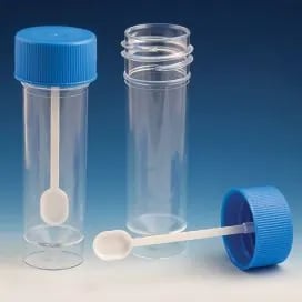 Figure: Fecal specimen collecting tubes
Caution: Specimens should not be collected from bedpans, because the bedpans may contain residual disinfectant or other contaminants.
Stool collection is one part of the broader pre-analytical process. For the general principles of specimen collection, transport, and rejection, see Specimen Collection and Transport in Microbiology.
Passed stool or rectal swab, and when a swab is acceptable
Freshly passed stool is the preferred specimen for almost every stool investigation. A rectal swab is the compromise, used only when passed stool cannot be obtained.
| Rank | Specimen | When to use it | How to collect it | What you trade off |
|---|---|---|---|---|
| 1 (ideal) | Freshly passed stool | Any patient who can pass a sample into a container | Into a clean wide-mouth container; sample any blood or mucus | None, provided it reaches the lab in time and in the right medium |
| 2 (compromise) | Rectal swab | Neonates, severely ill or debilitated patients, or when stool cannot be obtained | Insert the swab 1 to 1.5 inches (2.5 to 4 cm) past the anal sphincter, rotate gently, withdraw; the swab must be visibly stained with feces, then place straight into Cary-Blair | Smaller sample; acceptable for bacterial culture but poor for parasites and toxin testing |
A rectal swab is a bacterial-culture specimen, not a parasitology specimen. It carries too little material for reliable ova-and-parasite examination or for C. difficile toxin testing. If a swab is all you can get and parasites are the question, note the limitation on the request so the lab interprets a negative result cautiously.
One rule makes the rectal swab work or fail: it must be visibly stained with stool. A clean-looking swab has not sampled the feces and will usually be rejected. Place it into Cary-Blair immediately so it does not dry out.
How many samples, and when
The number of samples depends on what you are looking for.
- Bacterial culture (Salmonella, Shigella, Vibrio, Campylobacter): a single fresh sample collected in the acute phase is usually enough. A second sample raises yield if the first is negative and suspicion is high.
- Ova and parasite (O&P) examination: parasites are shed intermittently, so a single stool can miss them. The classic approach is three samples collected on separate days (often over 7 to 10 days). One negative stool does not exclude a parasitic infection.
- Typhoid (enteric fever): stool cultures are most often positive from the second to third week of illness onward, later than blood cultures, which are the better test in the first week.
Storage
Storage rules depend on the target organism, and this is where mistakes happen.
- Bacterial culture: if processing is delayed beyond 2 hours, refrigerate at 2 to 8 degrees C, or hold in Cary-Blair. Refrigeration slows overgrowth of commensal flora.
- Trophozoites (parasitology): do not refrigerate. Examine fresh within about 30 minutes, or fix immediately in an appropriate preservative.
- Cysts and ova: tolerate refrigeration and fixation well.
- C. difficile toxin: refrigerate if not tested promptly; the toxin degrades at room temperature.
The rule to carry: refrigeration is a friend to bacteria and cysts, and an enemy to trophozoites.
Transport media for fecal specimens
Media appropriate for the transport of fecal specimens that are suspected of containing Shigella, Vibrio cholerae, or Salmonella (including serotype Typhi) specimens are:
- Cary-Blair transport medium High pH (8.4): Medium of choice for transport and preservation of V. cholerae. Cary-Blair transport medium can transport many bacterial enteric pathogens, including Shigella, Salmonella, and Vibrio cholerae.
- Amies and Stuart's transport media: acceptable for Shigella and Salmonella (including serotype Typhi), but inferior to Cary-Blair for Vibrio cholerae.
- Alkaline peptone water: This medium may be used to transport V. cholerae, but this medium is inferior to Cary-Blair and should be used only when the Cary-Blair medium is unavailable. Alkaline peptone water should not be used if the subculture will be delayed more than 6 hours from the time of collection because other organisms will overgrow vibrios after 6 hours.
- Buffered glycerol saline (BGS) It’s a liquid medium that can be used for Shigella, but this transport medium is unsuitable for the transport of V. cholerae.
Rejection of fecal Specimens
Stool specimens are unacceptable if any of the following conditions apply;
- The information on the label does not match the information in the requisition
- The specimen has not been transported in the proper medium
- The quantity of specimens is insufficient for testing
Preservatives for parasitology (a different set of media)
If the request is for ova and parasites, the transport chemistry is different from bacterial culture. The choice depends on what the lab will do with the sample.
| Preservative | What it is for | Key point |
|---|---|---|
| None (fresh stool) | Wet mount for motile trophozoites | Must be examined within about 30 minutes; do not refrigerate, cold kills and distorts trophozoites |
| 10% formalin | Concentration technique and preservation of cysts, ova, and larvae | Good morphology for many stages; does not preserve trophozoite motility |
| PVA (polyvinyl alcohol) | Permanent stained smears | Preserves trophozoites for staining; traditionally contains mercury, so handle and dispose of per local rules |
| SAF or single-vial fixatives | Both concentration and permanent stain from one vial | Mercury-free alternatives now widely used |
The single most common parasitology error is refrigerating an unpreserved liquid stool meant for trophozoite examination. For bacteria, refrigeration protects the sample. For trophozoites, it destroys them. When both bacteria and parasites are requested, send separate portions handled the right way for each.
Processing of Stool Sample in Microbiology Lab
Stool is the opposite of a sterile specimen: it is packed with normal flora, so the entire laboratory approach is built on selective and differential media that suppress the commensals and make the pathogen stand out. This is why the request matters so much. The media set up depends on which pathogen is suspected, and a stool processed for routine bacteria will not reveal a parasite, and vice versa.
Direct examination.
- A saline and iodine wet mount is examined when a parasitic cause is suspected, looking for motile trophozoites, cysts, ova, and larvae. The full parasite workup, including concentration methods and permanent stains, is covered on the intestinal parasitic infections page; because the preservatives and technique differ from bacterial culture, that side is handled separately.
- A methylene blue or Gram stain of the stool can show sheets of pus cells (fecal leukocytes), which point toward an invasive, inflammatory diarrhea (such as Shigella, invasive Escherichia coli, or Campylobacter) rather than a toxin-mediated watery diarrhea.
- A hanging-drop or wet preparation showing darting motility raises Vibrio or Campylobacter.
Culture media, chosen by the suspected pathogen. This is the heart of stool processing. Enteric pathogens each have preferred selective and differential media, so telling the lab what is suspected changes what is set up:
| Suspected pathogen | Media commonly used | What it shows |
|---|---|---|
| Salmonella, Shigella | MacConkey, XLD, and a moderately selective medium such as DCA or SS agar | Non-lactose-fermenting (pale) colonies; XLD shows Salmonella with black centers from H2S |
| Salmonella (enrichment) | Selenite F broth, then subculture | Enriches small numbers of Salmonella before plating |
| Vibrio cholerae | TCBS agar; alkaline peptone water as enrichment | Yellow (sucrose-fermenting) colonies on TCBS |
| Campylobacter | Selective Campylobacter medium (for example Skirrow), microaerophilic, 42 degrees C | Curved gram-negative rods, darting motility |
| E. coli O157 (STEC) | Sorbitol MacConkey (SMAC) | Sorbitol non-fermenting (pale) colonies, unlike most E. coli |
Routine stool culture typically includes MacConkey and a selective medium plus an enrichment broth, with the more specialized media (TCBS, SMAC, Campylobacter medium) added when the history points that way.
Incubation. Most enteric plates are incubated at 35 to 37 degrees C for 24 to 48 hours. Campylobacter is the exception, needing a microaerophilic atmosphere and incubation at 42 degrees C.
Reading and interpretation.
- Against the dense normal flora, the read hunts for the pathogen's signature: non-lactose-fermenting colonies on MacConkey, black-centered colonies on XLD, yellow colonies on TCBS, or sorbitol-negative colonies on SMAC.
- Normal flora is expected and is not reported as a pathogen; the point of the selective media is to let the pathogen be recognized within that background.
- Fecal leukocytes on the direct smear support an inflammatory, invasive cause and help interpret which pathogen to pursue.
- Remember the collection-side rule that three or more organisms in large numbers usually means a contaminated or poorly handled specimen rather than a true polymicrobial enteric infection.
Identification and antimicrobial susceptibility testing
The organisms commonly sought from stool, and the first approach to each:
- Salmonella species: non-lactose fermenter, H2S positive (black on XLD); confirmed biochemically and serologically. See the Salmonella page.
- Shigella species: non-lactose fermenter, non-motile, H2S negative; confirmed biochemically and serologically. See the Shigella page.
- Vibrio cholerae: oxidase-positive curved gram-negative rod, yellow on TCBS; confirmed by serotyping. See the Vibrio cholerae page.
- Campylobacter jejuni: curved or seagull-shaped gram-negative rod, oxidase positive, grown microaerophilically at 42 degrees C. See the Campylobacter page.
- Diarrheagenic Escherichia coli (including O157 STEC): separated on sorbitol MacConkey and by molecular or serological methods. See the Diarrheagenic E. coli page.
- Parasites (Entamoeba histolytica, Giardia lamblia, helminths): sought by microscopy, not culture; the full workup, including concentration and staining, is on the intestinal parasitic infections page.
For the full identification scheme and, where relevant, antimicrobial susceptibility testing of the organism grown, visiting the detailed article for that organism. Note that for many enteric infections susceptibility testing is done selectively, since a number are self-limiting and managed supportively; testing is guided by severity and local guidance.
How to Remember
Three questions decide a stool sample: right part, right time, right medium. Sample the blood and mucus (right part), get it there fast and in the acute phase (right time), and match the medium to bacteria versus parasites (right medium). Almost every failed stool investigation breaks one of these three.
Cary-Blair is the bacterial workhorse; formalin and PVA are the parasite pair. For culture, think Cary-Blair. For O&P, think two vials: formalin for concentration and PVA (or a single-vial fixative) for permanent stains. Different question, different chemistry.
Refrigeration: friend to bacteria, enemy to trophozoites. Cold protects a stool going for culture and kills the trophozoites in a stool going for parasites. If you remember which is which, you avoid the commonest storage error on the page.
Three samples for parasites, one for bacteria. Parasites are shed on and off, so one stool can miss them; three on separate days is the classic rule. Bacteria are shed heavily in the acute phase, so one good sample usually does.
The rectal swab must be dirty to be useful. A clean swab has not sampled the stool. It must be visibly stained with feces, and it goes for culture, not parasites.
Key exam facts in one table
| Point | Fact |
|---|---|
| Ideal specimen | Freshly passed stool |
| Compromise specimen | Rectal swab (neonates, debilitated, or when stool cannot be obtained) |
| Rectal swab depth | 1 to 1.5 inches past the sphincter, visibly stained with feces |
| Rectal swab limits | Bacterial culture only; poor for O&P and C. difficile |
| Volume needed | About 5 mL if liquid, 2 g if formed |
| Sample which part | The bloody or mucoid portion in dysentery |
| Bacterial transport time | Process within 2 hours, or Cary-Blair and refrigerate |
| Medium of choice (enteric bacteria) | Cary-Blair (best for V. cholerae; also Shigella, Salmonella, Campylobacter) |
| APW limit | Vibrio only; subculture within 6 hours before overgrowth |
| O&P sampling | Three samples on separate days (intermittent shedding) |
| Trophozoites | Examine fresh within about 30 min; do NOT refrigerate |
| Parasite preservatives | Formalin (concentration), PVA or single-vial (permanent stain) |
| Typhoid stool culture | Positive mainly from week 2 to 3 onward |
| C. difficile 3-day rule | Do not do routine stool culture after 3 days of admission for non-diarrheal patients; test for C. difficile instead |
| Do not collect from | Bedpans, or stool mixed with urine |
Where Students Get Confused
"Why send three samples for parasites but only one for bacteria?" Parasites are shed intermittently, so a single stool can be falsely negative. Three samples over several days catch the shedding. Bacterial pathogens are shed heavily during the acute phase, so one good sample is usually enough.
"Cary-Blair is the stool transport medium, so it works for everything?" No. Cary-Blair is a bacterial-culture transport medium. It does nothing useful for a parasitology request, which needs formalin, PVA, or a single-vial fixative. The word "stool transport medium" hides the fact that bacteria and parasites need different chemistry.
"Refrigerate the delayed stool, always safe?" Only for bacteria and cysts. An unpreserved liquid stool going for trophozoites must not be refrigerated, cold kills the motile trophozoites you are looking for. Examine it fresh or fix it.
"A rectal swab is basically the same as stool?" For bacterial culture, close enough when passed stool cannot be obtained. For parasites and C. difficile toxin, no, a swab carries too little material. And a swab that is not visibly stained with feces has not sampled anything useful.
"Bloody stool means culture, watery means cholera, done?" Only roughly. Shigella dysentery is bloody only about half the time, and many organisms cause watery diarrhea. Stool appearance guides suspicion but does not replace culture.
References
- Tille PM. Bailey & Scott's Diagnostic Microbiology. 15th ed. St. Louis: Elsevier; 2022.
- Leber AL, editor. Clinical Microbiology Procedures Handbook. 4th ed. Washington, DC: ASM Press; 2016. DOI: 10.1128/9781683670438.CMPH
- Garcia LS, et al. Diagnostic Medical Parasitology. (O&P specimen collection, preservatives, and the three-sample rule.) ASM Press.
- Cheesbrough M. District Laboratory Practice in Tropical Countries, Part 2. 2nd ed. Cambridge: Cambridge University Press; 2006.
Frequently Asked Questions
How many stool samples are needed to look for parasites?
How many stool samples are needed to look for parasites?
Three samples collected on separate days, because parasites are shed intermittently and a single stool can be falsely negative. For bacterial culture, one fresh sample in the acute phase is usually enough.
When is a rectal swab acceptable instead of passed stool?
When is a rectal swab acceptable instead of passed stool?
When passed stool cannot be obtained, for example in neonates or severely debilitated patients. The swab must be inserted past the anal sphincter and be visibly stained with feces, then placed in Cary-Blair. It is suitable for bacterial culture but not for parasite examination or C. difficile toxin testing.
Which transport medium should a stool sample go into?
Which transport medium should a stool sample go into?
For enteric bacteria, Cary-Blair is the medium of choice. For parasites, use a preservative instead: formalin for concentration methods and PVA or a single-vial fixative for permanent stains. Bacteria and parasites need different chemistry.
Can a delayed stool sample always be refrigerated?
Can a delayed stool sample always be refrigerated?
Only for bacterial culture and for cysts and ova. An unpreserved liquid stool intended for trophozoite examination must not be refrigerated, since cold kills the motile trophozoites. Examine it fresh within about 30 minutes or fix it immediately.
How much stool should be collected?
How much stool should be collected?
About 5 mL if the stool is liquid, or 2 grams (roughly two bean-sized portions) if formed. Sample any blood or mucus specifically, since pathogens concentrate there in dysentery.
Why should stool not be collected from a bedpan?
Why should stool not be collected from a bedpan?
Bedpans may contain residual disinfectant that kills pathogens, and the sample can be contaminated with urine. Collect directly into a clean, dry, wide-mouth container.
When are stool cultures positive in typhoid fever?
When are stool cultures positive in typhoid fever?
Mainly from the second to third week of illness onward. In the first week, blood culture is the higher-yield test.

Tankeshwar Acharya, MSc (Medical Microbiology)
Tankeshwar Acharya is an Assistant Professor in the Department of Microbiology at Patan Academy of Health Sciences (PAHS), Nepal, where he has been teaching and practicing clinical microbiology for over 14 years. He is the founder of Microbe Online, one of the leading free microbiology education resources on the web, covering bacteriology, mycology, parasitology, immunology, and clinical laboratory diagnostics written from direct experience in both the classroom and the diagnostic laboratory.
Comments
No comments yet. Be the first to share your thoughts.
Leave a comment
All comments are reviewed before they appear.