

The shape of a bacterium is determined by its rigid cell wall. Bacteria that lack a cell wall (Mycoplasma and L-forms) display a great diversity of unusual shapes. Bacteria having various shapes are said to be pleomorphic (heterogeneous shape or many-shaped).
Bacteria are classified by shape into three primary groups: cocci, bacilli, and spiral-shaped. The cocci are round, the bacilli are rods, and the spiral-shaped bacteria are either rigid (spirilla) or flexible (spirochetes).
These three primary bacteria shapes determine the bacteria’s morphological characteristics. This is one of the most important criteria used in identifying organisms.
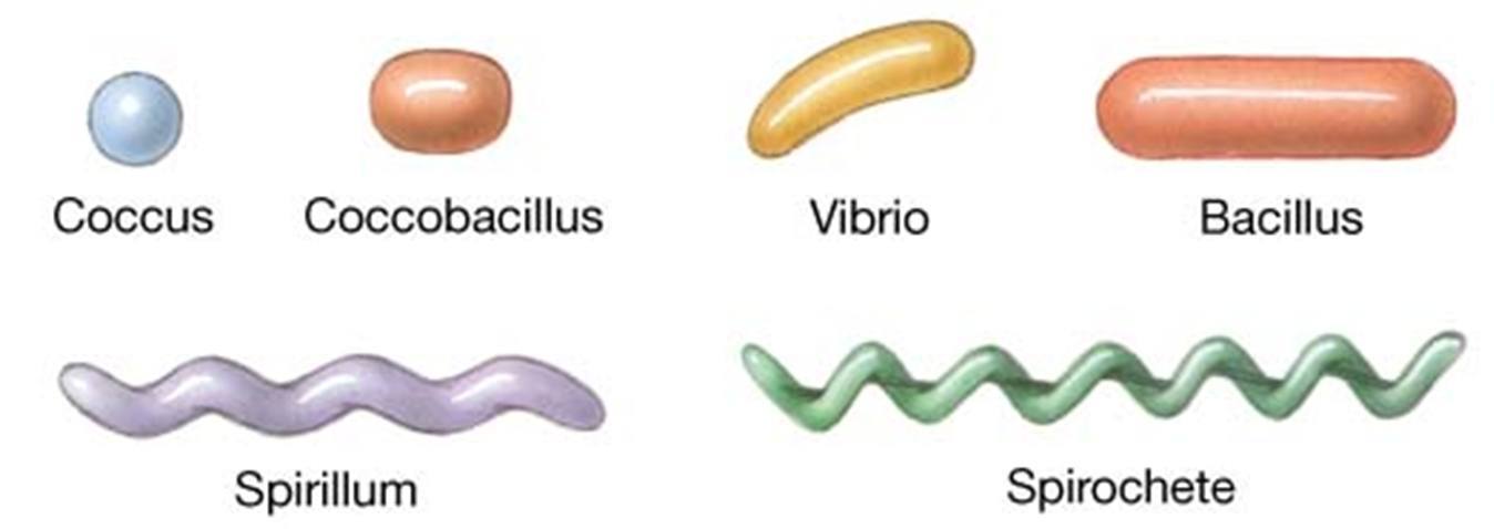 Figure: Various Shapes of Bacteria
Picture a clinician at 2 AM who has just received a Gram stain result from a blood culture: 'Gram-positive cocci in clusters.' No organism name, no sensitivity results, just morphology and Gram reaction. That clinician starts antistaphylococcal coverage right there, and they're right to do it. A Gram stain takes 15 minutes. A blood culture takes 24-72 hours. The entire clinical value of learning bacterial shapes is compressed into that gap.
Why bacterial morphology matters clinically
Knowing the shape and arrangement of bacteria is not just an academic exercise. It is the first step in clinical bacterial identification. When a gram-stained smear is examined from a clinical specimen, the combination of gram reaction, shape, and arrangement gives the microbiologist and clinician an immediate presumptive identification within minutes, before any culture results are available.
This rapid presumptive identification directly guides empirical antibiotic therapy, the treatment a clinician starts before definitive culture and sensitivity results return. A gram stain showing gram-positive cocci in clusters from a wound swab suggests Staphylococcus aureus and guides antistaphylococcal treatment. Gram-negative diplococci in a urethral discharge smear is virtually diagnostic of Neisseria gonorrhoeae and guides immediate gonococcal therapy.
Understanding morphology therefore has direct patient care implications.
- Spherical or oval bacteria are called cocci (singular: coccus)
- Rods are called bacilli (singular: bacillus)
Very short rods that can sometimes almost be mistaken for cocci are called coccobacilli (singular: coccobacillus). Rod-shaped bacteria having tapered ends are called fusiform, whereas others are characteristically club-shaped and may be curved or comma-shaped (Vibrios) or bent.
- Spiral-shaped bacteria are called spirilla if the cells are rigid and spirochetes if they are more flexible and undulating.
In addition to their characteristic shapes, the arrangement of bacteria is essential. For example, certain cocci occur in pairs (diplococci), some in chains (streptococci), and others in grapelike clusters (staphylococci). These arrangements are determined by the bacteria’s orientation and degree of attachment at the time of cell division. The arrangement of rods and spirochetes is medically less important.
Cocci
Cocci appear in several characteristic arrangements such as diplococci (cocci in pairs), streptococci (cocci in chains), tetrads, staphylococci (cocci in grape-like clusters), and sarcinae. These arrangements are determined by whether the daughter cells stay together following division.
Bacteria don't choose their arrangement. It's determined by two things: the plane of division (one plane = pairs/chains, two planes = tetrads, three planes = packets of eight), and whether daughter cells separate after dividing. Streptococci stay attached after dividing in one plane, so they form chains. Staphylococci divide in random planes and don't always separate cleanly, so they form irregular clusters. The arrangement is the division history made visible.
| Name of the Bacteria | Characteristics Shape |
|---|---|
| Staphylococcus | Spherical, Gram-positive cocci arranged in irregular, grape-like clusters. |
| Streptococcus | Spherical, Gram-positive cocci arranged in chains or pairs |
| Streptococcus pneumoniae | Gram-positive lancet-shaped cocci arranged in pairs (diplococci) or short chains. |
| Merismopedia | Cocci arranged in a tetrad, formed by division into two planes |
| Sarcina lutea | Cocci arranged in a sarcina, formed by a division in three planes. |
| Neisseria meningitidis (meningococci) | Gram-negative diplococci with adjacent flattened sides, giving a kidney-bean or coffee-bean pair appearance. |
| Neisseria gonorrhoeae (gonococci) | Gram-negative diplococci with adjacent flattened sides, giving a kidney-bean or coffee-bean pair appearance. |
Cocci can be found in pairs, chains, squares of four, cubes of eight, or grapelike clusters.
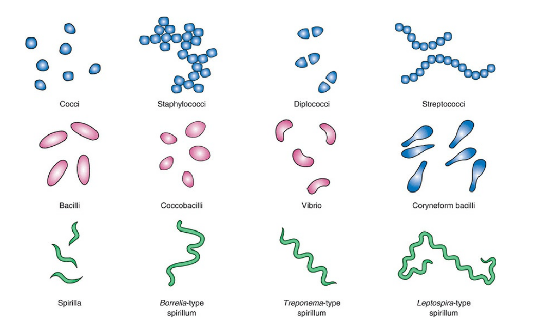 Figure: Basic morphological shapes of bacteria
Clinical significance of coccal arrangements
The arrangement of cocci on a gram stain is as diagnostically important as the gram reaction itself:
| Gram stain appearance | Most likely organism(s) | Clinical context |
|---|---|---|
| Gram-positive cocci in grape-like clusters | Staphylococcus aureus, CoNS | Wound infections, bacteremia, abscess, pneumonia, endocarditis |
| Gram-positive cocci in pairs and chains | Streptococcus pyogenes (GAS), S. agalactiae (GBS), Enterococcus spp. | Pharyngitis, cellulitis, neonatal sepsis, UTI, endocarditis |
| Gram-positive lancet-shaped diplococci | Streptococcus pneumoniae | Pneumonia, meningitis, otitis media, sinusitis |
| Gram-negative kidney-bean shaped diplococci (intracellular) | Neisseria gonorrhoeae | Urethral discharge, cervicitis, PID, conjunctivitis |
| Gram-negative diplococci (extracellular + intracellular) | Neisseria meningitidis | Meningitis, septicemia (meningococcemia) |
| Gram-negative diplococci (catarrhalis) | Moraxella catarrhalis | COPD exacerbations, otitis media, sinusitis |
| Gram-positive cocci in tetrads | Micrococcus spp. | Usually contaminant; rarely causes opportunistic infections |
How to Remember
"CDSS" as an observation sequence at the bench: Clusters → Staphylococcus. Diplococci (pairs) → narrow it by Gram reaction: Gram-positive lancet-shaped = pneumococcus, Gram-negative kidney-bean = Neisseria. Streptococci/chains → Gram-positive = Strep/Enterococcus. Squares of four (tetrads) → Micrococcus.
See also: Lancefield Classification of Streptococci for serological grouping of beta-hemolytic streptococci
Diplococci
Diplococci (division in one plane, daughter cells remain paired): Neisseria gonorrhoeae, N. meningitidis, Streptococcus pneumoniae, Moraxella catarrhalis.
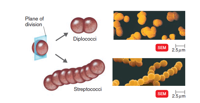 Figure: Diplococci and Streptococci (Image source: Ref-3)
Streptococci
Long chains of cocci (streptococci) result when cells adhere after repeated divisions in one plane; this pattern is seen in the genera Streptococcus agalactiae, Streptococcus pyogenes, Enterococcus faecalis, and Lactococcus.
Staphylococci
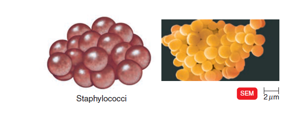 Figure: Staphylococci (grape-like cluster) (Image source: Ref-3)
Staphylococcus divides into random planes to generate irregular grapelike clumps. Divisions in two or three planes can produce symmetrical clusters of cocci. Examples, Staphylococcus aureus, Staphylococcus saprophyticus, Staphylococcus epidermidis, etc.
Tetrad
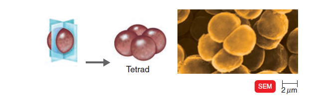 Figure: Tetrad (Image source: Ref-3)
Members of the genus Micrococcus often divide into two planes to form square groups of four cells called tetrads.
Sarcina
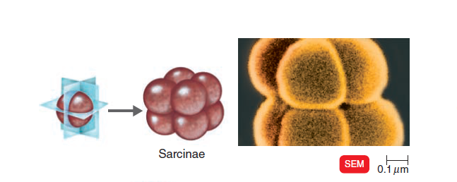 Figure: Sarcinae (Image source: Ref-3)
In the genus Sarcina, cocci divide into three planes producing cubical packets of eight cells.
Bacilli
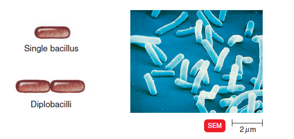 Figure: Single bacillus and Diplobacilli (Image source: Ref-3)
| Name of the Bacteria | Characteristics Shape |
|---|---|
| Bacillus anthracis | Large, Gram-positive rod with square ends, frequently found in chains. |
| Clostridium tetani | Gram-positive rod with characteristics “tennis racket shaped” or “drum stick appearance” due to presence of terminal spore. |
| Clostridium perfringens | Large, Gram-positive, “boxcar” shaped bacilli. |
| Corynebacteria | Gram-positive rods are club-shaped and arranged in palisades or V or L-shaped formations, also called “Chinese-Letter” appearance. |
| Bacillus megaterium | Gram-positive, streptobacilli (bacillus arranged in chains). |
| Listeria monocytogenes | Small Gram-positive rods arranged in V or L-shaped formations similar to corynebacteria. |
| Vibrio cholerae | Comma-shaped, curved, Gram-negative bacilli |
| Campylobacter | Curved Gram-negative rods that appear as comma or S-shaped |
| Haemophilus influenzae | Small, Gram-negative coccobacilli |
| Fusobacterium spp. | These pleomorphic, long Gram-negative rods, often with tapered “pointy” ends. |
Clinical significance of rod-shaped bacteria
| Gram stain appearance | Most likely organism(s) | Clinical context |
|---|---|---|
| Large gram-positive rods with square ends, in chains | Bacillus anthracis | Anthrax (cutaneous, inhalation, gastrointestinal) |
| Gram-positive rods with terminal spore ("drumstick") | Clostridium tetani | Tetanus |
| Large gram-positive "boxcar" rods | Clostridium perfringens | Gas gangrene, food poisoning, necrotizing fasciitis |
| Gram-positive club-shaped rods in palisades/"Chinese letters" | Corynebacterium diphtheriae | Diphtheria |
| Small gram-positive rods in V or L shapes | Listeria monocytogenes | Neonatal meningitis, listeriosis in pregnancy, immunocompromised |
| Gram-negative comma-shaped (curved) rods | Vibrio cholerae | Cholera (rice-water diarrhea) |
| Gram-negative S-shaped or comma-shaped rods | Campylobacter jejuni | Bloody diarrhea, gastroenteritis |
| Small gram-negative coccobacilli | Haemophilus influenzae | Meningitis, pneumonia, epiglottitis, otitis media |
| Small gram-negative coccobacilli | Moraxella catarrhalis | Respiratory tract infections |
| Long gram-negative rods with tapered ends | Fusobacterium nucleatum | Oral infections, aspiration pneumonia, Lemierre's syndrome |
| Tiny gram-negative rods (poorly staining) | Brucella spp. | Brucellosis (undulant fever) |
| Small gram-negative rods | Francisella tularensis | Tularaemia |
| Gram-positive branching filaments | Actinomyces spp. | Actinomycosis; draining sinus tracts with sulfur granules |
These are not arranged in patterns as complex as cocci; most occur singly or in pairs (diplobacilli). But some species, such as Bacillus subtilis, form chains (streptobacilli); others, such as Beggiatoa and Saprospira species, form trichomes (which are similar to chains but have a much larger area of contact between the adjacent cells).
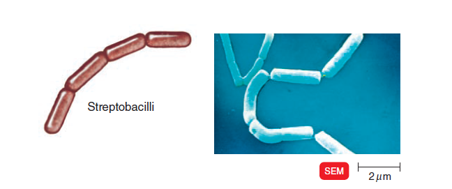 Figure: Streptobacilli (Image source: Ref-3)
Rod-shaped organisms may be regular in morphology, maybe somewhat shorter (i.e., “coccobacillary”), or may appear club- or dumbbell-shaped (“coryneform”).
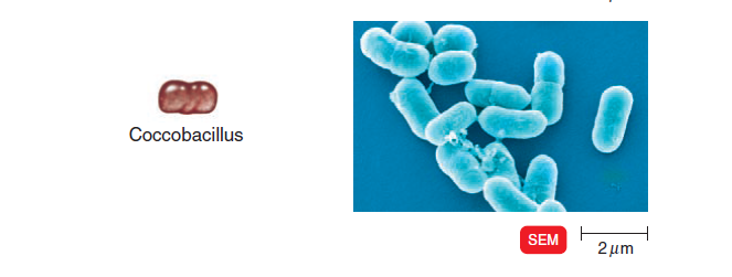 Figure: Coccobacillus (Image source: Ref-3)
Comma-shaped cells generally define a basic characteristic of certain species (e.g., Vibrio species).
In some bacillus, the cells are lined side by side like matchsticks and at angles to one another. Such arrangement is called palisade arrangement and is found in Corynebacterium diphtheriae. Streptomyces species form long, branched, multinucleate filaments called hyphae, which collectively form a mycelium.
Spiral-Shaped Bacteria
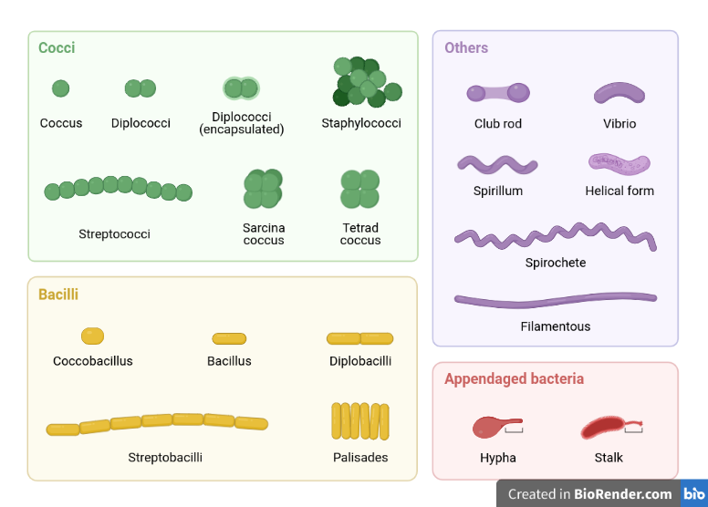 Figure: Various morphology of bacteria
Spiral bacteria have a variety of curved shapes. Bacteria with less than one complete twist or turn have a vibriod shape, whereas those with one or more complete turns have a helical shape.
Spirilla are rigid helical bacteria, whereas spirochetes are highly flexible. Spirilla (singular: spirillum) are rigid, wavy-shaped curved bacteria, and spirochete is curved corkscrew-shaped bacteria.
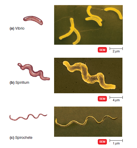 Figure: Spiral-shaped bacteria (Image source: Ref-3)
- Spirochetes (Treponema, Leptospira, and Borrelia): Thin-walled, flexible, spiral rods (Corkscrew shaped) seen only by darkfield microscope and generally not seen in a standard light microscope.
- Borrelia: They are corkscrew-shaped and are larger than the Treponema; they can be viewed under a light microscope with Giemsa or Wright stains.
Clinical significance of spiral-shaped bacteria
| Gram stain appearance | Organism | Clinical context | Special stain needed |
|---|---|---|---|
| Thin, tightly coiled spirochetes (not visible on gram stain) | Treponema pallidum | Syphilis (primary, secondary, tertiary, congenital) | Dark-field microscopy |
| Loosely coiled large spirochetes (visible with Giemsa/Wright) | Borrelia burgdorferi | Lyme disease | Giemsa or Wright stain |
| Loosely coiled large spirochetes (visible with Giemsa/Wright | Borrelia recurrentis | Relapsing fever (louse-borne, tick-borne) | Giemsa stain |
| Hooked-end spirochetes | Leptospira interrogans | Leptospirosis (Weil's disease) | Dark-field microscopy |
Note on classification: Vibrio and Campylobacter are curved or S-shaped gram-negative rods, not true spirochetes, and are covered in the Bacilli section above. They are included in discussions of "spiral" morphology only because of their curved appearance, but unlike spirochetes they stain and are read on a standard gram stain.
Note on visibility: True spirochetes (Treponema, Leptospira, Borrelia) are too thin to be visualized on a standard gram stain.
Pleomorphic Bacteria
Some bacteria are variable in shape and are said to be pleomorphic (heterogeneous shape).
Pleomorphic bacteria are particularly challenging to identify on gram stain because their variable shape does not give a consistent morphological clue. The key to identifying them lies in the clinical context: the specimen type, the patient's history, and the combination of gram reaction and growth characteristics.
| Organism | Why pleomorphic | Clinical clue on gram stain |
|---|---|---|
| Corynebacterium diphtheriae | Variable club and rod shapes depending on growth phase | Gram-positive rods in palisades or "Chinese letter" arrangement; metachromatic granules (Babes-Ernst bodies) with methylene blue stain |
| Haemophilus influenzae | Range from coccobacilli to short rods depending on growth conditions | Tiny gram-negative coccobacilli; found intracellularly in CSF in meningitis |
| Francisella tularensis | Very small, variable rods | Tiny, poorly-staining gram-negative rod; biosafety concern, notify lab if tularemia suspected |
| Bartonella henselae | Curved to rod-shaped | Gram-negative rod; associated with cat scratch disease and bacillary angiomatosis |
| Cardiobacterium hominis | Variable rod shapes | Gram-negative rod; part of HACEK group; associated with endocarditis |
Other unusual shapes of bacteria
Some bacteria do not fit in any of the above-mentioned categories and have spindle or irregular, lobed shapes. Some of these bacterial shapes are;
Bacillus arranged in a rosette attached by stalks to a substrate, for example, Caulobacter.
Star-shaped bacteria, for example, Stella
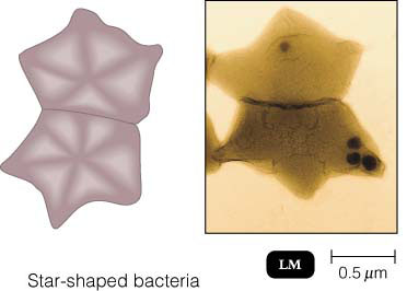 Square-shaped bacteria, for example, Haloarcula (a salt-loving member of the Archaea)
Pear-shaped cells, e.g., Pasteuria
Lobed spheres e.g., Sulfolobus
Disks arranged like stacks of coins, e.g., Caryophanon
Rods with helically sculptured surfaces, e.g., Seliberia
Key exam facts
| Shape | Arrangement | Genus | Gram reaction | Clinical hook |
|---|---|---|---|---|
| Cocci | Grape-like clusters | Staphylococcus | Positive | Abscesses, wound infections |
| Cocci | Chains or pairs | Streptococcus, Enterococcus | Positive | Pharyngitis, endocarditis, UTI |
| Cocci | Lancet-shaped pairs | S. pneumoniae | Positive | Pneumonia, meningitis |
| Cocci | Kidney-bean pairs (intracellular) | N. gonorrhoeae | Negative | Urethral discharge |
| Cocci | Pairs | N. meningitidis | Negative | Meningitis |
| Cocci | Tetrads | Micrococcus | Positive | Usually contaminant |
| Bacilli | Chains, square ends | B. anthracis | Positive | Anthrax |
| Bacilli | Drumstick (terminal spore) | C. tetani | Positive | Tetanus |
| Bacilli | Palisades/"Chinese letters" | C. diphtheriae | Positive | Diphtheria |
| Bacilli | Comma/S-shaped | V. cholerae, Campylobacter | Negative | Cholera, gastroenteritis |
| Spiral | Corkscrew, not visible on Gram stain | Treponema, Leptospira | N/A | Syphilis, leptospirosis |
References and further readings
- Procop, G. W., et al. (2017). Koneman's Color Atlas and Textbook of Diagnostic Microbiology (7th ed.). Wolters Kluwer.
- Levinson, W., et al. (2022). Review of Medical Microbiology and Immunology (16th ed.). McGraw-Hill.
- Tortora, G. J., Funke, B. R., & Case, C. L. (2019). Microbiology: An Introduction (13th ed.). Pearson Education.

Comments
No comments yet. Be the first to share your thoughts.
Leave a comment
All comments are reviewed before they appear.