
Casein Hydrolysis Test: Principle, Procedure, and Uses
The casein hydrolysis test detects caseinase on skim milk agar, shown by a clear zone around growth. Learn the principle, how to read the result, positive and negative organisms, and how it differs from gelatin hydrolysis.

On this page
A student streaks two organisms across one skim milk agar plate. After a day, a clear halo has spread around one line of growth, while the other sits on milk-white agar with no halo at all. The plate has answered a single question: which organism can break down protein it cannot swallow whole. That clear zone is the whole point of the test, and learning why it appears is how one plate sorts proteolytic bacteria from the rest.
The physiological reactions of microorganisms are central to identifying and differentiating species. Because enzymes drive these reactions, detecting a specific enzyme is a useful way to tell organisms apart. The casein hydrolysis test is one such method. It identifies organisms by detecting the activity of an extracellular enzyme, caseinase, that breaks down the milk protein casein.
Casein hydrolysis is the degradation of casein protein present in an opaque milk agar due to metabolic activity of organism into soluble end products like amino acids, peptides, etc. This results in a transparent zone around the growth Thus, the test performed to detect the breakdown of casein is known as the casein hydrolysis test.
Casein, a dominant milk protein, is a macromolecule comprising amino acid subunits linked together by a CO-NH bond. The microorganism cannot uptake casein as it is a large molecule. Therefore, it must undergo step-by-step degradation into peptides and a small chain of amino acids for assimilation. These reactions are catalyzed by proteinases (caseinase) and peptidase enzymes. The proteinase cleaves the peptide (CO-NH ) bond of adjacent amino acids by introducing water molecules to release polypeptides and amino acids.
The overall reaction; Casein → small chain of amino acids, dipeptides, and polypeptides; in the presence of caseinase and water molecules.
Principle of Casein Hydrolysis Test
Microorganisms cannot take in large protein molecules directly. Some organisms overcome this by secreting an extracellular enzyme that digests casein into small, absorbable products. This enzyme is called caseinase (also written casease). It is a protease, meaning an enzyme that cleaves the peptide bonds of a protein.
Skim milk agar is used to demonstrate this activity. The medium contains skim milk powder, tryptone, yeast extract, dextrose, and agar. The casein in the skim milk is what makes the agar opaque and white, because the intact protein scatters light rather than letting it pass through. This is the key to reading the test. When an organism digests the casein around its growth, the protein that was scattering light is gone, so the agar there turns clear and colorless. The tryptone, yeast extract, and dextrose supply amino acids, B vitamins, and carbon for growth.
Following inoculation and incubation of the culture agar plate, proteolytic bacteria will form a zone of proteolysis, demonstrating a transparent area surrounding the bacterial growth. The formation of the transparent zone is due to the formation of soluble molecules by the degradation of casein protein, and it represents a positive reaction.
Without proteolytic activity, the whole medium remains opaque even around the growth area, representing a negative reaction.
Objectives of Casein Hydrolysis Test
- To determine the capability of specific microorganisms to break down the casein protein.
- To differentiate microorganisms based on their capability to produce exoenzymes.
Media used in casein hydrolysis test
Composition of Skim Milk Agar
| Ingredients | Gram per liter |
|---|---|
| Skim milk powder | 28.0 |
| Trypton | 5.0 |
| Yeast extract | 2.50 |
| Dextrose | 1.0 |
| Agar | 15.0 |
Procedure of Casein Hydrolysis test
The procedure of the test includes the following steps:
- Take a skim milk agar plate and inoculate it in a straight line or zigzag line with the help of a sterile inoculating loop.
- Incubate the inoculated plate at 25 to 37ºC for 18-24 hours.
- Then, observe the presence or absence of a clear zone around the growth of the test organism in the agar plate culture.
Results with Examples
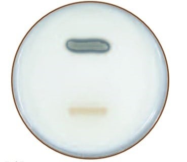 Figure: CASEIN HYDROLYSIS TEST RESULTS: This Milk Agar plate was inoculated with Bacillus megaterium (caseinase-positive) above and Micrococcus roseus (caseinase-negative) below.
- Positive result: a clear, transparent zone appears around the line of growth, and sometimes directly beneath the colonies. The clearing means the organism secreted caseinase and digested the casein, removing the opacity from that part of the agar. Read the plate against the light. The zone may be easier to see when the plate is tilted. Positive organisms include Bacillus subtilis, Bacillus cereus, Pseudomonas aeruginosa, Serratia marcescens, and the aerobic actinomycetes such as Streptomyces and Nocardia.
- Negative result: the agar stays opaque and milk-white right up to the growth, with no clearing. This means no caseinase was produced. Negative organisms include Escherichia coli and Enterococcus faecalis.
Do not report a negative result too early. Casein hydrolysis can be slow in some organisms. Incubate for at least 3 days at 35°C, and up to 14 days at 25°C for slow growers such as actinomycetes, before recording a negative. A common mistake is calling a plate negative at 24 hours when the zone simply has not developed yet.
Reading around pigmented organisms. Some positive organisms produce pigment that can mask the clear zone. Pseudomonas aeruginosa produces a green pigment (pyocyanin) and Serratia marcescens a red pigment (prodigiosin), either of which can sit over the clearing. Look for the loss of opacity in the agar rather than a simple change in color.
How to read the result
The test asks one thing: did the organism clear the casein around its growth?
Look for loss of opacity, not just a color change. The milk-white agar turning clear and see-through is the positive signal. Reading the plate against a light source, or tilting it, makes a faint zone easier to see.
Measure the zone from the edge of growth outward if you need to compare organisms, but remember the test is qualitative. A larger zone suggests stronger or faster caseinase activity, but the result is reported simply as positive or negative.
The zone can appear beneath the colonies, not only around them. If growth is heavy, scrape or look under the growth line, because some organisms clear the casein directly under themselves before the zone spreads outward.
Give slow organisms time. A negative call at 24 hours is unreliable for slow proteolytic organisms. Hold the plate for the full incubation period before reporting negative.
Trust the agar, not the pigment. When a pigmented organism grows, judge the result by whether the agar has lost its opacity, not by the color of the colony or the diffusing pigment.
How casein hydrolysis differs from gelatin hydrolysis
Both tests detect proteolysis, the ability to break down protein, which is why students often confuse them. They differ in the substrate and in how the result is read.
| Feature | Casein hydrolysis | Gelatin hydrolysis |
|---|---|---|
| Protein tested | Casein (milk protein) | Gelatin (from collagen) |
| Medium | Skim milk agar | Nutrient gelatin |
| Enzyme detected | Caseinase (a protease) | Gelatinase (a protease) |
| Positive result | Clear zone around growth | Medium stays liquid after chilling |
| How it is read | Loss of opacity on the plate | Failure of the medium to solidify at 4°C |
Both are proteolysis tests, but they are not interchangeable. An organism can be positive for one and negative for the other, because caseinase and gelatinase are different enzymes. For the gelatin test in full, see the separate article on the gelatin hydrolysis test.
Uses of Casein Hydrolysis Test
The uses of the casein hydrolysis test include;
- It helps to identify the organism that produces caseinase.
- The casein hydrolysis test helps determine the microorganisms that grow in milk.
- It also helps in differentiating Bacillaceae, Enterobacteriaceae, and other families.
- The identification of aerobic actinomycetes is easily possible by this test.
Limitations of Casein Hydrolysis Test
The limitation of the casein hydrolysis test includes:
- Further, biochemical, immunological, molecular, or mass spectrometry tests are required to identify species.
- Some strains might show less growth due to variable nutrient requirements.
Quality control
Bacillus subtilis subsp. spizizenni ATCC 6633-positive reaction, clear zone surrounding the colonies.
Pseudomonas aeruginosa ATCC 27853-positive reaction, clear zone surrounding the colonies
Serratia marcescens ATCC 8100-positive reaction, clear zone surrounding the colonies.
Escherichia coli ATCC 25922-negative reaction, no clear zone around the colonies
Enterococcus fecalis ATCC 29212-negative reaction, no clear zone around the colonies.
How to Remember
Casein makes milk white, so clearing means it is gone. The white of the agar is the casein. A clear halo means the protein has been digested away right there.
Caseinase is a protease with a job title. It is simply a protease that happens to act on casein. If an organism makes it, the organism can eat protein it cannot swallow whole.
Clear halo, positive. Milk-white to the edge, negative. The reader is looking for a see-through zone against a milk-white background.
Wait before you write negative. Slow proteolysis is real. An early negative is often just an undeveloped zone.
Key exam facts
| Item | Fact |
|---|---|
| Detects | Caseinase (casease), an extracellular protease |
| Medium | Skim milk agar (casein is the opaque, white component) |
| Positive | Clear zone around or beneath growth (loss of opacity) |
| Negative | Agar stays opaque and milk-white to the edge of growth |
| Positive organisms | Bacillus spp., Pseudomonas aeruginosa, Serratia marcescens, aerobic actinomycetes |
| Negative organisms | Escherichia coli, Enterococcus faecalis |
| Incubation | 25 to 37°C; hold up to 3 days (35°C) or 14 days (25°C) before reporting negative |
| Nature | Qualitative |
| Main uses | Detecting proteolytic bacteria; differentiating families; identifying aerobic actinomycetes |
| Key limitation | Presumptive only; confirm with further tests |
Where Students Get Confused
"A positive result means the milk changed color." No. The signal is loss of opacity, not a color change. The milk-white agar turns clear and see-through where the casein has been digested.
"No zone at 24 hours means the organism is negative." Not reliably. Casein hydrolysis can be slow. Hold the plate for the full incubation period, up to several days, before calling it negative.
"Casein hydrolysis and gelatin hydrolysis are the same test." No. Both detect proteolysis, but they use different substrates (casein versus gelatin) and different readouts (a clear zone versus a medium that stays liquid). An organism can be positive for one and negative for the other.
"The pigment is covering the zone, so I cannot read it." Judge the agar, not the colony. With Pseudomonas or Serratia, look for the agar losing its opacity underneath and around the pigment, rather than expecting a colorless colony.
"Caseinase and casease are different enzymes." No. They are two names for the same enzyme, a protease that digests casein. The term proteinase is also used loosely for it.
Reference
- Tille, P. M. (2022). Bailey and Scott's Diagnostic Microbiology (15th ed.). Elsevier.
- Cappuccino, J. G., and Welsh, C. T. (2020). Microbiology: A Laboratory Manual (11th ed.). Pearson.
- Brown, A. E. (2009). Benson's Microbiological Applications: Laboratory Manual in General Microbiology (11th ed.). McGraw-Hill.
Frequently Asked Questions
What does a positive casein hydrolysis test look like?
What does a positive casein hydrolysis test look like?
A clear, transparent zone around the line of growth on skim milk agar. The clearing means the organism produced caseinase and digested the casein, removing the white opacity from that part of the agar.
What does a negative casein hydrolysis test look like?
What does a negative casein hydrolysis test look like?
The agar stays opaque and milk-white right up to the growth, with no clearing. This means the organism did not produce caseinase.
Why does the clear zone form?
Why does the clear zone form?
Casein is the protein that makes skim milk agar white and opaque. When caseinase digests the casein into soluble peptides and amino acids, the protein that was scattering light is gone, so the agar becomes clear there.
Which organisms are positive and which are negative?
Which organisms are positive and which are negative?
Positive organisms include Bacillus species, Pseudomonas aeruginosa, Serratia marcescens, and aerobic actinomycetes such as Streptomyces. Escherichia coli and Enterococcus faecalis are negative.
What enzyme does the test detect?
What enzyme does the test detect?
Caseinase, also called casease. It is a protease, an enzyme that breaks the peptide bonds of casein.
How long should the plate be incubated before reporting negative?
How long should the plate be incubated before reporting negative?
Hold it for at least 3 days at 35°C, and up to 14 days at 25°C for slow growers, because casein hydrolysis can be delayed. Reporting negative at 24 hours is unreliable.
How is casein hydrolysis different from gelatin hydrolysis?
How is casein hydrolysis different from gelatin hydrolysis?
Both detect the ability to break down protein, but casein hydrolysis uses skim milk agar and shows a clear zone, while gelatin hydrolysis uses nutrient gelatin and shows the medium staying liquid after chilling. They detect different enzymes.
Is the test enough to identify an organism?
Is the test enough to identify an organism?
No. It is presumptive. It narrows the possibilities but must be combined with other biochemical tests for a definitive identification.

Tankeshwar Acharya, MSc (Medical Microbiology)
Tankeshwar Acharya is an Assistant Professor in the Department of Microbiology at Patan Academy of Health Sciences (PAHS), Nepal, where he has been teaching and practicing clinical microbiology for over 14 years. He is the founder of Microbe Online, one of the leading free microbiology education resources on the web, covering bacteriology, mycology, parasitology, immunology, and clinical laboratory diagnostics written from direct experience in both the classroom and the diagnostic laboratory.
Comments
No comments yet. Be the first to share your thoughts.
Leave a comment
All comments are reviewed before they appear.