
Acetamide Utilization Test: Principle, Procedure, and Its Role in Identifying Pseudomonas
The acetamide utilization test detects acylamidase, the enzyme that deaminates acetamide to release ammonia. How to read the green-to-blue result and why it helps identify Pseudomonas aeruginosa among non-fermenters.

On this page
A wound swab from an ICU patient grows a Gram-negative rod that does not ferment glucose, smells faintly grape-like, and produces a greenish pigment. The clinical suspicion is Pseudomonas aeruginosa, but a handful of other non-fermenters can look similar, and getting the identification right matters because P. aeruginosa is often multidrug-resistant and drives its own infection-control response.
The acetamide test adds one confirmatory piece: P. aeruginosa makes an enzyme, acylamidase, that splits the acetamide molecule and releases ammonia, alkalinizing the medium to a striking royal blue. Combined with a fluorescent pigment and a positive oxidase test, a positive acetamide reaction is enough to call P. aeruginosa
Acetamide (ethanamide) is the simplest amide derived from acetic acid. The formula of this organic compound is CH3CONH2.
Acetamide utilization test is recommended for differentiating Pseudomonas aeruginosa from other non-glucose-fermenting, gram-negative rods. It differentiates microorganisms based on utilizing acetamide as the sole carbon source.
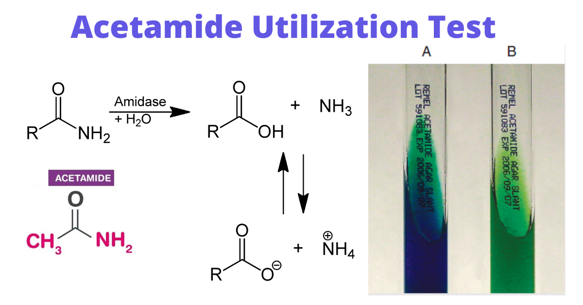 Figure: Acetamide utilization test (A: positive, B: negative)
Acetate vs acetamide: don't mix them up
These two tests look almost the same and are easy to confuse, but they answer different questions about different organisms.
| Acetate utilization | Acetamide utilization | |
|---|---|---|
| Substrate | Sodium acetate (a salt of acetic acid) | Acetamide (the amide, CH₃CONH₂) |
| What it tests | Can the organism grow using acetate as its sole carbon source? | Does the organism make acylamidase to deaminate acetamide and release ammonia? |
| Type of test | A growth/utilization test (same family as Simmons citrate) | An enzyme (deamination) test |
| Source of the ammonia that raises pH | Ammonium salts in the medium, used as nitrogen | The acetamide substrate itself, deaminated by the enzyme |
| Indicator | Bromothymol blue, green → blue (positive) | Bromothymol blue, green → blue (positive) |
| Main diagnostic use | Separate Shigella (negative) from E. coli (positive) | Help identify Pseudomonas aeruginosa among non-fermenters |
Memory hook: acet-AMIDE has an amide to chop (enzyme, Pseudomonas); acet-ATE is just a plate to grow on (carbon source, enteric split).
This article covers the acetamide test. For the growth test used in differentiating E.coli from Shigella, see the Acetate Utilization Test.
Principle
Acetamide agar (or broth) contains acetamide (CH₃CONH₂) as the sole carbon source and, in the standard formulation, ammonium salts and phosphates. Organisms that can use acetamide produce the enzyme acylamidase (an amidase), which hydrolyzes acetamide, cleaving off the amide group and releasing ammonia. The ammonia raises the pH, turning the bromothymol blue indicator from green to a deep royal blue.
The key mechanistic point that separates this from the acetate test: here the ammonia comes from the substrate itself being deaminated by the enzyme, not merely from ammonium salts being used as nitrogen. Growth together with the shift to blue indicates a positive test.
Materials and Methods
- Sterile inoculating loops or sticks
- Incubator
- Acetamide Agar: The composition of acetamide agar is tabulated below.
| Ingredients | Amount (gram/liter) |
|---|---|
| Sodium chloride | 5.0 g |
| Magnesium sulfate | 0.2 g |
| Ammonium phosphate (NH 4 H2PO 4 ), monobasic | 1.0 g |
| Potassium phosphate (K 2 HPO 4 ), dibasic | 1.0 g |
| Acetamide | 10.0 g |
| Agar | 15.0 g |
| Bromothymol blue | 0.08 g |
| Final pH: 6.8 |
Note: Acetamide broth can be used for acetamide utilization test.
Test organisms
- Isolated colonies of fluorescent pigment-producing, non-glucose-fermenting, gram-negative rods that are suggestive of Pseudomonas aeruginosa.
- Unusual non-glucose-fermenting, gram-negative rods, as part of the identification.
Quality Control
Perform quality control (QC) on each new lot or media shipment before using them. Inspect agar for evidence of prior freezing, contamination, cracks, dehydration, and bubbles before using or storing them. Discard any blue tubes.
- Acetamide Positive: Pseudomonas aeruginosa (ATCC 27853), growth; blue color
- Acetamide Negative: Escherichia coli(ATCC 25922), no growth; green color
Method
Expected Results
- Positive: Growth is seen in the slant, and the color of the slant changes from green to intense blue
- Negative: No growth, no color change, and the slant remain green.
Delftia (Comamonas) acidovorans, P. aeruginosa, and Alcaligenes faecalis can perform acetamide deamination. A combination of fluorescent pigment, acetamide deamination, and positive oxidase test results is sufficient to identify Pseudomonas aeruginosa.
Limitations
- Growth without a color change may indicate a positive test result. Repeat the test with less inoculum if further incubation results in no color change.
- A negative test does not rule out the identification of P. aeruginosa, and 6% of Pseudomonas fluorescens organisms have been reported to give a false positive reaction. Other fluorescent Pseudomonas spp. do not give positive reactions.
How to remember
Acet-AMIDE has an amide to chop. The enzyme acylamidase splits the acetamide molecule and frees ammonia. The ammonia comes from the substrate itself, which is what makes this an enzyme test rather than just a growth test.
Pigment + oxidase + acetamide = Pseudomonas aeruginosa. No single one is enough (other non-fermenters like Delftia and Alcaligenes can deaminate acetamide too), but the three together seal the identification.
Key exam facts
| Question | Answer | The reason behind it |
|---|---|---|
| What does the test detect? | Acylamidase (amidase) activity | Deaminates acetamide → ammonia |
| Substrate | Acetamide, CH₃CONH₂ | The amide is the sole carbon source |
| Enzyme | Acylamidase | Cleaves the amide, releasing ammonia |
| Source of ammonia | The acetamide substrate itself | Distinguishes it from acetate/citrate tests |
| Indicator | Bromothymol blue | Green → royal blue (positive) |
| Positive | Growth + green-to-blue | Ammonia raises pH |
| Negative | No growth, stays green | No acylamidase |
| Main use | Help identify Pseudomonas aeruginosa | Among non-glucose-fermenting GNRs |
| Confirmatory combo | Fluorescent pigment + oxidase + acetamide | No single test is definitive |
| Not unique to Pseudomonas | Delftia acidovorans, Alcaligenes faecalis also deaminate acetamide | Why the combo is needed |
| Don't confuse with | Acetate utilization | Growth test, enteric split, different organism |
Where students get confused
Confusing it with the acetate test. The central trap of this pair. Acetamide is an enzyme (acylamidase) test where the ammonia comes from deaminating the substrate; acetate is a carbon-source growth test. Different organisms (Pseudomonas vs the E. coli/Shigella split). See the boundary box above.
Thinking a positive acetamide test alone identifies Pseudomonas. It does not. Delftia acidovorans and Alcaligenes faecalis also deaminate acetamide. Identification of P. aeruginosa rests on the combination of fluorescent pigment, positive oxidase, and acetamide deamination.
Misreading carryover growth as positive. As with acetate, a heavy inoculum can carry over nutrients and produce apparent growth. Use a light inoculum and repeat with less if the color does not develop.
Treating the two "amide/ate" reactions as reading yellow-vs-blue. The meaningful result is green-to-blue for positive and green for negative. Do not over-interpret a transient yellow.
References and further readings
- Tille PM. Bailey and Scott's Diagnostic Microbiology. 15th ed. St. Louis: Elsevier; 2022.
- Leber AL, editor. Clinical Microbiology Procedures Handbook. 4th ed. Washington, DC: ASM Press; 2016. doi:10.1128/9781683670438
- Procop GW, Church DL, Hall GS, et al. Koneman's Color Atlas and Textbook of Diagnostic Microbiology. 7th ed. Philadelphia: Wolters Kluwer; 2017.

Tankeshwar Acharya, MSc (Medical Microbiology)
Tankeshwar Acharya is an Assistant Professor in the Department of Microbiology at Patan Academy of Health Sciences (PAHS), Nepal, where he has been teaching and practicing clinical microbiology for over 14 years. He is the founder of Microbe Online, one of the leading free microbiology education resources on the web, covering bacteriology, mycology, parasitology, immunology, and clinical laboratory diagnostics written from direct experience in both the classroom and the diagnostic laboratory.
Comments
No comments yet. Be the first to share your thoughts.
Leave a comment
All comments are reviewed before they appear.