
Visual Analogies in Microbiology: Food, Objects, and Everyday Things That Aid Identification

On this page
One of the best-kept secrets of experienced microbiologists is that they do not always think in Latin binomials when they look down a microscope. They think in pictures. They think: that looks like a drumstick. That looks like spaghetti and meatballs. Those are coffee beans kissing.
These food and object analogies were not invented by students desperate to pass exams. They were coined by the founding generation of clinical microbiologists, people who spent their careers at the microscope and reached for the most vivid, unforgettable description they could find. The fact that many of these descriptions involve food says something about microbiologists.

This article collects the most clinically important visual analogies in microbiology, organized by organism group, with the identification context that makes each one useful rather than merely amusing.
Three companion articles cover other sensory identification clues: characteristic bacterial smells, pigment production, and bacterial shapes and arrangements.
Bacteria
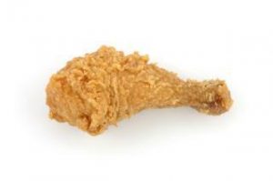
Drumstick Shaped Clostridium tetani
Clostridium tetani produces a spherical, terminal spore, a spore at the very end of the bacillus, wider than the cell body itself. Under the microscope this produces an unmistakable drumstick or lollipop shape: a thin rod with a round bulge at one end.
The appearance is pathognomonic enough that seeing drumstick-shaped gram-positive rods in a wound specimen should immediately raise suspicion for tetanus. Note that C. tetani is an obligate anaerobe and fastidious (it may not grow easily on routine culture) making the morphological clue on direct smear particularly valuable.
 Contrast: Clostridium perfringens also forms spores but they are rarely seen in clinical specimens (spores are suppressed in rich culture media). C. perfringens is therefore identified by its other characteristics (double zone of haemolysis on blood agar, lecithinase on egg yolk agar, stormy fermentation in litmus milk) rather than spore morphology.
Memory hook: Drumstick = C. tetani = tetanus = the disease that causes muscle rigidity and spasms. The drumstick spore is as rigid and terminal as the disease outcome if untreated.
Coffee Bean (Kidney Bean) shaped Neisseria Species
Neisseria gonorrhoeae and Neisseria meningitidis appear as gram-negative diplococci with flattened adjacent sides, giving each pair the appearance of two coffee beans (or kidney beans) facing each other, with a central flattening where they meet.
This appearance is among the most diagnostically important in all of microbiology:
- Gram-negative intracellular diplococci in a urethral or cervical smear = presumptive N. gonorrhoeae, sufficient to begin treatment immediately in the right clinical context
- Gram-negative diplococci in CSF (intracellular within neutrophils) = presumptive N. meningitidis, a medical emergency requiring immediate antibiotic therapy before culture confirmation.

Memory hook: Two coffee beans facing each other = Neisseria; the "kissing" diplococci. The gonococci "kiss" like two coffee beans; the meningococci can be remembered the same way.
Staphylococcus aureus: Grape-Like Clusters
Staphylococcus aureus appears on Gram stain as gram-positive cocci arranged in irregular grape-like clusters — a pattern arising from cell division occurring in multiple random planes, so daughter cells remain attached in three dimensions rather than forming chains or pairs.
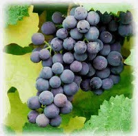 The name Staphylococcus encodes this directly: from the Greek staphyle (bunch of grapes) + kokkos (berry). The founders named it exactly what it looks like.
Diagnostic significance: Gram-positive cocci in grape-like clusters from a clinical specimen (blood culture, wound swab, sputum, pus) is strong presumptive evidence for Staphylococcus. Coagulase testing then distinguishes S. aureus (coagulase-positive) from coagulase-negative staphylococci (CoNS) such as S. epidermidis and S. saprophyticus.
Memory hook: Staphyle = grapes in Greek. The genus name is the memory hook. If you know what the word means, you never forget the morphology.
Safety Pin shaped Yersinia pestis (Bipolar Staining)
Yersinia pestis, the causative agent of plague, exhibits bipolar staining when stained with Wayson's stain or methylene blue, the organism stains more intensely at both poles than in the centre, giving a safety pin or closed safety pin appearance. This is also described as a "closed safety pin" or "bipolar ovoid."
Bipolar staining occurs because the poles of the cell accumulate more stain due to the distribution of intracellular granules (metachromatic granules or volutin granules).
Other organisms showing bipolar staining: Burkholderia pseudomallei (melioidosis), Pasteurella multocida, Francisella tularensis (less prominent).
Clinical context: Y. pestis is a Tier 1 select agent. Any laboratory receiving a specimen from a patient with suspected plague (fever, painful lymphadenopathy, bubo, in a person from an endemic area) must follow strict biosafety protocols. The safety pin appearance on Wayson's stain from lymph node aspirate is a classic exam finding.
Sulfur Granules of Actinomyces israelii
Actinomyces israelii causes actinomycosis, which is a slowly progressive suppurative infection typically affecting the cervicofacial region, thorax, or abdomen. The organism forms distinctive sulfur granules in tissue and pus: yellowish, gritty granules (0.1–5 mm) visible to the naked eye and on microscopy appearing as a central mass of gram-positive filaments surrounded by radiating club-shaped projections (the so-called "ray fungus" appearance).
The name refers to their color, a sulfur yellow, rather than to any chemical composition. They contain no sulfur.
Diagnostic significance: Finding sulfur granules in pus or tissue is essentially diagnostic of actinomycosis. They should be gently crushed on a slide and Gram stained revealing the gram-positive branching filaments.
Memory hook: Sulphur granules + draining sinus tract + jaw/cervical swelling = actinomycosis. Actinomyces is a normal oral commensal, and infection typically follows dental procedures, trauma, or tooth extraction.
Chinese Letters / Cuneiform Arrangement of Corynebacterium diphtheriae
Corynebacterium diphtheriae appears as club-shaped gram-positive rods arranged in characteristic patterns, described variously as Chinese letters, cuneiform (wedge-shaped) script, or a "picket fence" arrangement. This occurs because the bacteria snap apart at angles after cell division (snapping division), creating V, L, and Y arrangements rather than chains.
On Albert's stain or toluidine blue, C. diphtheriae reveals metachromatic granules (Babes-Ernst granules / volutin granules), polar granules that stain blue-black against a blue-green cell body, giving a "Chinese dumbbell" or "beads in a row" appearance.
Diagnostic significance: The combination of club-shaped rods in Chinese letter arrangement + metachromatic granules on Albert's stain from a throat swab = presumptive C. diphtheriae. Culture on Löffler's serum medium and tellurite media confirms identification; toxigenicity testing (Elek test) confirms virulence.
Boxcar / Rectangular Rods shaped Bacillus anthracis
Bacillus anthracis is a large, gram-positive bacillus with squarely-cut ends, described as boxcar-shaped or rectangular. In clinical specimens and culture, the organisms form long chains of these rectangular rods, like a train of boxcars.
In clinical smears from cutaneous anthrax lesions or from blood in systemic anthrax, the appearance is striking; large, square-ended rods in long parallel chains, often without a clear Gram-positive background due to the density of organisms.
Memory hook: Boxcar = large, rectangular = B. anthracis — the largest of the common bacterial pathogens. Spore-forming, non-motile (unlike other Bacillus species), non-haemolytic on blood agar (in contrast to B. cereus).
Seagull / Comma Shaped Campylobacter and Vibrio
Campylobacter jejuni appears as curved, S-shaped, or seagull-shaped gram-negative rods under microscopy. The spiral or gull-wing appearance on Gram stain of stool (or direct wet preparation under dark-field/phase contrast microscopy showing corkscrew motility) is characteristic.
Vibrio cholerae is a comma-shaped gram-negative rod. Under dark-field microscopy, the organism shows characteristic "shooting star" motility.
Rice-water stool (the term for the massive, colorless, watery diarrhea of cholera), is one of the most evocative clinical descriptions in infectious disease. The stool has the appearance of water in which rice has been washed, with white flecks of mucus. The volume can reach 20 liters per day in severe cases. This clinical finding, not any microscopy result, is what makes the immediate clinical diagnosis.
School of Fish / Parallel Rods: Haemophilus ducreyi
Haemophilus ducreyi, the causative agent of chancroid, appears on Gram stain from ulcer specimens as gram-negative coccobacilli arranged in parallel chains, classically described as a "school of fish" or "railroad tracks" pattern. Organisms tend to align parallel to each other in groups.
Clinical context: Chancroid presents as a painful genital ulcer (unlike the painless ulcer of primary syphilis) with tender inguinal lymphadenopathy. It is an important cause of genital ulcer disease in Africa, Asia, and Latin America.
Tumbling Motility of Listeria monocytogenes
At room temperature (25°C), Listeria monocytogenes demonstrates characteristic tumbling motility, an end-over-end or tumbling motion visible on wet preparation. At 37°C motility is reduced. This is due to peritrichous flagella that are expressed more at lower temperatures.
Practical significance: Motility at room temperature but not 37°C is a useful differentiating feature distinguishing Listeria from Corynebacterium (non-motile) and other gram-positive rods.
Fungi
Spaghetti and Meatballs: Malassezia furfur
Malassezia furfur, the causative agent of tinea versicolor (pityriasis versicolor), produces a classic spaghetti and meatball appearance on KOH preparation of skin scrapings: short, curved hyphal fragments (the spaghetti) mixed with clusters of round yeast cells (the meatballs).
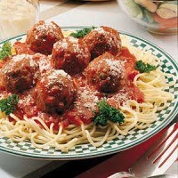 This appearance on KOH mount is essentially diagnostic; culture is rarely needed for tinea versicolor. The organism requires lipid-supplemented media (Sabouraud agar with olive oil overlay) to grow in culture because it is lipophilic and cannot synthesize its own fatty acids.
Clinical note: Tinea versicolor causes hypopigmented or hyperpigmented patches on the trunk and upper arms which are more visible in darker skin. It is not a true "infection" in the pathological sense; Malassezia is a normal skin commensal that overgrows under certain conditions (warm, humid climate, oily skin, immunosuppression).
Bunch of Bananas: Epidermophyton floccosum
Epidermophyton floccosum, a dermatophyte causing tinea pedis (athlete's foot) and tinea cruris (jock itch), produces large, smooth-walled, club-shaped macroconidia in clusters, described as resembling a bunch of bananas, and sometimes as beaver-tail or bread-loaf shaped. It is the only dermatophyte that produces no microconidia.
Watering Can: Aspergillus Conidiophore
The conidiophore of Aspergillus fumigatus has a distinctive architecture: a swollen vesicle at the top covered with flask-shaped phialides (sterigmata) only on the upper two-thirds, giving a flask or club shape when stained. The overall conidiophore is sometimes described as resembling a watering can (aspergillum, the Latin root of the genus name refers to a holy water sprinkler).
A. fumigatus conidia are blue-grey/green, giving colonies on Sabouraud agar their characteristic color. The conidial pigment (melanin-like) contributes to immune evasion, as discussed in the pigments of microorganisms article.
Steering Wheel / Ship's Wheel: Paracoccidioides brasiliensis
Paracoccidioides brasiliensis, the causative agent of paracoccidioidomycosis (the most common systemic mycosis in Latin America), produces a "steering wheel" or "ship's wheel" appearance in tissue specimens and culture: a large central yeast cell surrounded by multiple smaller budding daughter cells attached around the periphery. Also described as a "Mickey Mouse" or "mariner's wheel."
This multiply-budding yeast appearance is pathognomonic and distinguishes Paracoccidioides from Blastomyces dermatitidis, which produces broad-based single buds.
Germ tube: Candida albicans
Candida albicans produces a germ tube when incubated in serum at 37°C for 2–3 hours. Germ tube is a short hyphal projection emerging from the yeast cell without a constriction at the base. The germ tube test is a rapid, inexpensive presumptive identification of C. albicans (and C. dubliniensis). No other clinically important Candida species routinely produce true germ tubes under these conditions.
On cornmeal agar with Tween 80, C. albicans also produces thick-walled chlamydospores at the end of pseudohyphae.
Parasites
Banana-Shaped Gametocyte: Plasmodium falciparum
The gametocytes of P. falciparum are crescent- or banana-shaped which is a morphology unique among the Plasmodium species that infect humans. The gametocytes of P. vivax, P. malariae, P. ovale, and P. knowlesi are all round or oval.

This single feature on a blood film is diagnostically critical:
- Crescent/banana gametocyte = P. falciparum: the species causing the most severe and potentially fatal malaria
- Round gametocyte = one of the other species (requires further morphological assessment)
Additional P. falciparum morphological features: Only ring forms and gametocytes are seen in peripheral blood (mature trophozoites and schizonts are sequestered in deep capillaries). Multiple infections per red cell are common. Appliqué (accolé) forms which are the ring forms pressed against the RBC membrane, are characteristic.
Pear-Shaped Trophozoite: Giardia lamblia
The trophozoite of Giardia lamblia (also G. intestinalis or G. duodenalis) is pear-shaped. It is broad anteriorly and tapered posteriorly with two nuclei giving an "owl face" appearance, a concave ventral suction disc, and four pairs of flagella. On fixed preparations the paired nuclei can look like spectacles.
Identification: On wet mount of stool or duodenal aspirate, the trophozoite shows a characteristic tumbling or "falling leaf" motility due to its flagella. Cysts are oval with four nuclei.
Clinical context: Giardia is the most common intestinal protozoan infection globally. Foul-smelling, greasy, non-bloody diarrhea + bloating + flatulence in a traveler or child from an endemic area = Giardia until proven otherwise.
Maltese Cross (Tetrad): Babesia Species
Babesia parasites infecting red blood cells occasionally form a tetrad of four merozoites, the "Maltese cross" or "cross of Lorraine" appearance on blood film. This tetrad formation is pathognomonic of Babesia and distinguishes it from P. falciparum ring forms, which can look similar but never form tetrads.
Clinical context: Babesiosis is transmitted by Ixodes ticks (the same vector as Lyme disease), primarily in the northeastern USA. Severe disease occurs in asplenic patients; the spleen normally clears parasitized RBCs, so splenectomy removes this defense.
Barrel-Shaped Egg with Polar Plugs: Trichuris trichiura
The egg of Trichuris trichiura (whipworm) is barrel- or lemon-shaped with a distinctive plug at each pole. The plugs are pale and refractile, sitting like stoppers at both ends of the barrel.

This morphology is unmistakable on direct stool microscopy and makes Trichuris one of the easiest helminths to identify.
Cartwheel: Taenia Scolex
The scolex (head) of Taenia solium has four suckers plus a rostellum with two rows of hooklets which gives it a cartwheel or flower-like appearance. Taenia saginata has four suckers but no rostellum and no hooks. It is unarmed, as opposed to the armed T. solium. This distinction matters clinically: T. solium causes cysticercosis (larval migration to CNS), while T. saginata does not.
Viruses
Owl Eye Inclusion: Cytomegalovirus (CMV)
CMV-infected cells in tissue sections show a pathognomonic large intranuclear inclusion surrounded by a clear halo which is the classic "owl eye" appearance. The inclusion is the viral factory within the nucleus; the halo represents displacement of nuclear chromatin to the periphery.
Diagnostic context: Owl eye inclusions are seen in lung biopsy (CMV pneumonitis), colonic biopsy (CMV colitis), and retinal biopsy/ophthalmoscopy (CMV retinitis) in immunocompromised patients. They can be identified on H&E stain and confirmed by immunohistochemistry or in situ hybridization.
Cowdry Type A Inclusions: Herpes Simplex Virus / Varicella-Zoster
Cells infected with HSV or VZV show Cowdry Type A inclusions which are eosinophilic intranuclear inclusions with a surrounding clear halo, similar to CMV but typically in multinucleated giant cells. The multinucleated giant cell (Tzanck cell) with intranuclear inclusions is the basis of the Tzanck smear. The smear is a rapid, inexpensive test for HSV/VZV infection obtained by scraping the base of a vesicle.
Visual Analogies at a Glance
| Analogy | Organism | Type | Identification Context |
|---|---|---|---|
| Drumstick | Clostridium tetani | Bacteria | Terminal spore on Gram stain |
| Coffee/kidney bean | Neisseria spp. | Bacteria | Gram-negative diplococci on smear |
| Safety pin | Yersinia pestis | Bacteria | Bipolar staining on Wayson's stain |
| Sulphur granules | Actinomyces israelii | Bacteria | In pus/tissue from draining sinus |
| Chinese letters | Corynebacterium diphtheriae | Bacteria | Gram stain + Albert's stain |
| Boxcar/rectangular | Bacillus anthracis | Bacteria | Large square-ended rods in chains |
| Seagull/S-shape | Campylobacter jejuni | Bacteria | Gram stain or dark-field microscopy |
| Comma shape | Vibrio cholerae | Bacteria | Gram stain; "shooting star" motility |
| Rice-water stool | Vibrio cholerae | Clinical | Voluminous pale watery diarrhoea |
| School of fish | Haemophilus ducreyi | Bacteria | Gram stain from chancroid ulcer |
| Tumbling motility | Listeria monocytogenes | Bacteria | Wet preparation at room temperature |
| Spaghetti and meatballs | Malassezia furfur | Fungi | KOH mount of skin scraping |
| Bunch of bananas like macroconidia | Epidermophyton floccosum | Fungi | LPCB mount from culture |
| Steering/ship's wheel | Paracoccidioides brasiliensis | Fungi | Tissue/culture; multiply-budding yeast |
| Germ tube | Candida albicans | Fungi | Serum at 37°C, 2–3 hours |
| Banana/crescent gametocyte | Plasmodium falciparum | Parasite | Peripheral blood film |
| Pear shape + owl face nuclei | Giardia lamblia | Parasite | Stool wet mount |
| Barrel + polar plugs | Trichuris trichiura | Parasite | Stool microscopy |
| Maltese cross/tetrad | Babesia spp. | Parasite | Peripheral blood film |
| Owl eye inclusion | CMV | Virus | Tissue biopsy (H&E stain) |
| Cowdry A inclusion + giant cells | HSV/VZV | Virus | Tzanck smear, tissue biopsy |
How to Remember
Food analogies (the most memorable):
- Spaghetti and meatballs = Malassezia KOH
- Coffee beans = Neisseria
- Drumstick = C. tetani
- Banana = P. falciparum gametocyte
- Barrel with plugs = Trichuris
- Rice-water = cholera stool
Object analogies:
- Safety pin = Y. pestis (bipolar staining)
- Sulphur granules = Actinomyces
- Steering wheel = Paracoccidioides
- Maltese cross = Babesia
- Owl eye = CMV
Movement analogies:
- Tumbling = Listeria at room temperature
- Shooting star/darting motility = Vibrio cholerae
- Falling leaf = Giardia trophozoite
References
- Procop GW, Koneman EW. Koneman's Color Atlas and Textbook of Diagnostic Microbiology. 7th ed. Wolters Kluwer; 2017.
- Garcia LS. Diagnostic Medical Parasitology. 6th ed. Washington, DC: ASM Press; 2016.
- Larone DH. Larone's Medically Important Fungi: A Guide to Identification. 6th ed. ASM Press; 2018.
- Murray PR, Rosenthal KS, Pfaller MA. Medical Microbiology. 9th ed. Elsevier; 2020.
- Ryan KJ, Ray CG, editors. Sherris Medical Microbiology. 7th ed. McGraw-Hill; 2018.

Tankeshwar Acharya, MSc (Medical Microbiology)
Tankeshwar Acharya is an Assistant Professor in the Department of Microbiology at Patan Academy of Health Sciences (PAHS), Nepal, where he has been teaching and practicing clinical microbiology for over 14 years. He is the founder of Microbe Online, one of the leading free microbiology education resources on the web, covering bacteriology, mycology, parasitology, immunology, and clinical laboratory diagnostics written from direct experience in both the classroom and the diagnostic laboratory.
Comments
No comments yet. Be the first to share your thoughts.
Leave a comment
All comments are reviewed before they appear.