

When DNA is copied or read, the double helix has to be pulled apart. But DNA is a long, twisted molecule, and unwinding one part forces the rest to coil up more tightly, the same way pulling apart a twisted phone cord makes the tangle worse further along. If nothing fixed this, the mounting strain would stop replication and transcription in their tracks.
The enzymes that solve this problem are the topoisomerases. They cut the DNA, let it untwist or pass through the break, and then reseal it, releasing the strain. This makes them essential for life. It also makes them one of the most important drug targets in medicine: some antibiotics kill bacteria by jamming their topoisomerases, and several anticancer drugs kill tumor cells the same way. This article explains what topoisomerases do, the two main types and how they differ, and why they matter as drug targets.
Topoisomerases are a class of enzymes that play a crucial role in DNA metabolism. Their primary function is to regulate the topological state of DNA by introducing temporary breaks in the DNA strands.
This activity allows them to relieve the torsional strain that builds up during processes such as DNA replication, transcription, recombination, and chromosome condensation.
In the early 1970s, James C. Wang and collaborators made a significant breakthrough when they isolated an enzyme from Escherichia coli (E. coli) that was capable of introducing negative supercoiling into DNA molecules. They named this enzyme DNA gyrase and demonstrated its role in modulating DNA topology.
Topoisomerases are essential for maintaining the structural integrity of DNA and ensuring proper cellular function. Inhibition of topoisomerase activity has been exploited as a strategy for cancer treatment, as many anticancer drugs, such as etoposide and doxorubicin, target these enzymes to interfere with DNA replication and cell division in rapidly dividing cancer cells.
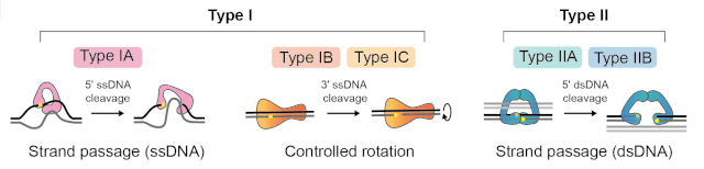 Figure: Illustration showing the Catalytic mechanisms of Topoisomerases
What do topoisomerases do?
To understand topoisomerases, first understand the problem they solve: supercoiling.
DNA is a double helix, wound like a twisted rope. When an enzyme such as DNA polymerase or RNA polymerase unwinds one region to read it, the twisting does not simply disappear. It is pushed ahead, forcing the DNA in front to over-wind and coil upon itself. This over-winding is called positive supercoiling. If it is not relieved, the DNA becomes too tight to open, and replication or transcription stalls.
Topoisomerases relieve this strain. They do it by cutting the DNA backbone, letting the strain release, and then resealing the cut. The cut is always temporary and precise: the enzyme holds onto the broken ends the whole time, so no genetic information is lost.
There are two main types, and the difference between them is simple to state:
- Type I topoisomerases cut one strand of the double helix.
- Type II topoisomerases cut both strands, and they use energy from ATP to do their work.
That single difference, one strand versus two, drives most of the differences between them, including which drugs target which type.
Types of Topoisomerase
Topoisomerases are enzymes classified into two main types based on their mechanisms of action and the number of strands they cleave during their catalytic cycle. Topoisomerases are therefore classified into four subclasses: IA and IB (both type I, cutting one strand) and IIA and IIB (both type II, cutting both strands).
Type I Topoisomerases
Type I topoisomerases are a class of enzymes involved in regulating DNA topology by introducing transient single-strand breaks in the DNA double helix. These enzymes are essential in DNA replication, transcription, recombination, and chromatin remodeling. Type I topoisomerases are further subdivided into two subclasses: Type IA and Type IB.
- Type IA Topoisomerases:
- Type IA topoisomerases are characterized by their ability to change DNA topology by introducing transient single-strand breaks in one DNA strand.
- These enzymes pass another segment of DNA through the break before resealing it, thereby relaxing the DNA supercoiling.
- Type IA topoisomerases typically act on negatively supercoiled DNA and are involved in DNA replication and transcription processes.
- Examples of Type IA topoisomerases include bacterial DNA topoisomerase I and archaeal topoisomerase III.
- Type IB Topoisomerases:
- Type IB topoisomerases also act on negatively supercoiled DNA but are structurally and mechanistically distinct from Type IA enzymes.
- These enzymes cleave one strand of the DNA double helix, allowing the other strand to rotate freely around the intact phosphodiester bond.
- Type IB topoisomerases are involved in DNA replication, transcription, and repair processes.
- Human topoisomerase I is a well-known example of a Type IB topoisomerase, and it plays essential roles in DNA metabolism and as a target for anticancer drugs such as camptothecin derivatives.
Type II Topoisomerases
These enzymes regulate DNA topology through the introduction of transient double-strand breaks in the DNA double helix. During cell division, these enzymes are vital for DNA replication, transcription, recombination, and chromosome segregation. Type II topoisomerases are subdivided into two subclasses: Type IIA and Type IIB.
- Type IIA Topoisomerases:
- Type IIA topoisomerases cleave both strands of the DNA double helix simultaneously, generating a double-strand break.
- They pass another segment of DNA through the break before resealing it, thereby altering DNA topology.
- Type IIA topoisomerases are involved in DNA replication, transcription, recombination, and chromosome segregation processes.
- Examples of Type IIA topoisomerases include bacterial DNA gyrase and eukaryotic topoisomerase II (also known as DNA topoisomerase II).
- DNA gyrase, found in bacteria, is essential for supercoiling bacterial DNA and is a target for antibacterial drugs such as fluoroquinolones.
- Bacteria have a second type II enzyme, topoisomerase IV, which separates the two linked daughter chromosomes after replication. Both gyrase and topoisomerase IV are targets of the fluoroquinolone antibiotics.
- Type IIB Topoisomerases:
- Type IIB topoisomerases are less common and have unique structural and mechanical features than Type IIA enzymes.
- They also cleave both strands of the DNA double helix but differ in their mode of action and regulation.
- The main example of a Type IIB topoisomerase is topoisomerase VI, found in archaea and in plants. Humans do not have a type IIB topoisomerase.
- Type IIB topoisomerases are less well-characterized than Type IIA enzymes, and their exact biological functions still need to be studied.
Topoisomerase I versus Topoisomerase II
| Feature | Type I | Type II |
|---|---|---|
| Strands cut | One strand | Both strands |
| ATP needed? | No (type IA needs no ATP; works by strand passage or swivel) | Yes, requires ATP |
| Change in linking number | By 1 at a time | By 2 at a time |
| Main job | Relaxes supercoils | Relaxes supercoils, and untangles/separates whole DNA molecules |
| Can separate interlinked DNA circles (catenanes)? | No | Yes |
| Key bacterial example | Topoisomerase I | DNA gyrase and topoisomerase IV |
| Key human example | Topoisomerase I | Topoisomerase II |
| Important drug target | Human topo I: camptothecin, irinotecan (anticancer) | Bacterial gyrase/topo IV: fluoroquinolones (antibacterial). Human topo II: etoposide, doxorubicin (anticancer) |
The most important practical difference: because type II cuts both strands and passes a whole DNA duplex through the gap, only type II can untangle two separate DNA molecules or separate the two linked circles produced at the end of replication. Type I can loosen twists but cannot pass one whole DNA molecule through another.
Structure of Topoisomerase
Topoisomerases are proteins with complex structures crucial for their functions in DNA metabolism. While the specific structures can vary between different types and subclasses of topoisomerases, they generally share several standard features:
- Catalytic Core: The catalytic core of topoisomerases contains the active site responsible for cleaving and rejoining DNA strands. This core typically consists of conserved amino acid residues that coordinate metal ions required for catalysis.
- DNA-binding Domains: Topoisomerases have domains or regions that specifically bind to DNA. These domains recognize and interact with the DNA substrate, facilitating the cleavage and rejoining reactions.
- Gate Domains: Gate domains are protein regions that undergo conformational changes to open and close the active site, allowing access to the DNA substrate. These conformational changes are essential for the catalytic cycle of topoisomerases.
- Linker Regions: Linker regions connect different domains of the topoisomerase protein and play a role in coordinating their movements and interactions during catalysis.
- Dimerization Interfaces: Many topoisomerases function as dimers, with two protein subunits coming together to form an active enzyme complex. Dimerization interfaces facilitate the formation of these complexes.
- Regulatory Domains: Some topoisomerases contain regulatory domains that modulate their activity in response to cellular signals or interactions with other proteins. These domains can regulate factors such as DNA binding, catalysis, and subcellular localization.
- Additional Structural Elements: Depending on the specific type and subclass of topoisomerase, additional structural elements may be present, such as ATP-binding domains in Type IIA topoisomerases or unique insertion domains in particular subclasses.
The structures of topoisomerases have been studied extensively using techniques like cryo-electron microscopy (cryo-EM), nuclear magnetic resonance (NMR) spectroscopy, and X-ray crystallography.
Function of Topoisomerase
Topoisomerases play crucial roles in various cellular processes by modulating the topological state of DNA. Some of the critical roles of topoisomerases include:
- DNA Replication: During DNA replication, the double helix must unwind to expose the template strands for DNA polymerase. This unwinding generates positive supercoiling ahead of the replication fork. Both type I and type II topoisomerases relieve this positive supercoiling. In bacteria, DNA gyrase (a type II enzyme) is especially important for removing the supercoiling ahead of the fork. Type II topoisomerases also untangle DNA knots and separate catenanes (interlinked DNA circles) that form during replication.
- Transcription: Transcription involves the synthesis of RNA from a DNA template. As RNA polymerase moves along the DNA strand, it generates positive supercoiling ahead and negative supercoiling behind. Topoisomerases help relieve this torsional stress by introducing transient breaks in the DNA strands.
- Chromosome Segregation: Chromosomes condense and segregate into daughter cells during cell division. Type II topoisomerases are particularly important for this process, as they help to resolve the interwound DNA strands (chromatids) that form during chromosome condensation. They also play a role in unlinking sister chromatids at the onset of anaphase.
- DNA Repair and Recombination: Topoisomerases participate in DNA repair and recombination processes by resolving DNA knots, tangles, and supercoils that can impede these processes. They facilitate the unwinding and recombination of DNA strands, promoting accurate repair of damaged DNA and genetic recombination.
- Regulation of Gene Expression: Topoisomerases can regulate gene expression through modulation of the accessibility of DNA to transcription factors and other regulatory proteins. By altering DNA topology, they can influence the binding of proteins to specific DNA sequences, thereby controlling gene expression levels.
- Response to Cellular Stress: Topoisomerases play a role in cellular responses to various stresses, including DNA damage, oxidative stress, and replication stress. They help maintain genomic stability by resolving DNA lesions and preventing the accumulation of DNA damage.
Overall, topoisomerases are essential enzymes that contribute to the maintenance of DNA structure and integrity and the regulation of various cellular processes critical for cell viability and function. Their dysregulation or inhibition can lead to genomic instability, cell death, and disease, making them important targets for therapeutic intervention.
Topoisomerases as drug targets
Because topoisomerases are essential and must cut DNA to work, they are excellent drug targets. A drug that traps a topoisomerase while the DNA is cut turns the enzyme into a weapon against its own cell: the DNA is left broken, and the cell dies. Two important drug groups work this way.
Fluoroquinolone antibiotics (for example ciprofloxacin and levofloxacin) target the bacterial type II enzymes, DNA gyrase and topoisomerase IV. By trapping these enzymes on cut DNA, the drug leaves the bacterial chromosome broken and the bacterium dies. Because human topoisomerases are different enough from the bacterial ones, the drug harms the bacterium far more than the host. This selectivity is what makes fluoroquinolones useful antibiotics.
Anticancer drugs target human topoisomerases in rapidly dividing tumor cells. Camptothecin and its derivatives (such as irinotecan and topotecan) trap human topoisomerase I. Etoposide and doxorubicin trap human topoisomerase II. In each case, the cancer cell is left with broken DNA and dies. Because cancer cells divide quickly, they rely heavily on topoisomerases and are especially vulnerable to these drugs.
This is why topoisomerases matter well beyond the textbook: the same enzyme that keeps DNA usable is also one of medicine's most valuable targets, in both infection and cancer. The role of gyrase in bacterial replication is covered further in the article on the enzymes involved in DNA replication.
How to Remember
Type I cuts one, Type II cuts two. The number in the name equals the number of strands cut. Type I = one strand. Type II = two strands. Everything else follows from this.
Type II needs ATP (it does more work). Cutting both strands and passing a whole DNA molecule through is heavy lifting, so type II uses ATP. Type I does the lighter job and (in the IA form) needs none. "More strands, more energy."
Only type II can untangle. Because type II passes one whole DNA duplex through another, only it can separate interlinked DNA circles and untangle knots. Type I can loosen twists but cannot pass one molecule through another. "Two strands to pass two molecules apart."
The drug-target split. Fluoroquinolones hit the bacterial type II enzymes (gyrase and topo IV), which is why they are antibiotics. Etoposide, doxorubicin, and camptothecin hit human topoisomerases, which is why they are anticancer drugs. "Quinolones for bacteria, the others for cancer."
Gyrase is the supercoiling one. Bacterial DNA gyrase is special: it actively introduces negative supercoils. Remember gyrase by its unique trick, "gyrase gyrates the DNA into negative supercoils."
Key exam facts in one table
| Fact | Detail |
|---|---|
| What topoisomerases do | Relieve supercoiling strain by cutting and resealing DNA |
| Type I | Cuts one strand; no ATP (type IA); changes linking number by 1 |
| Type II | Cuts both strands; needs ATP; changes linking number by 2 |
| Only type II can | Untangle knots and separate catenanes (linked DNA circles) |
| Bacterial type I | Topoisomerase I |
| Bacterial type II | DNA gyrase and topoisomerase IV |
| DNA gyrase special role | Introduces negative supercoils |
| Human type I drug | Camptothecin, irinotecan (anticancer) |
| Human type II drug | Etoposide, doxorubicin (anticancer) |
| Bacterial type II drug | Fluoroquinolones (antibacterial) |
| Discovered | DNA gyrase, by James Wang's group (early 1970s) |
Where Students Get Confused
Type I versus Type II: count the strands. Type I cuts one strand; type II cuts both. The Roman numeral matches the number of strands cut. This one fact anchors the whole topic.
Only type II uses ATP and only type II can untangle whole molecules. Type I relaxes supercoils by nicking one strand, needing no ATP (in the type IA form). Type II cuts both strands and passes a whole DNA duplex through, which needs ATP and is the only way to separate two interlinked DNA circles.
Gyrase is unusual: it adds negative supercoils. Most topoisomerases relax supercoiling. Bacterial DNA gyrase does the opposite as its special job: it actively introduces negative supercoils, which helps compact the bacterial chromosome and prepares DNA for unwinding.
Fluoroquinolones and anticancer topoisomerase drugs work the same way but on different enzymes. Both trap a topoisomerase on cut DNA, leaving the DNA broken. Fluoroquinolones do this to bacterial enzymes (so they are antibiotics); etoposide, doxorubicin, and camptothecin do it to human enzymes in tumor cells (so they are anticancer drugs). The selectivity comes from which enzyme is targeted.
Topoisomerase VI is not human. Topo VI (type IIB) is found in archaea and plants. Humans have type I and type II (specifically type IIA) topoisomerases, not type IIB.
References
- Champoux JJ. DNA topoisomerases: structure, function, and mechanism. Annu Rev Biochem. 2001;70:369-413. https://doi.org/10.1146/annurev.biochem.70.1.369
- Nelson DL, Cox MM. Lehninger Principles of Biochemistry. 8th ed. W.H. Freeman; 2021.
- Watson JD, Baker TA, Bell SP, Gann A, Levine M, Losick R. Molecular Biology of the Gene. 7th ed. Pearson; 2013.
- Reece RJ, Maxwell A. DNA gyrase: structure and function. Crit Rev Biochem Mol Biol. 1991;26(3-4):335-375. https://doi.org/10.3109/10409239109114072

Comments
No comments yet. Be the first to share your thoughts.
Leave a comment
All comments are reviewed before they appear.