
Starch Hydrolysis Test: Reading the Clear Halo, and Why It Flags a Blood Culture Worth Worrying About
Amylase-positive organisms clear a halo in starch agar that only appears when you flood the plate with iodine. This is a test you read by where the color fails to develop. It also helps identify Streptococcus bovis, the organism whose presence in a blood culture sends clinicians looking at the colon. Here is how to read the halo and why it matters.
The blood culture that points at the colon
A 68-year-old man is admitted with fever and a new heart murmur. Blood cultures grow a Gram-positive coccus in chains. It is catalase negative, it grows in bile esculin, it does not grow in 6.5% salt, and it is PYR negative. On a starch plate flooded with iodine, it clears a halo.
That halo helps name the organism: Streptococcus bovis, now often called Streptococcus gallolyticus.
And naming it changes what the physicians do next. Because there is one of the most reliable associations in clinical microbiology waiting behind it: S. bovis bacteremia and endocarditis are strongly linked to occult colorectal carcinoma. A significant fraction of patients with S. bovis endocarditis turn out to have a colonic tumor that has not yet declared itself. The teaching phrase is blunt and it sticks: S. bovis in the blood, look for cancer in the colon.
So this patient will get a colonoscopy, not because of his heart, but because of what grew in his blood. The starch hydrolysis test is one of the small reactions that got him there.
Most of the time the starch test is a quiet identification step for environmental Bacillus and a handful of other organisms. But it earns its place in a clinical panel partly because it helps pin down an organism whose name is a warning. This article covers how to read the test, which turns out to be less obvious than it looks, and where it matters.
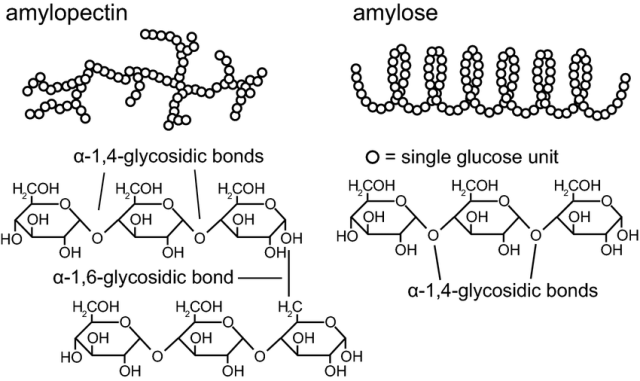
Starch hydrolysis test is used to determine if the organism is capable of breaking down starch into maltose through the activity of the extra-cellular α-amylase enzyme. Starch, the most important source of carbohydrates for humans, is a polysaccharide mixture of two polymers, amylose, and amylopectin, the latter being predominant.
Amylose is a linear polysaccharide of several thousand α-D-glucose linked by 1,4-α-glycosidic bonds. Amylopectin is a branched-chain polysaccharide composed of glucose units linked primarily by α-1,4-glycosidic bonds but with occasional α-1,6-glycosidic bonds, which are responsible for the branching.
 ## Principle
Starch molecules are too large to enter the bacterial cell, so only bacteria that secrete exoenzymes (α -amylase and oligo-1,6-glucosidase) are able to hydrolyze starch into subunits (dextrin, maltose, or glucose). These molecules are readily transported into the bacterial cell to be used in metabolism.
In starch hydrolysis test (also known as amylase test), we use starch agar, which is a differential nutritive medium. The test organisms are inoculated onto a starch plate and incubated at 30°C until growth is seen (i.e. up to 48 hours). The Petri plate is then flooded with an iodine solution.
If there is no enzyme present, and therefore no hydrolysis, the amylose, and iodine react together to form a blue color. Depending on the concentration of the iodine used, iodine turns blue, purple, or black in the presence of starch.
When bacteria capable of producing α-amylase and oligo-1,6-glucosidase are grown on starch agar, they secrete enzymes into the surrounding areas and hydrolyze the starch. As no amylose is present in the medium surrounding the bacterial colony, clearing around the bacterial growth is seen (there is no color development).
Why starch has to be digested outside the cell
Starch is a huge molecule, a polymer of thousands of glucose units. It is far too large to cross the bacterial cell membrane, so an organism cannot simply swallow starch and digest it internally. To use starch as food, the organism must break it down outside the cell first, into fragments small enough to import.
That is the job of an exoenzyme: an enzyme secreted into the surrounding medium that works on a substrate the cell cannot bring inside. For starch, the exoenzyme is α-amylase, often working alongside oligo-1,6-glucosidase to handle the branch points. Together they chop starch into dextrins, maltose, and glucose, which the cell then transports in and metabolizes.
This is why the test reports what it does. When an amylase-positive organism grows on starch agar, it does not just grow; it clears the starch out of the agar in a zone around itself, because its secreted enzyme has been digesting the medium. That cleared zone is invisible while the plate looks uniform, until you add the reagent that makes starch visible.
The organism's nutritional strategy and the test's readout are the same event seen from two sides: the bacterium is eating the plate, and the test shows you where it has eaten.
Materials
- Heart infusion agar with 2% starch or Mueller Hinton agar (MHA). MHA contains starch as a standard ingredient, so it can double as a starch-hydrolysis medium without purchasing additional plates.
- Gram’s iodine
- Sterile sticks or inoculating loops
- Incubator
Quality Control
Inspect starch agar for freezing, contamination, cracks, and dehydration prior to storage and before use. Perform QC on each new lot of starch agar prior to using them. Test the performance of the agar using the following test organisms.
- S. bovis ATCC 33317—clear halo around colony with the addition of iodine (starch positive)
- Enterococcus faecalis ATCC 29212—blue-black color right up to the colony with the addition of iodine (starch negative)
Uses
Starch hydrolysis test is used
- To differentiate members of various genera including Bacillus, Clostridium, Corynebacterium, Fusobacterium, Enterococcus, Pseudomonas, and Streptococcus. These genera have both amylase-positive and amylase-negative species.
- To separate Streptococcus bovis (S. gallolyticus), which is starch positive, from other viridans group streptococci. This separation matters clinically: S. bovis bacteremia and endocarditis are strongly associated with occult colorectal carcinoma, so identifying it prompts a search for a colonic tumor. The full presumptive profile is bile-esculin positive, 6.5% NaCl negative, PYR negative, and starch positive.
Test Procedure
- Pick a few colonies of the test organism using a sterile swab or loop.
- Streak a starch plate in the form of a line across the width of the plate. Several cultures can be tested on a single agar plate, each represented by a line or the plate may be divided into four quadrants for this purpose.
- Incubate plate at 37 °C for 48 hours.
- Add 2-3 drops of 10% iodine solution directly onto the edge of colonies. Wait 10-15 minutes and record the results. Read plates immediately after the addition of iodine, as the blue color fades.
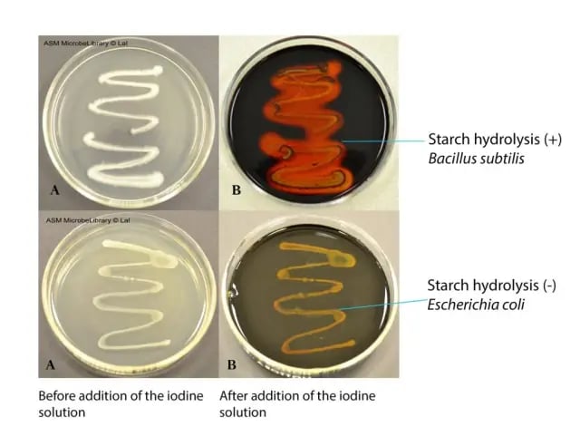 Figure: Starch hydrolysis test (Image source: ASM)
Interpretation
- Positive ("+"): the agar turns blue-black everywhere except for a clear or pale halo immediately around the growth. That halo is the zone where secreted amylase digested the starch, leaving nothing for the iodine to stain.
- Negative ("−"): the agar turns blue-black right up to the edge of the colony, with no clearing. The organism produced no amylase, so the starch is intact everywhere.
Applied to the clinical picture: a catalase-negative, PYR-negative, Gram-positive coccus that is bile-esculin positive, does not grow in 6.5% salt, and clears a starch halo is presumptively Streptococcus bovis (S. gallolyticus).
Starch hydrolysis test results of selected organisms
| Starch hydrolysis (+ve) | Starch hydrolysis (−ve) |
|---|---|
| Bacillus subtilis | Streptococcus agalactiae |
| Bacillus cereus | Staphylococcus epidermidis |
| Bacillus megaterium | Escherichia coli |
| Streptococcus bovis (S. gallolyticus) | Enterococcus faecalis |
| Clostridium perfringens | most other viridans streptococci |
Why the clear halo is the whole test
Iodine binds intact starch and turns it blue-black. That is a reaction most students already know from a school chemistry class. The trap in this test is what that means for reading it.
When you flood a starch plate with iodine after incubation, the entire plate turns blue-black, except where the starch has been destroyed. So you are not looking for a color to appear at the colony. You are looking for a color to fail to appear, a clear halo standing out against a dark background.
- Amylase positive: a clear or pale halo around the growth, surrounded by blue-black agar. The organism digested the starch there, so there is nothing left for the iodine to stain.
- Amylase negative: blue-black agar right up to the edge of the colony. The starch is intact everywhere; the organism cleared nothing.
This is negative-space reading, and it inverts the usual instinct. In most biochemical tests, a color change at the organism is the positive. Here, the positive is a color absence at the organism, framed by color everywhere else. A student expecting "positive equals colored colony" will read every plate backwards.
Two practical consequences follow:
- Read immediately. The blue-black fades as iodine evaporates and diffuses. A halo that was crisp at two minutes can be ambiguous at fifteen. Record the result as soon as the color develops.
- A red-violet zone is not a clean positive. It means partial hydrolysis: the starch has been broken to intermediate dextrins that stain red-violet rather than blue-black or clearing completely. Reincubate and repeat rather than call it.
Limitations
- Avoid using a glucose starch medium, since the metabolism of glucose may interfere with the assay.
- Once the iodine is added, the organisms are nonviable.
- A red-violet color is due to partial hydrolysis, and the test should be repeated after further incubation.
How to remember
Read the hole, not the colony.
This test is the photographic negative of most biochemical tests. Everywhere the starch survives goes blue-black; the positive is the gap in the dark, the clear halo where the organism ate the starch. Before you read any starch plate, remind yourself: I am looking for where the color is missing, not where it is. That one sentence prevents the most common misread.
S. bovis in the blood, look in the colon.
The single highest-yield fact attached to this test. A starch-positive, bile-esculin-positive, PYR-negative, salt-negative Gram-positive coccus from a blood culture is S. bovis, and S. bovis endocarditis points to occult colorectal cancer. The starch halo is one of the reactions that gets you to that name. If you remember nothing else about the clinical use of this test, remember this.
Amylase is an outside eater.
Starch is too big to import, so the organism secretes α-amylase to digest it in the agar and then absorbs the pieces. The clear halo is literally the organism's dinner plate: the area it has already eaten. Positive organisms are the ones that can feed on starch from a distance.
Key exam facts in one table
| Question | Answer | The reason behind it |
|---|---|---|
| What does the test detect? | Production of α-amylase (with oligo-1,6-glucosidase) | Starch is too large to import; it must be digested outside the cell |
| What kind of enzyme? | An exoenzyme, secreted into the medium | It works on a substrate the cell cannot bring inside |
| What does amylase produce? | Dextrins, maltose, glucose | Small enough to transport and metabolize |
| Medium | Starch agar (or MHA, which contains starch) | Starch is the differential substrate |
| Reagent | Gram's iodine | Iodine binds intact starch → blue-black |
| How is the plate read? | By the clear halo where color fails to develop | Amylase digested the starch, leaving nothing to stain |
| Positive result | Clear halo around growth, blue-black elsewhere | Starch destroyed locally |
| Negative result | Blue-black up to the colony edge | Starch intact everywhere |
| Red-violet zone? | Partial hydrolysis; reincubate and repeat | Intermediate dextrins stain red-violet |
| Why read immediately? | The blue-black fades as iodine diffuses/evaporates | A crisp halo becomes ambiguous with time |
| Incubation | 30–37°C, up to 48 hours | Amylase-positive growth and clearing take time |
| QC positive | Streptococcus bovis ATCC 33317 | Clears a halo; also the clinical anchor organism |
| QC negative | Enterococcus faecalis ATCC 29212 | Blue-black to the colony edge (note: not ATCC 25922, which is E. coli) |
| Key clinical use | Identify S. bovis / S. gallolyticus | S. bovis bacteremia/endocarditis flags occult colorectal carcinoma |
| S. bovis presumptive profile | Bile-esculin +, 6.5% NaCl −, PYR −, starch + | The four-reaction fingerprint |
| Common positives | Bacillus, Clostridium perfringens, S. bovis | Broad among environmental and some clinical organisms |
| Common negatives | E. coli, S. epidermidis, S. agalactiae, E. faecalis | No amylase |
| Why avoid glucose in the medium? | Glucose metabolism interferes with the assay | The organism uses glucose preferentially |
Where students get confused
Looking for a colored colony instead of a clear halo. The defining error, and it comes from applying the usual biochemical-test instinct. In most tests, color appearing at the organism is the positive. Here it is the reverse: the whole plate goes blue-black and the positive is the clear gap around the growth. You read the absence of color, not its presence.
Reading the plate too late. The blue-black color fades as iodine evaporates and diffuses into the agar. A halo that was obvious at two minutes can be washed out and unreadable at fifteen. Flood, read, and record immediately.
Calling a red-violet zone positive. Red-violet is not a clean positive. It signals partial hydrolysis, where starch has been broken only as far as intermediate dextrins. The correct response is to reincubate and repeat, not to record a positive.
Forgetting why the medium must be glucose-free. If glucose is present, the organism will use it preferentially and may not express or need amylase, so starch is left undigested and a true positive reads negative. Use a glucose-free starch medium.
Treating this as a purely environmental test. It is easy to file starch hydrolysis under "Bacillus identification" and forget it has a clinical edge. Its role in identifying S. bovis connects it to one of the highest-yield associations in medicine: S. bovis endocarditis and occult colorectal cancer. The environmental use is common; the clinical use is what makes it worth knowing well.
Assuming halo size grades the result. The test is qualitative. A large clear halo and a small one are both simply positive. Do not read strength into halo diameter.
References and further reading
- Archana Lal, Naowarat Cheeptham. 2012. Starch agar protocol.
- Madigan MT, Martinko JM, Stahl DA, Clark DP. 2012. Brock biology of microorganisms, 13th ed. Benjamin Cummings, San Francisco, CA.
- Clinical Microbiology Procedures Handbook, Fourth Edition. (2016). In Clinical Microbiology Procedures Handbook, Fourth Edition. American Society of Microbiology. https://doi.org/10.1128/9781555818814
Frequently Asked Questions
Do I look for a colored colony or a clear zone in the starch test?
Why does the plate turn blue-black when I add iodine?
What does a red-violet zone around the colony mean?
Why must I read the starch plate immediately after adding iodine?
Why is the starch hydrolysis test clinically important for Streptococcus bovis?
Which organisms are starch hydrolysis positive?
Why can't the starch medium contain glucose?

Tankeshwar Acharya, MSc (Medical Microbiology)
Tankeshwar Acharya is an Assistant Professor in the Department of Microbiology at Patan Academy of Health Sciences (PAHS), Nepal, where he has been teaching and practicing clinical microbiology for over 14 years. He is the founder of Microbe Online, one of the leading free microbiology education resources on the web, covering bacteriology, mycology, parasitology, immunology, and clinical laboratory diagnostics written from direct experience in both the classroom and the diagnostic laboratory.