
Simple Staining: Principle, Procedure, Results, and Uses in Microbiology
Simple staining uses a single basic dye to reveal bacterial cell morphology, size, and arrangement. Learn the methylene blue procedure, when simple staining is used over Gram staining, and how to interpret results.
Simple staining is rarely the first choice in a clinical microbiology laboratory — Gram staining provides far more information in the same time. But there are specific situations where the simplicity of a single dye is exactly what is needed: when you want to confirm bacterial morphology quickly, when you suspect a culture contaminant, or when a mixed colony plate needs rapid triage before more elaborate testing.
Understanding simple staining also builds the foundation for understanding differential staining — knowing what a single dye does to all bacteria equally makes it easier to appreciate what a differential stain does differently.
The simple stain can be used as a quick and easy way to determine the cell shape, size, and arrangement of bacteria. True to its name, the simple stain is a very simple staining procedure involving a single stain solution. Any basic dye, such as methylene blue, safranin, or crystal violet, can be used to color the bacterial cells.
These stains will readily give up a hydroxide ion or accept a hydrogen ion, which leaves the stain positively charged. Since most bacterial cells and cytoplasm surface is negatively charged, these positively charged stains adhere readily to the cell surface. After staining, bacterial cell morphology (shape and arrangement) can be appreciated.
Procedure
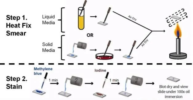 Figure: Simple Staining Procedure
Preparation of a smear
- Using a sterilized inoculating loop, transfer a loopful of liquid suspension containing bacteria to a slide (clean grease-free microscopic slide) or transfer an isolated colony from a culture plate to a slide with a water drop.
- Disperse the bacteria on the loop in the drop of water on the slide and spread the drop over an area the size of a dime. It should be a thin, even smear.
- Allow the smear to dry thoroughly.
- Heat-fix the smear cautiously by passing the underside of the slide through the burner flame two or three times. It fixes the cell in the slide. Do not overheat the slide as it will distort the bacterial cells.
Staining
- Cover the smear with methylene blue and allow the dye to remain in the smear for approximately one minute (Staining time is not critical here; somewhere between 30 seconds to 2 minutes should give you an acceptable stain, the longer you leave the dye in it, the darker will be the stain).
- Using distilled water wash bottle, gently wash off the excess methylene blue from the slide by directing a gentle stream of water over the surface of the slide.
- Wash off any stain that got on the bottom of the slide as well.
- Saturate the smear again but this time with Iodine. Iodine will set the stain
- Wash any excess iodine with gently running tap water. Rinse thoroughly. (You may not get a mention of steps 4 and 5 in some textbooks)
- Wipe the back of the slide and blot the stained surface with bibulous paper or with a paper towel.
- Place the stained smear on the microscope stage smear side up and focus the smear using the 10X objective.
- Choose an area of the smear in which the cells are well spread in a monolayer. Center the area to be studied, apply immersion oil directly to the smear, and focus the smear under oil with the 100X objective.
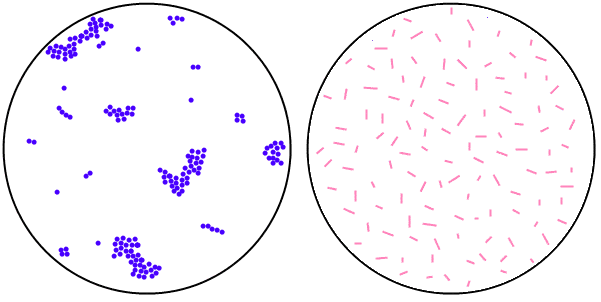 Figure: Left: Cocci in Cluster; Right: Bacilli (Image source:microrao.com)
Results
The bacterial cells usually stain uniformly and the color of the cell depends on the type of dye used. If methylene blue is used, some granules in the interior of the cells of some bacteria may appear more deeply stained than the rest of the cell, which is due to the presence of different chemical substances.
Uses
Diagnostic microbiology laboratory generally does not perform simple staining methods. Differential staining such as Gram staining and AFB staining are commonly used to identify and differentiate bacterial isolates. Simple staining can be useful in the following circumstances.
- To differentiate bacteria from yeast cells: When endocervical swab culture is done in blood agar both Staphylococcus spp and yeast cells may give similar-looking colonies in Blood agar (a common error for a new technologist or microbiologist with less experience). Performing the wet mount technique or simple staining from the isolate can be helpful.
- To presumptively identify the bacterial isolate
Due to their ubiquitous presence, Bacillus spp may present as a contaminant in the culture plates. In some circumstances (e.g. growth in Blood agar but no growth in MacConkey agar), identifying the shape of the bacteria (rod or cocci) may help to eliminate the isolate as possible contaminants (e.g., Bacillus spp) or further process as a potential pathogen (cocci). - Rapid morphology check before reporting: When a Gram stain result is ambiguous — for example, when over-decolourisation has made gram-positive organisms appear gram-negative — a simple stain can confirm the basic morphology (cocci vs rods, arrangement) independently of the Gram reaction, helping the microbiologist decide whether to repeat the Gram stain or proceed with further testing.
Simple Staining vs Differential Staining — When to Use Each
| Feature | Simple Staining | Differential Staining (Gram, ZN) |
|---|---|---|
| Number of dyes | One | Two or more |
| Information provided | Morphology, size, arrangement | Morphology + cell wall type (GP/GN) or acid-fastness |
| Time | ~5 minutes | ~10–20 minutes |
| Used in clinical labs? | Rarely — occasional triage use | Routinely on all clinical specimens |
| Best use | Quick morphology confirmation; teaching; research | Standard clinical specimen examination |
| Dyes used | Methylene blue, safranin, crystal violet, basic fuchsin | Crystal violet + safranin (Gram); carbol fuchsin + malachite green (ZN) |
How to Remember: Simple Staining
"One dye, one colour, one question answered": Simple staining answers only: what shape and arrangement are these bacteria? It cannot tell you gram-positive or gram-negative, acid-fast or non-acid-fast. When you need more than morphology, reach for a differential stain.
The methylene blue granule point: If methylene blue is used and some granules stain more deeply than the rest of the cell, this is not an artifact — it reflects the presence of different chemical substances (e.g., volutin/metachromatic granules, lipid inclusions). This observation can itself be informative and is one reason methylene blue is preferred for simple staining over crystal violet.
Key Exam Facts in One Table
| Feature | Detail |
|---|---|
| Definition | Single basic dye applied to heat-fixed smear; all cells stain same colour |
| Dyes used | Methylene blue (most common), safranin, crystal violet, basic fuchsin |
| Why basic dyes work | Cationic (positively charged) — binds negatively charged bacterial cell surface |
| Information gained | Cell shape, size, arrangement only — no differentiation between organism types |
| Heat fixation required? | Yes — kills organisms, adheres cells to slide |
| Used routinely in clinical labs? | No — Gram stain preferred; simple staining used for specific situations |
| When useful clinically | Bacteria vs yeast differentiation; contaminant triage; morphology confirmation |
| Methylene blue granules | Deeper staining of granules indicates volutin/metachromatic granules (e.g., C. diphtheriae) |
| Limitation vs Gram stain | Provides no information about cell wall type — all bacteria appear same colour |
References
- Moyes, R. B., Reynolds, J., & Breakwell, D. P. (2009). Preliminary staining of bacteria: simple stains. Current protocols in microbiology, Appendix 3, . https://doi.org/10.1002/9780471729259.mca03es15
- Tripathi, N., & Sapra, A. (2023). Gram Staining. In StatPearls. StatPearls Publishing.
- Dagnall, G. J., & Wilsmore, A. J. (1990). A simple staining method for the identification of chlamydial elementary bodies in the fetal membranes of sheep affected by ovine enzootic abortion. Veterinary microbiology, 21(3), 233–239. https://doi.org/10.1016/0378-1135(90)90034-s
Frequently Asked Questions
When would a clinical microbiology laboratory use simple staining instead of Gram staining?
What is the significance of deeply stained granules seen on methylene blue simple staining?

Tankeshwar Acharya, MSc (Medical Microbiology)
Tankeshwar Acharya is an Assistant Professor in the Department of Microbiology at Patan Academy of Health Sciences (PAHS), Nepal, where he has been teaching and practicing clinical microbiology for over 14 years. He is the founder of Microbe Online, one of the leading free microbiology education resources on the web, covering bacteriology, mycology, parasitology, immunology, and clinical laboratory diagnostics written from direct experience in both the classroom and the diagnostic laboratory.