
Real-time PCR (qPCR): Principles and Applications
Real-time PCR (qPCR) amplifies and quantifies DNA simultaneously using fluorescent probes. Learn SYBR Green vs TaqMan, Ct values, and clinical uses in viral load testing.
A 34-year-old man newly diagnosed with HIV starts antiretroviral therapy. Before treatment, his HIV viral load is 450,000 copies/mL — a number that comes directly from a real-time PCR assay run on his plasma. Three months after starting therapy, his viral load is 1,200 copies/mL. Six months in, it is below 50 copies/mL — undetectable by the assay's lower limit. His CD4 count is rising. His clinician continues the same regimen.
This monitoring — the decision to continue, switch, or intensify antiretroviral therapy — rests entirely on quantitative PCR numbers. Not on culture (HIV cannot be cultured in routine laboratories). Not on serology (antibodies remain positive regardless of treatment). Only real-time PCR can tell you how much virus is present, and whether therapy is working.
This is what distinguishes real-time PCR from conventional PCR. Conventional PCR answers a yes/no question: is the target present? Real-time PCR answers a quantitative question: how much target is present? In viral load monitoring, that number is the clinical result.
Real-time PCR also called quantitative PCR (qPCR), is a variant of standard polymerase chain reaction in which amplification and simultaneous quantitation of a target DNA is done in the same PCR machine, using commercially available fluorescence-detecting thermocyclers. Fluorescent dyes specifically label DNA of interest, and the amount of fluorescence generated is proportional to the quantity of DNA present.
Although various models of real-time PCR are available, all share common features: a standard thermocycler platform coupled with an excitation source (usually a laser or tungsten lamp), a camera for fluorescence detection, and computer and software for data processing.
Depending on the available excitation source and detection filters, a variety of fluorescent dyes may be used in qPCR. The two most commonly used real-time PCR methods are SYBR green (a dye that binds to double-stranded DNA but not to single-stranded DNA, and, when so bound, fluoresces) and TaqMan probes, respectively.
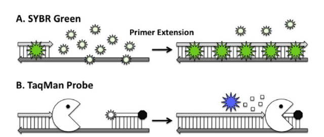 Figure: Popular Real-time detection methods
Some essential features of real-time PCR are
- Both amplification and product detection are accomplished in one reaction vessel without ever opening. This dramatically reduces the chances of cross-contamination of samples with the amplified product.
- Compared to conventional PCR assays, real-time PCR instruments are not only able to measure amplified product (amplicon) as it is made but are also able to quantitate the amount of product and thereby determine the number of copies of the target in the original specimen.
- The amount of time required to complete a real-time PCR assay is significantly less compared to conventional PCR-based assays because the time needed for the post-PCR detection of amplified product is eliminated by the use of fluorescent probes. Also, some systems are able to perform rapid thermal cycling based on instrument design, detecting product in as little as 20 to 30 minutes.
In general, a conventional PCR assay would require a minimum of at least 4 to 6 hours from the time that extracted nucleic acid is placed into a thermal cycler to begin amplification to subsequent product detection.
Principle
Real-time PCR is accomplished in the same manner as conventional PCR-based assays (denaturation of double-stranded DNA followed by primer annealing and extension). However, it is the detection process that discriminates real-time PCR from traditional PCR assays. In real-time PCR assays, the accumulation of amplicon is monitored as it is generated using the labeling of primers with dyes capable of fluorescence. These labels produce a change in fluorescent signal measured by the instrument following their direct interaction with or hybridization to the amplicon. This signal is related to the amount of amplified product present during each cycle and increases as the number of specific amplicons increases.
Currently, a range of fluorescent chemistries are used for amplicon detection; the more commonly used chemistries can be divided into two categories: (1) those that involve the nonspecific binding of a fluorescent dye (e.g., SYBER Green I) to double-stranded DNA and (2) fluorescent probes that bind specifically to the target of interest.
The Ct Value: What Real-time PCR Actually Measures
Understanding real-time PCR requires understanding the Ct value (cycle threshold) — the single number that the instrument generates and the clinician interprets.
During PCR amplification, the fluorescent signal from the reaction starts near zero and rises exponentially as more amplicon accumulates. At some point, the signal crosses a pre-set fluorescence threshold — the point where signal reliably rises above background noise. The cycle number at which this crossing occurs is the Ct value.
The Ct value has one critical property: it is inversely proportional to the amount of starting template. A sample with a high viral load reaches the threshold early — low Ct value. A sample with a low viral load takes more cycles to accumulate enough product to cross the threshold — high Ct value.
Practical interpretation:
- HIV viral load 450,000 copies/mL → Ct ~20 (reaches threshold early)
- HIV viral load 50 copies/mL → Ct ~34 (takes many more cycles)
- No target present → signal never crosses threshold → "undetectable" or "negative"
In COVID-19 PCR testing, Ct values became widely discussed during the pandemic. A Ct value below 25 generally indicates high viral load and is associated with early symptomatic infection or high infectivity. A Ct value above 35 indicates very low viral load — the clinical significance depends on context (early infection, late recovery, or background noise).
Important caveat: Ct values are not directly comparable between different assays, instruments, or laboratories. A Ct of 30 on one platform does not equal a Ct of 30 on another. Clinical interpretation always requires the reference range from the specific assay used.
Detection Methods
A number of real-time PCR methods have been described, but two have emerged as the most popular.
PCR using SYBR
SYBR green is a dye that binds to double-stranded DNA but not to single-stranded DNA, and, when so bound, fluoresces. During PCR cycle, as more and more double-stranded product is generated to which SYBR green dye attach and fluoresces, an increasing amount of fluorescent signal is generated. The amount of fluorescence in the reaction at any particular time is directly related to the number of double-stranded DNA molecules in the reaction.
However, the downside of SYBR green is that it will bind and fluoresce all double-stranded products in the reaction, whether they are specific products, nonspecific products, primer dimers, or other amplification artefacts.
TaqMan probes were named after the popular videogame Pac-Man (Taq polymerase + PacMan = TaqMan) as the authors noticed that the exonuclease activity of the Taqpolymerase had similarities to the classic Pac-Man game.
TaqMan PCR (5’ nuclease assay)
TaqMan PCR uses dye-labelled nucleic-acid probe complementary to an internal segment of the target DNA. This dye-labelled probe anneals to one of the template strands close to and downstream from one of the two PCR primers. The probe is labeled with two fluorescent moieties, the reporter (fluorophore) is attached to the probe’s 5’ end and the quencher is attached to its 3’ end.
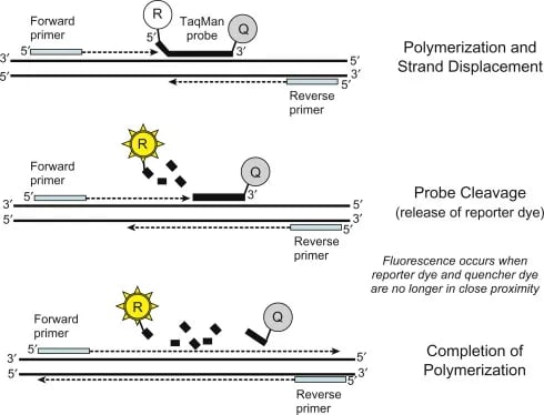 Figure: Schematic of TaqMan (5′ nuclease) assay(Image source: Ref-2)
When the reporter and the quencher are connected, the quencher reduces the fluorescent signal of the reporter dye as it absorbs the energy via a fluorescence resonance energy transfer (FRET). However, during PCR, Taq polymerase, extending the primer on the probe’s target strand, displaces and degrades the annealed probe through the action of its 5’ to 3’ exonuclease function. The fluorophore is thereby released from its molecular attachment to the quencher and fluoresces.
As more PCR products are generated, the more dye-labeled probe will find target regions to join which eventually leads to release of the reporter molecule during the subsequent amplification process. The release of reporter dyes is reflected as increased intensity of the fluorescent signal which is proportional to the amount of amplicon synthesized.
Whether using SYBR green or TaqMan probes, the relationship between signal intensity and the amount of template in a real-time PCR reaction provides a reliable means both to quantitate nucleic acids and to assay for the presence or absence of specific gene sequences.
SYBR Green vs. TaqMan: Choosing the Right Chemistry
| Feature | SYBR Green | TaqMan Probes |
|---|---|---|
| Mechanism | Dye binds all double-stranded DNA and fluoresces | Sequence-specific probe is cleaved by Taq; reporter dye released |
| Specificity | Lower — detects all dsDNA including primer dimers and non-specific products | Higher — only the specific target sequence releases signal |
| Verification needed | Yes — melting curve analysis required to confirm specific product | No — probe binding itself confirms specificity |
| Cost | Lower — no probe synthesis required | Higher — custom probe designed and synthesised for each target |
| Best for | Gene expression studies, research applications, low-budget settings | Diagnostic assays, viral load testing, clinical laboratories |
| Risk | False positives from primer dimers or non-specific amplification | Minimal — requires both primer binding AND probe hybridisation |
| Multiplex capability | Limited — all products give same signal colour | Yes — different probes labelled with different fluorophores |
The practical rule: For diagnostic clinical assays (HIV viral load, HCV quantitation, COVID-19 PCR), TaqMan is the standard — specificity is non-negotiable in patient care. For research gene expression work where cost matters and melting curve verification is feasible, SYBR Green is a reasonable choice.
Applications
Real-time PCR enables calculation of the starting template concentration and is a frequently used analytical tool in evaluating DNA copy number, viral load, SNP detection, and allelic discrimination. When preceded by reverse-transcription PCR, qPCR is a powerful tool to measure mRNA expression and is the gold standard for microarray gene expression data confirmation.
How to Remember
Ct value = cycles to threshold = inversely proportional to viral load. High viral load → reaches threshold in fewer cycles → low Ct number. Low viral load → needs more cycles → high Ct number. Think of it as a race: a sample with lots of virus gets to the finish line (threshold) faster. Fewer laps = lower Ct = more virus.
SYBR Green = simple but non-specific; TaqMan = specific but expensive. SYBR Green binds any double-stranded DNA — it cannot distinguish your target from a primer dimer. It is the budget option, suitable for research. TaqMan requires a specific probe for your target — it will only signal when the right sequence is amplified. It is the clinical standard. Simple rule: research = SYBR; diagnostics = TaqMan.
TaqMan = Pac-Man eating the probe. The article already contains this origin story — and it is exactly right. Taq polymerase's 5'→3' exonuclease activity chews up the probe as it extends, separating the reporter from the quencher and releasing fluorescence. Every PCR cycle where the target is amplified, more probes are chewed, more fluorescence released. The signal accumulates in direct proportion to amplicon.
Real-time PCR vs. conventional PCR — the key distinction: Conventional PCR: amplify first, detect after (open the tube, run a gel). Real-time PCR: amplify and detect simultaneously, in a closed tube. Closed tube = no post-PCR handling = no carry-over contamination. This is why real-time PCR replaced conventional PCR in most clinical diagnostic laboratories.
Real-time PCR vs. RT-PCR — they are not the same: Real-time PCR = detection method (fluorescence during cycling; can be quantitative). RT-PCR = template type (RNA converted to cDNA before amplification). COVID-19 PCR is technically RT-qPCR — it uses reverse transcriptase (because SARS-CoV-2 is RNA) AND real-time detection (for sensitivity and closed-tube safety). See the RT-PCR article for the distinction in full.
Advantages
Significant advantages of real-time PCR include
- its ability to measure DNA concentrations over a large range,
- its high sensitivity,
- its ability to process multiple samples simultaneously and providing immediate information.
A disadvantage is the machines are more expensive than traditional PCR machines.
Key exam facts in one table
| Topic | Key fact |
|---|---|
| Full name | Real-time PCR = quantitative PCR = qPCR |
| Key difference from conventional PCR | Detection is simultaneous with amplification; closed tube; quantitative |
| Ct value definition | Cycle threshold — the PCR cycle number at which fluorescence crosses the detection threshold |
| Ct value interpretation | Low Ct = high viral load (reaches threshold early); High Ct = low viral load |
| Ct comparability | Ct values are NOT directly comparable between different assays or platforms |
| SYBR Green mechanism | Dye binds all double-stranded DNA and fluoresces; non-specific |
| SYBR Green limitation | Cannot distinguish specific product from primer dimers; requires melting curve verification |
| TaqMan mechanism | Sequence-specific probe with reporter (5' end) and quencher (3' end); Taq exonuclease cleaves probe during extension; reporter dye released and fluoresces |
| TaqMan advantage | Sequence-specific; no post-PCR verification needed; suitable for multiplex |
| TaqMan name origin | Taq polymerase + Pac-Man = TaqMan (exonuclease activity resembles Pac-Man eating) |
| Clinical applications | HIV viral load, HCV viral load, HBV viral load, COVID-19 (SARS-CoV-2), CMV monitoring, TB quantification |
| Advantage over conventional PCR | Quantitative; closed tube (no contamination risk); faster (no gel electrophoresis step) |
| RT-qPCR | Combination of RT-PCR (RNA template) and real-time detection; used for RNA virus quantification |
| Instrument components | Thermocycler + excitation source (laser/lamp) + fluorescence detector + software |
References and further reading
- Arya, M., Shergill, I. S., Williamson, M., Gommersall, L., Arya, N., & Patel, H. R. H. (2005). Basic principles of real-time quantitative PCR. Expert Reviews of Molecular Diagnostics, 5(2), 209–219. https://doi.org/10.1586/14737159.5.2.209
- Holland, P. M., Abramson, R. D., Watson, R., & Gelfand, D. H. (1991). Detection of specific polymerase chain reaction product by utilizing the 5'→3' exonuclease activity of Thermus aquaticus DNA polymerase. Proceedings of the National Academy of Sciences, 88(16), 7276–7280. https://doi.org/10.1073/pnas.88.16.7276
- Gibson, U. E., Heid, C. A., & Williams, P. M. (1996). A novel method for real-time quantitative RT-PCR. Genome Research, 6(10), 995–1001. https://doi.org/10.1101/gr.6.10.995
- Bustin, S. A., Benes, V., Garson, J. A., et al. (2009). The MIQE guidelines: Minimum information for publication of quantitative real-time PCR experiments. Clinical Chemistry, 55(4), 611–622. https://doi.org/10.1373/clinchem.2008.112797
- Mahon, C. R., Lehman, D. C., & Manuselis, G. (2018). Textbook of Diagnostic Microbiology (6th ed.). Elsevier.
Frequently Asked Questions
What is the Ct value in real-time PCR and how is it interpreted?
What is the difference between SYBR Green and TaqMan probes in real-time PCR?
How does real-time PCR differ from conventional PCR?
What are the clinical applications of real-time PCR in microbiology?
Why is real-time PCR preferred over conventional PCR in clinical diagnostic laboratories?

Tankeshwar Acharya, MSc (Medical Microbiology)
Tankeshwar Acharya is an Assistant Professor in the Department of Microbiology at Patan Academy of Health Sciences (PAHS), Nepal, where he has been teaching and practicing clinical microbiology for over 14 years. He is the founder of Microbe Online, one of the leading free microbiology education resources on the web, covering bacteriology, mycology, parasitology, immunology, and clinical laboratory diagnostics written from direct experience in both the classroom and the diagnostic laboratory.