
Polymerase Chain Reaction (PCR): Steps, Types, and Applications

On this page
Polymerase chain reaction (PCR) is a laboratory technique that makes millions to billions of copies of a specific DNA (or RNA) sequence, by repeatedly cycling the sample through three temperatures. It lets scientists detect and study a tiny amount of genetic material that would otherwise be impossible to find.
In December 2019, a cluster of patients in Wuhan, China, developed severe pneumonia of unknown cause. Within two weeks, a novel coronavirus had been identified, its genome sequenced, and a PCR diagnostic test designed. By January 2020, laboratories across the world were running that test. The speed of that response, moving from unknown pathogen to diagnostic tool in under a month, was possible because of one technique: polymerase chain reaction.
PCR does not just detect pathogens. It amplifies a specific DNA or RNA sequence from a complex biological sample (blood, sputum, CSF, urine, tissue) until enough copies exist to be detected and characterized. A single copy of a viral genome in a milliliter of blood, invisible to culture and undetectable by serology, becomes a confirmed positive after PCR amplification. This is why PCR has become the diagnostic gold standard for tuberculosis, HIV, hepatitis C, COVID-19, and dozens of other infections.
 Figure: I was too excited when our lab got the Real-Time PCR first time
Principle of PCR
PCR works by copying one short stretch of DNA over and over until there are enough copies to see and study. It combines two natural processes. The first is base pairing (hybridization): short pieces of DNA called primers find and stick to the exact target sequence. The second is DNA replication: a DNA polymerase enzyme extends those primers to build a new copy of the target.
The clever part is that each new copy becomes a template for the next round. So the number of copies doubles with every cycle: 1 becomes 2, 2 becomes 4, 4 becomes 8, and so on. This doubling is why PCR is called an exponential amplification. After about 30 cycles, a single starting molecule becomes roughly a billion copies.
Polymerase chain reaction (PCR) is an in vitro molecular technique that amplifies a specific target DNA or RNA sequence exponentially, producing up to 10⁷ copies from a single starting template within a few hours. It was developed in 1983 by American biochemist Kary Mullis, who was awarded the Nobel Prize in Chemistry in 1993 for this discovery.
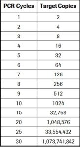 Figure: PCR Cycle and Target copies
Why PCR Changed Diagnostic Microbiology
Before PCR, detecting a pathogen required either growing it in culture (which takes days and fails for fastidious or non-cultivable organisms) or detecting the host's antibody response (which takes weeks to develop and cannot distinguish current from past infection). Both approaches have fundamental limitations that PCR bypasses.
PCR directly detects the pathogen's nucleic acid (its DNA or RNA) regardless of whether the organism is alive, dead, cultivable, or present in tiny quantities. This gives PCR four critical advantages in clinical microbiology:
Speed: Results in hours rather than days. Culture of Mycobacterium tuberculosis takes 6–8 weeks; PCR on sputum can confirm TB in a single working day.
Sensitivity: Can detect as few as 1–10 copies of target DNA per reaction. Culture may miss organisms present in low numbers; PCR does not.
Specificity: Primers are designed to bind only the target sequence. A positive result identifies the specific organism or gene.
Versatility: Works on organisms that cannot be cultured (viruses, some parasites), on degraded or archival samples (formalin-fixed tissue, dried blood spots), and on specimens with mixed flora where culture is uninterpretable.
Components of PCR
Every PCR reaction requires five essential components:
| Component | Role |
|---|---|
| DNA template | The sample containing the target sequence to be amplified (extracted from patient specimen) |
| Primers (forward and reverse) | Short synthetic oligonucleotides (18–24 bp) that flank and define the target sequence; forward primer binds the sense strand, reverse primer binds the antisense strand |
| Taq polymerase | Thermostable DNA polymerase isolated from Thermus aquaticus; extends new DNA strands from primers; survives the 94°C denaturation step |
| dNTPs | Deoxyribonucleoside triphosphates (dATP, dCTP, dGTP, dTTP) — the building blocks incorporated into the new DNA strand during extension |
| PCR buffer | Maintains optimal pH and ionic conditions for Taq polymerase activity; usually contains MgCl₂, which is a required cofactor for Taq |
Primer: A short, synthetic, single-stranded DNA sequence complementary to one end of the target region. Two primers are used: the forward primer binds upstream of the target; the reverse primer binds downstream. Together they define the exact sequence to be amplified.
Taq polymerase: Named after Thermus aquaticus, a thermophilic bacterium isolated from hot springs in Yellowstone National Park. Its heat stability (remaining active at 72°C and surviving 94°C) is what makes automated PCR cycling possible.
Steps of PCR
PCR is performed in a thermocycler; an instrument that raises and lowers temperature in programmed cycles automatically. Each cycle consists of three steps:
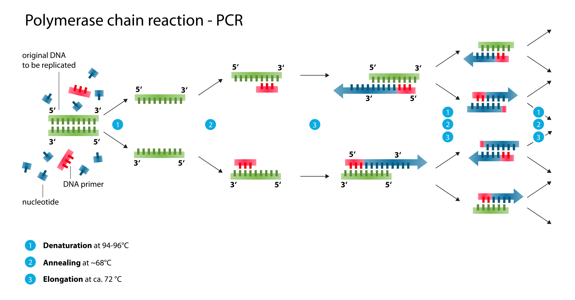 Figure: Steps of PCR (denaturation, annealing, extension)
Step 1: Denaturation (94–96°C)
The double-stranded DNA template is heated to 94–96°C. At this temperature, the hydrogen bonds between complementary base pairs break, separating the two strands into single-stranded templates. This makes each strand accessible for primer binding in the next step.
Step 2: Annealing (45–65°C)
The temperature is lowered to 45–65°C (the exact temperature depends on the primer design and is typically 5°C below the primer melting temperature). The primers bind to their complementary sequences on each single-stranded template. The forward primer binds to one strand; the reverse primer binds to the other, flanking the target region.
Step 3: Extension (72°C)
The temperature is raised to 72°C which is the optimal temperature for Taq polymerase activity. Starting from each primer, Taq polymerase reads the template strand and synthesizes a new complementary DNA strand by incorporating dNTPs. Extension proceeds in the 5' to 3' direction at approximately 1,000 bases per minute.
After the first cycle, two double-stranded DNA copies exist where one existed before. After 30 cycles, the target sequence is amplified by a factor of 2³⁰ = approximately 10⁹ copies. However, practically, amplification efficiency is never 100%, and yields of 10⁶–10⁷ copies are typical after 30–40 cycles.
Detection of PCR Products
Amplified products (amplicons) are detected by one of two methods:
Gel electrophoresis: The PCR product is run on an agarose gel stained with ethidium bromide. A band at the expected size confirms amplification of the correct target. This method is simple and inexpensive, but qualitative only, as it cannot quantify the original amount of target.
Labelled probe detection: A probe complementary to an internal sequence of the amplicon hybridizes to the product and generates a signal (fluorescent, colorimetric, or chemiluminescent). Probe-based detection adds specificity by confirming the amplicon is the correct target rather than a non-specific product. This is the basis of real-time PCR.
Types of PCR
Several modifications of standard PCR have been developed for specific diagnostic needs. Understanding which type to use and why, is more useful than memorizing definitions.
Real-time PCR (Quantitative PCR, qPCR)
Amplification and detection occur simultaneously in a closed tube using fluorescent probes or dyes. The instrument measures fluorescence after each cycle, generating a real-time amplification curve. This enables precise quantitation by calculating the exact number of target copies in the original sample, rather than simply detecting presence or absence.
Clinical use: HIV viral load, HCV viral load, HBV viral load, COVID-19 PCR, TB quantification, CMV monitoring in transplant patients.
Key advantage: No post-PCR handling required as closed tube eliminates carry-over contamination. Faster and more quantitative than conventional PCR.
See full article: Real-time PCR: Principles and Applications
Reverse Transcriptase PCR (RT-PCR)
Standard PCR amplifies DNA. RT-PCR adds a reverse transcription step before amplification. The enzyme reverse transcriptase converts the RNA template into complementary DNA (cDNA), which is then amplified by standard PCR.
Clinical use: Detection of RNA viruses; HIV, hepatitis C, dengue, SARS-CoV-2, influenza, enteroviruses. Also used to detect bacterial and parasitic rRNA (indicating viable organisms) and to study mRNA gene expression.
Key advantage: The only PCR method that can amplify RNA targets directly.
See full article: Reverse Transcriptase PCR: Principles and Applications
Nested PCR
Two successive PCR reactions use two sets of primers. The first reaction (outer primers) amplifies a large fragment; the second reaction (inner or nested primers) amplifies a smaller region within that fragment. The double primer system dramatically increases both sensitivity and specificity.
Clinical use: Detection of organisms present in very low quantities: Rickettsia, Bartonella, M. tuberculosis in paucibacillary samples, herpesvirus and enterovirus in CSF, Leishmania in tissue.
Key advantage: Higher sensitivity than single-round PCR for samples with very low pathogen load.
See full article: Nested PCR: Principle and Applications
Multiplex PCR
Multiple primer pairs targeting different sequences are included in a single PCR reaction, allowing simultaneous amplification of several targets at once.
Clinical use: Detection of multiple meningitis pathogens (S. pneumoniae, H. influenzae, N. meningitidis) from a single CSF sample; respiratory panel testing; BioFire FilmArray syndromic panels.
Key advantage: Multiple pathogens detected from one reaction, reducing cost, time, and sample volume.
See full article: Multiplex PCR: Principle, Applications, and Limitations
Other PCR Types
Several additional PCR modifications are used in research and specialized diagnostic settings:
| Type | Key feature | Use |
|---|---|---|
| Hot-start PCR | Taq polymerase is chemically inhibited at room temperature and activated only at high temperature | Reduces non-specific amplification during reaction setup |
| High-fidelity PCR | Uses proofreading polymerase (e.g., Pfu) instead of Taq | Cloning and sequencing where base-pair accuracy is critical |
| Fast PCR | Shortened cycle times using optimized enzymes and thermocyclers | High-throughput settings requiring rapid turnaround |
| Long-range PCR | Amplifies fragments >10 kb | Amplifying large genomic regions |
| GC-rich PCR | Modified buffer and additives for templates with high GC content | Amplifying GC-rich regions that form secondary structures |
RT-PCR vs. Real-time PCR
These two terms are routinely confused including in clinical settings during the COVID-19 pandemic, when "RT-PCR" was used by media and health authorities to mean different things simultaneously.
| Feature | RT-PCR (Reverse Transcriptase PCR) | Real-time PCR (qPCR) |
|---|---|---|
| Full name | Reverse transcriptase PCR | Real-time quantitative PCR |
| Target | RNA (converted to cDNA first) | DNA (or RNA if combined with RT) |
| What it adds to standard PCR | A reverse transcription step before amplification | Fluorescent detection during amplification |
| Quantitative? | No, detects presence/absence | Yes, measures copy number |
| Can they be combined? | Yes. RT-qPCR combines both: RNA target + real-time detection | Yes |
| COVID-19 test | SARS-CoV-2 is RNA; test uses RT-PCR for the RNA → cDNA step, then real-time detection. This is technically RT-qPCR | Same assay |
The practical rule: RT-PCR refers to the template type (RNA). Real-time PCR refers to the detection method (fluorescence during cycling). A test can be both, and most modern viral load assays are.
Applications of PCR
PCR is used across medicine, research, and forensics. Its applications fall into a few broad areas.
Diagnosis of infectious disease. This is the largest clinical use. PCR detects the DNA or RNA of a pathogen directly in a patient sample, which is faster and more sensitive than culture. It is the standard method for detecting tuberculosis, HIV, hepatitis B and C, SARS-CoV-2, and many other infections, including organisms that cannot be grown in the laboratory.
Measuring how much pathogen is present (viral load). Real-time PCR does not just say whether a virus is present; it counts the copies. This viral load is used to monitor HIV, hepatitis B and C, and CMV, and to judge whether treatment is working.
Detecting resistance genes. PCR can find the specific gene that makes a bacterium resistant to a drug, such as the mecA gene in MRSA or the genes behind rifampicin resistance in tuberculosis. This guides treatment faster than waiting for culture-based susceptibility testing.
Genetic testing and research. PCR is used to detect inherited disease-causing mutations, to study which genes are switched on (through RT-PCR of mRNA), and to prepare DNA for sequencing and cloning.
Forensic science and identity testing. Because PCR can amplify the tiny amount of DNA in a hair, a bloodstain, or a cheek swab, it is central to DNA fingerprinting, criminal investigation, and paternity testing.
Other uses. PCR is also used in food safety testing (detecting contaminating organisms), in checking blood and tissue donations for infections, and in tracking outbreaks by identifying and comparing pathogen strains.
How to Remember
PCR = molecular photocopier with a temperature dial. A photocopier takes one document and makes many copies. PCR takes one DNA sequence and makes millions. The thermocycler is the machine; the three temperatures are the three steps: high heat separates (94°C), cool down for primers to bind (50–65°C), warm up for extension (72°C).
"Denature, Anneal, Extend": DAE, like the name. The three steps in order: Denaturation (94°C) → Annealing (50–65°C) → Extension (72°C). DAE. Each step has one job: separate, bind, build.
Taq polymerase = hot spring survivor. Thermus aquaticus lives in boiling hot springs. Its polymerase survives 94°C without denaturing — the property that makes automated PCR cycling possible. Without a heat-stable polymerase, fresh enzyme would have to be added after every denaturation step.
PCR type selection: the three clinical questions:
- Is the target RNA? → Need RT-PCR
- Do I need a number (viral load), not just yes/no? → Need real-time (qPCR)
- Is the target present in tiny amounts or is sensitivity critical? → Consider nested PCR
- Are multiple pathogens possible from one sample? → Consider multiplex PCR
Key exam facts in one table
| Topic | Key fact |
|---|---|
| PCR invented by | Kary Mullis, 1983; Nobel Prize in Chemistry 1993 |
| What PCR amplifies | Specific target DNA sequence; RNA requires RT-PCR first |
| Amplification factor | ~10⁷ copies from a single template after 30–40 cycles |
| Denaturation step | 94–96°C; breaks hydrogen bonds; separates double-stranded DNA |
| Annealing step | 45–65°C; primers bind complementary sequences on template |
| Extension step | 72°C; Taq polymerase synthesizes new DNA strand from primers |
| Taq polymerase source | Thermus aquaticus is thermophilic bacterium from hot springs |
| Why Taq, not other polymerases | Heat-stable; survives 94°C denaturation; active at 72°C |
| dNTPs | Building blocks (dATP, dCTP, dGTP, dTTP) incorporated during extension |
| Gel electrophoresis detection | Qualitative as it confirms band at expected size; cannot quantify |
| Real-time PCR advantage | Quantitative + closed tube (no post-PCR contamination risk) |
| RT-PCR — what it adds | Reverse transcriptase converts RNA → cDNA before amplification |
| RT-PCR vs real-time PCR | RT-PCR = RNA target; real-time = fluorescent detection method; COVID test = both (RT-qPCR) |
| Clinical use of Nested PCR | Very low pathogen load: TB paucibacillary, Rickettsia, Bartonella, CSF viruses |
| Clinical use of Multiplex PCR | Simultaneous detection of multiple pathogens from one sample |
| Filter tips in PCR setup | Mandatory as it prevents aerosol carry-over contamination between reactions |
Frequently Asked Questions
What is polymerase chain reaction (PCR) and what does it do?
Polymerase chain reaction (PCR) is an in vitro molecular technique that amplifies a specific DNA or RNA sequence exponentially, producing up to 10 million copies from a single starting template within a few hours. It works by repeatedly cycling through three temperature-controlled steps (denaturation, annealing, and extension), using a heat-stable DNA polymerase (Taq polymerase) and short synthetic primers that define the target sequence. In clinical microbiology, PCR directly detects a pathogen's nucleic acid in a patient specimen, regardless of whether the organism is alive, cultivable, or present in small quantities.
What are the three steps of PCR and what temperature is used for each?
PCR has three steps that repeat in each cycle. Denaturation occurs at 94–96°C, heat breaks the hydrogen bonds between the two DNA strands, separating them into single-stranded templates. Annealing occurs at 45–65°C, the temperature is lowered so primers can bind to their complementary sequences on each strand. Extension occurs at 72°C, Taq polymerase synthesizes a new complementary DNA strand starting from each primer. After 30–40 cycles, the target sequence is amplified by a factor of approximately 10 million.
What is Taq polymerase and why is it used in PCR?
Taq polymerase is a thermostable DNA polymerase originally isolated from Thermus aquaticus, a bacterium that lives in boiling hot springs. Its defining property is heat stability, it remains active at 72°C and survives the 94°C denaturation step without being destroyed. This allows automated PCR cycling without adding fresh enzyme after every cycle. Without a heat-stable polymerase, PCR as an automated process would not be possible.
What is the difference between RT-PCR and real-time PCR?
These two terms describe different aspects of PCR and are frequently confused. RT-PCR (reverse transcriptase PCR) refers to the template type: it adds a reverse transcription step that converts RNA into complementary DNA before amplification, making it possible to detect RNA viruses such as HIV, hepatitis C, dengue, and SARS-CoV-2. Real-time PCR (quantitative PCR or qPCR) refers to the detection method: fluorescence is measured during each amplification cycle, allowing quantitation of the target. A test can be both simultaneously: the COVID-19 PCR test is technically RT-qPCR, using reverse transcriptase for the RNA template and real-time detection for quantitation.
When should nested PCR be used instead of standard PCR?
Nested PCR should be used when the target organism is present in very low quantities, below the detection threshold of standard single-round PCR. It uses two successive PCR reactions with two primer sets: outer primers amplify a large fragment first, then inner (nested) primers amplify a smaller specific region within that product. The double amplification dramatically increases sensitivity. Clinical applications include detection of Rickettsia and Bartonella in blood, M. tuberculosis in paucibacillary samples, herpesviruses and enteroviruses in CSF, and Leishmania in tissue.
What are the advantages of PCR over culture in clinical microbiology?
PCR offers four key advantages over culture. Speed: results in hours rather than days: TB culture takes 6–8 weeks; PCR confirms TB the same day. Sensitivity: detects as few as 1–10 DNA copies per reaction, far below the threshold for culture positivity. Specificity: primers target a defined sequence, identifying the exact organism or resistance gene rather than just confirming growth. Versatility: works on organisms that cannot be cultured (many viruses, some parasites), on degraded specimens (formalin-fixed tissue, dried blood), and on samples with mixed flora where culture is uninterpretable.
References
- Mullis, K. B. (1990). The unusual origin of the polymerase chain reaction. Scientific American, 262(4), 56–65.
- Murray, P. R., Rosenthal, K. S., & Pfaller, M. A. (2021). Medical Microbiology (9th ed.). Elsevier.
- Mahon, C. R., Lehman, D. C., & Manuselis, G. (2018). Textbook of Diagnostic Microbiology (6th ed.). Elsevier.
- Tille, P. M. (2022). Bailey & Scott's Diagnostic Microbiology (15th ed.). Elsevier.
- Chang-Hui Shen. (2019). Amplification of Nucleic Acids. Diagnostic Molecular Biology. Academic Press. https://doi.org/10.1016/B978-0-12-802823-0.00009-2
- Clinical and Laboratory Standards Institute (CLSI). (2016). Clinical Microbiology Procedures Handbook (4th ed.). American Society of Microbiology.

Tankeshwar Acharya, MSc (Medical Microbiology)
Tankeshwar Acharya is an Assistant Professor in the Department of Microbiology at Patan Academy of Health Sciences (PAHS), Nepal, where he has been teaching and practicing clinical microbiology for over 14 years. He is the founder of Microbe Online, one of the leading free microbiology education resources on the web, covering bacteriology, mycology, parasitology, immunology, and clinical laboratory diagnostics written from direct experience in both the classroom and the diagnostic laboratory.
Comments
No comments yet. Be the first to share your thoughts.
Leave a comment
All comments are reviewed before they appear.