

A microbiologist spots bright red colonies on a MacConkey plate from a urinary catheter specimen. A burn nurse notices blue-green staining soaking through a wound dressing. A bacteriologist sees orange-tinged colonies from a neonatal blood culture. In each case, color is not cosmetic; it is a diagnostic clue, sometimes the first one available before any identification test is run.
Many pathogenic bacteria and fungi produce pigments as metabolic byproducts or as deliberate virulence tools. These pigments fall into two broad categories:
- Water-soluble pigments that diffuse into the surrounding medium and can color the agar, the specimen, or infected tissue (e.g. Pseudomonas pyocyanin staining wound dressings green)
- Water-insoluble pigments that remain within or on the colony itself, giving characteristic colony color (e.g. Serratia marcescens red colonies, S. aureus golden colonies)
Beyond their diagnostic utility, many bacterial pigments are active virulence factors that interfere with immune clearance, generate oxidative stress in host tissue, or protect the organism from phagocytosis. Understanding what a pigment is and what it does distinguishes a microbiologist who can identify an organism from one who understands why that organism behaves the way it does in a patient.
Pigment-Producing Pathogens
| Organism | Pigment Name | Color | Solubility | Medium Where Best Seen | Clinical Significance |
|---|---|---|---|---|---|
| Pseudomonas aeruginosa | Pyocyanin | Blue-green | Water-soluble | Mueller-Hinton, blood agar | Green wound dressings, blue-green sputum in CF; virulence factor |
| Pseudomonas aeruginosa | Pyoverdin (fluorescein) | Yellow-green fluorescent | Water-soluble | King's medium A/B | Fluorescence under UV; iron-scavenging siderophore |
| Serratia marcescens | Prodigiosin | Bright red | Water-insoluble | Nutrient agar, blood agar at room temp | Red colonies; hospital outbreaks; historical biowarfare agent |
| Staphylococcus aureus | Staphyloxanthin | Golden-yellow | Water-insoluble | Blood agar | Golden colonies; antioxidant virulence factor protecting from neutrophil killing |
| Chromobacterium violaceum | Violacein | Blue-violet | Water-insoluble | Blood agar, nutrient agar | Fatal septicemia in immunocompromised; tropical pathogen |
| Streptococcus agalactiae (GBS) | Granadaene | Orange-red | Water-insoluble | Granada medium (serum starch agar, anaerobic) | Neonatal sepsis pathogen; orange pigment aids identification |
| Porphyromonas gingivalis | Iron porphyrin (micro-oxo bis-hem) | Black | Water-insoluble | Blood agar (5–7 days) | Periodontal disease; black colonies after prolonged incubation |
| Prevotella melaninogenica | Melanin-like | Black-brown | Water-insoluble | Blood agar (prolonged incubation) | Oral and respiratory anaerobe; black pigment from hem degradation |
| Mycobacterium kansasii | Carotenoids | Yellow (light-induced) | Water-insoluble | Löwenstein-Jensen medium | Runyon Group I photochromogen; NTM lung disease |
| Mycobacterium scrofulaceum | Carotenoids | Yellow-orange (dark) | Water-insoluble | Löwenstein-Jensen medium | Runyon Group II scotochromogen; cervical lymphadenitis |
| Cryptococcus neoformans | Melanin | Brown-black | Water-insoluble | Birdseed (Niger seed) agar | Rapid ID on birdseed agar; melanin protects from phagocytosis |
| Aspergillus fumigatus | Melanin-like (conidial) | Blue-grey/green | Water-insoluble | Sabouraud agar | Conidial pigment aids immune evasion |
| Talaromyces marneffei | Unknown red pigment | Red, diffusible | Water-soluble | Sabouraud dextrose agar | Diffusible red pigment into agar = key ID feature; AIDS-defining infection |
Pseudomonas species
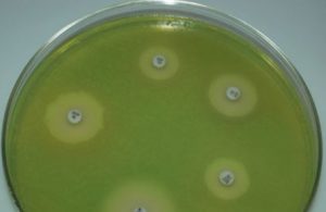 Figure: Pseudomonas aeruginosa (green color) growing on Mueller Hinton Agar
Pseudomonas aeruginosa is a leading bacterial pathogen in hospital settings and for immunocompromised patients (those with underlying conditions such as neutropenia, burns, or cystic fibrosis). P. aeruginosa (aeruginosa, which derives from a Latin word denoting the color of copper rust) produces a green fluorescent pigment (fluorescein/pyoverdin) and a blue pigment (pyocyanin). These pigments impart a greenish hue to the sputum of cystic fibrosis patients with a chronic lung infection, giving colonies and infected wound dressings a greenish-blue coloration.
P. aeruginosa is usually recognized by the pigments it produces and the distinctive smell of cultures. When the culture is left at room temperature, pigment color becomes more intense. A minority of strains are non-pigment producing. Other species of Pseudomonas (such as P. fluorescens and P. putida) also produce pigments.
P. aeruginosa produces at least four pigments, of which two are clinically most important:
Pyocyanin (blue-green) is a phenazine compound and a genuine virulence factor, not merely a color. It:
- Generates reactive oxygen species (ROS) directly in host tissue, causing oxidative damage to respiratory epithelium
- Impairs mucociliary clearance: relevant to why P. aeruginosa establishes chronic infection in cystic fibrosis lungs
- Inhibits the normal function of neutrophils and macrophages
- The name comes from Greek: pyo (pus) plus cyan (blue), literally blue pus.
Pyoverdin (yellow-green, fluorescent under UV) is a siderophore, an iron-chelating molecule that scavenges iron from the host environment. Since iron is essential for bacterial growth and the host actively withholds it as a defense mechanism (nutritional immunity), pyoverdin gives P. aeruginosa a significant survival advantage in iron-limited tissue.
Diagnostic note: King's medium A enhances pyocyanin production; King's medium B enhances pyoverdin. In resource-limited settings, Mueller-Hinton agar at room temperature is commonly used and usually shows adequate pigmentation. A minority of strains (mucoid strains in CF patients) produce little or no pigment.
Serratia marcescens
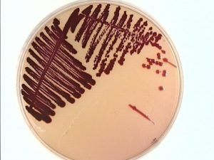 Figure: Red colored colonies of Serratia marcescens in MacConkey Agar
(Source University of Maryland)
Serratia marcescens (mnemonic: Mars is red) is notable for its production of a bright red pigment called prodigiosin, best seen on nutrient agar at room temperature.
Prodigiosin is a tripyrrole red pigment produced optimally at room temperature (25 to 28°C) but not at 37°C, which is why Serratia colonies from clinical specimens incubated at 37°C may appear white or pale pink, leading to misidentification. If you suspect Serratia and see non-pigmented colonies, subculture to room temperature incubation.
Clinical and historical significance:
- Serratia marcescens was historically considered a harmless saprophyte and its red pigment was used as a biological marker. In 1950, the US Navy conducted a secret experiment releasing S. marcescens over San Francisco Bay to simulate a biological warfare attack. This was subsequently linked to cases of Serratia bacteremia in local hospitals. The experiment was kept classified for decades.
- Today Serratia is a significant nosocomial pathogen, particularly in ICUs, causing UTIs, bacteremia, and pneumonia in catheterized and ventilated patients. It is intrinsically resistant to ampicillin and first-generation cephalosporins due to an inducible AmpC beta-lactamase.
- Red-pigmented colonies on MacConkey or blood agar from a hospital patient should prompt consideration of Serratia and a review of infection control implications.
Staphylococcus aureus
Staphylococcus aureus (aureus = “golden”, Latin) produces multiple carotenoid pigments, one being golden-yellow pigment (Staphyloxanthin). When cultured on sheep blood agar, S. aureus can be differentiated from other beta-hemolytic cocci by elaboration of golden pigment. The pigment is less pronounced in young colonies.
Staphyloxanthin is not merely a laboratory curiosity; it is a carotenoid virulence factor with a specific immune-evasion function. Its golden color is incidental to its primary role: neutralizing reactive oxygen species generated by neutrophils during the oxidative burst.
When neutrophils engulf S. aureus, they deploy hydrogen peroxide and hypochlorite to kill the bacterium. Staphyloxanthin acts as an antioxidant, quenching these ROS and allowing S. aureus to survive inside phagosomes. Mutant S. aureus strains lacking staphyloxanthin (and therefore appearing white rather than golden) are significantly less virulent in animal models.
Practical point: Golden pigment is best seen on blood agar after 24–48 hours at 37°C. Young colonies and some clinical strains may appear pale yellow rather than distinctly golden. MRSA strains do not lose their pigment — golden colonies with beta-hemolysis should still prompt MRSA screening in appropriate clinical contexts.
Chromobacterium violaceum
Chromobacterium violaceum is a Gram-negative, facultatively anaerobic bacillus found in soil and water in tropical and subtropical regions of Southeast Asia, South Asia, Central and South America, and parts of Africa. It is rare but memorable because of two features: its striking blue-violet pigment and its capacity to cause rapidly fatal septicemia.
Violacein is the purple-blue pigment responsible for the organism's name and its unmistakable colony appearance. It is produced from tryptophan via the vio biosynthetic pathway and has demonstrated antibacterial, antifungal, antiparasitic, and antitumor properties in laboratory studies, making it a subject of pharmaceutical research.
Clinical significance: Most infections follow skin exposure to contaminated water or soil, often through minor wounds. In healthy individuals, infection usually stays localized, presenting as skin nodules or lymphadenitis. In immunocompromised patients (particularly those with chronic granulomatous disease, CGD), C. violaceum can disseminate rapidly, causing multiple liver abscesses, lung involvement, and septicemia with high mortality. CGD patients are specifically vulnerable because C. violaceum resists killing by the defective NADPH oxidase pathway.
Laboratory recognition: Violet-pigmented colonies on blood agar are virtually diagnostic. Non-pigmented variants exist but are rare. The organism is oxidase-positive and produces HCN (hydrogen cyanide).
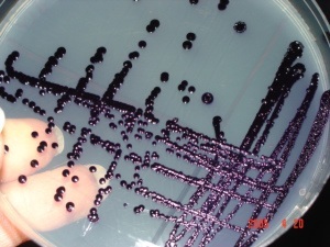 Figure: Characteristics pigment production by Chromobacterium violaceum
Group B Streptococcus
Group B Streptococcus (GBS), the leading etiologic cause of severe neonatal bacterial infection, expresses an orange-red pigment, called granadaene. This orange pigment is produced when Streptococcus agalactiae is cultured anaerobically on serum starch agar such as Granada medium. Granadaene is also the beta-hemolysin of GBS, so the pigment and the hemolytic activity are the same molecule, which is why pigment-negative strains are also non-hemolytic.
Iron porphyrin of Porphyromonas gingivalis
Porphyromonas gingivalis is a gram-negative anaerobic bacillus implicated in the pathogenesis of certain forms of periodontal disease. Arginine- and lysine-specific gingipain proteases of P. gingivalis degrade hemoglobin to release iron (III) protoporphyrin IX, which is dimerized to form the micro-oxo bis-hem-containing black pigment of the organism.
Nontuberculous Mycobacteria
The genus Mycobacterium includes many bacteria that produce pigments. The Runyon classification system groups mycobacteria based on growth rate and pigment production in the presence of light or dark. M. tuberculosis is a nonchromogen, meaning it produces no pigment in either light or darkness.
For the full Runyon classification of NTM based on pigment production and growth rate, see our detailed article on atypical mycobacterial infections.
Fungal Pathogens That Produce Pigments
Cryptococcus neoformans: Melanin
Cryptococcus neoformans produces melanin from diphenolic substrates (L-DOPA, catecholamines) via the enzyme laccase. This melanin production has direct clinical relevance:
- Virulence: Melanin protects the organism from oxidative killing by macrophages and from amphotericin B. The mechanism is thought to involve melanin binding to the drug, reducing its bioavailability within the cell
- Rapid identification: Birdseed (Niger seed) agar contains caffeic acid as a substrate; Cryptococcus colonies appear brown-black within 3–5 days, distinguishing them from non-melanin-producing yeasts. This is particularly useful in resource-limited settings where molecular identification is unavailable
- C. gattii also produces melanin and is identified the same way
Aspergillus fumigatus: Conidial Melanin
A. fumigatus conidia (spores) are coated with a melanin-like pigment that gives them their characteristic blue-grey color. This pigment:
- Inhibits complement activation on the conidial surface
- Impairs phagolysosomal acidification after phagocytosis by macrophages
- Confers resistance to UV damage
Mutant A. fumigatus strains with white (unpigmented) conidia are significantly less virulent in animal models.
Talaromyces marneffei: Diffusible Red Pigment
Formerly Penicillium marneffei, this thermally dimorphic fungus is an important AIDS-defining opportunistic infection in Southeast Asia and southern China. Its diffusible red pigment, which stains the surrounding agar red on Sabouraud dextrose agar at 25°C, is a key identification feature distinguishing it from other Talaromyces/Penicillium species, most of which do not produce this pigment.
At 37°C it grows as a yeast-like form (intracellular, found in macrophages on bone marrow or tissue biopsy); at 25°C it grows as a mold producing the red pigment. This thermal dimorphism plus the red pigment is essentially diagnostic.
Pigments as Virulence Factors
The diagnostic utility of pigments is well known. Less often taught is that several of these pigments are not metabolic waste products at all. They are active weapons.
| Pigment | Organism | Virulence Mechanism |
|---|---|---|
| Pyocyanin | P. aeruginosa | Generates ROS; impairs mucociliary clearance; inhibits phagocyte function |
| Pyoverdin | P. aeruginosa | Iron scavenging (siderophore); competes with host iron-binding proteins |
| Staphyloxanthin | S. aureus | Antioxidant; quenches neutrophil ROS; promotes survival in phagosomes |
| Melanin | C. neoformans | Resists oxidative killing; may reduce amphotericin B efficacy |
| Melanin | A. fumigatus | Inhibits complement; impairs phagolysosomal killing |
| Violacein | C. violaceum | Cytotoxic to host cells; may contribute to tissue destruction in septicemia |
| Granadaene | S. agalactiae | Hemolytic activity; damages host cell membranes |
The practical implication: organisms with pigment-based virulence mechanisms are often harder to clear with the immune system alone. C. neoformans meningitis requires prolonged antifungal therapy partly because melanin-coated cells in the CNS are well protected. S. aureus bacteremia recurs partly because staphyloxanthin-expressing organisms survive within phagocytes.
How to Remember: Bacterial Pigments
Color-organism pairs: the exam-tested ones:
- Green-blue = Pseudomonas aeruginosa (pyocyanin: think "pyo" = pus, "cyan" = blue)
- Bright red = Serratia marcescens (prodigiosin: mnemonic: Mars is red, Serratia = S, like Scarlet)
- Golden yellow = Staphylococcus aureus (aureus = gold in Latin)
- Blue-violet = Chromobacterium violaceum (violaceum = violet in Latin)
- Orange-red = Streptococcus agalactiae GBS (granadaene — think "Granada" in Spain, red terracotta)
- Black = Porphyromonas gingivalis and Prevotella melaninogenica (both oral anaerobes, both black on blood agar after prolonged incubation)
- Brown-black = Cryptococcus neoformans on birdseed agar (melanin)
- Diffusible red = Talaromyces marneffei on Sabouraud agar at 25°C
Runyon classification quick memory: "Photo, Scoto, Non, Fast":
- Photochromogen = color in LIGHT only (M. kansasii)
- Scotochromogen = color in DARK too (M. scrofulaceum)
- Nonchromogen = no color (M. avium complex, MTB)
- Fast grower = colonies in <7 days (M. fortuitum)
Key Exam Facts in One Table
| Organism | Pigment | Color | Key Exam Point |
|---|---|---|---|
| P. aeruginosa | Pyocyanin + pyoverdin | Blue-green | Pyocyanin is a virulence factor (ROS generation); pigment = water-soluble |
| Serratia marcescens | Prodigiosin | Bright red | Only at room temperature (25–28°C); absent at 37°C — white colonies possible |
| S. aureus | Staphyloxanthin | Golden-yellow | Carotenoid antioxidant; protects from neutrophil killing |
| Chromobacterium violaceum | Violacein | Blue-violet | Tropical pathogen; fatal septicemia in CGD; from tryptophan |
| S. agalactiae (GBS) | Granadaene | Orange-red | Seen on Granada/serum starch agar anaerobically; hemolytic |
| P. gingivalis | Iron porphyrin | Black | Needs prolonged incubation (5–7 days); periodontal pathogen |
| M. kansasii | Carotenoid | Yellow (light only) | Runyon Group I photochromogen |
| M. scrofulaceum | Carotenoid | Yellow-orange (dark and light) | Runyon Group II scotochromogen; cervical lymphadenitis |
| M. tuberculosis | None | Non-pigmented | Nonchromogen — important negative |
| C. neoformans | Melanin | Brown-black on birdseed agar | Laccase enzyme; virulence factor; rapid ID on Niger seed agar |
| A. fumigatus | Conidial melanin | Blue-grey conidia | Immune evasion; white mutants less virulent |
| T. marneffei | Unknown red | Diffusible red into agar | At 25°C only; AIDS-defining; dimorphic |
References
- Madigan MT, Bender KS, Buckley DH, Sattley WM, Stahl DA. Brock Biology of Microorganisms. 16th ed. Pearson; 2021.
- Lau YL, Yuen KY, Lee CW, Chan CF. Chromobacterium violaceum septicemia in children. Pediatr Infect Dis J. 1995;14(6):479–483.
- Clauditz A, Resch A, Wieland KP, Peschel A, Götz F. Staphyloxanthin plays a role in the fitness of Staphylococcus aureus and its ability to cope with oxidative stress. Infect Immun. 2006;74(8):4950–4953. https://doi.org/10.1128/IAI.00204-06
- Nosanchuk JD, Casadevall A. The contribution of melanin to microbial pathogenesis. Cell Microbiol. 2003;5(4):203–223. https://doi.org/10.1046/j.1462-5822.2003.00268.x
- Edenborough FM. Pseudomonas aeruginosa in cystic fibrosis: the battle continues. J R Soc Med. 2001;94(Suppl 40):3–8.
- Pitt TL. Serratia marcescens: clinical significance and epidemiology. J Hosp Infect. 1994;27(2):87–90.
- Tille PM. Bailey & Scott's Diagnostic Microbiology. 15th ed. St. Louis: Elsevier; 2022.
- Procop GW, Koneman EW. Koneman's Color Atlas and Textbook of Diagnostic Microbiology. 7th ed. Wolters Kluwer; 2017.

Comments
No comments yet. Be the first to share your thoughts.
Leave a comment
All comments are reviewed before they appear.