
Virus Neutralization Test: Why Some Antibodies Protect and Some Don't

On this page
Someone bitten by a potentially rabid animal months ago, who received the full vaccine series at the time, now needs to travel to a region with ongoing rabies risk and wants to know whether they are still protected or need a booster. The question is not "did they make antibodies," it is "do they still have neutralizing antibodies," and that distinction is the whole reason this test exists rather than a simpler antibody-detection test.
Not every antibody that binds a virus actually stops it from infecting a cell. An antibody can attach perfectly well to a viral protein and do nothing useful, while a different antibody, binding a different spot, physically blocks the exact step the virus needs to enter a cell. The neutralization test is built to measure that functional difference: it does not just ask "is antibody present," it asks "does this serum, at this concentration, actually stop the virus from infecting living cells." That is a harder and more clinically relevant question than a standard antibody-binding test answers, and it is why neutralization testing remains the gold standard for confirming protective immunity against rabies, even where faster antibody tests exist.
What is the neutralization test?
When a person is infected with a pathogen, antibodies are produced against many epitopes of that pathogen. A subset of these antibodies can block infection, a process called neutralization: they bind or distort the antigen enough that the pathogen can no longer carry out its biological activity.
The neutralization test (also called the neutralization assay, or serum neutralization test) measures this blocking activity. Neutralization reactions can occur in vitro or in vivo. Laboratory animals or tissue culture cells are used as "indicator systems." The toxin or virus being assayed must have a known, measurable effect on the indicator system, an effect that is then neutralized by adding a specific antibody. An antiserum whose antibody neutralizes a toxin is called an antitoxin.
Toxin Neutralization Assay
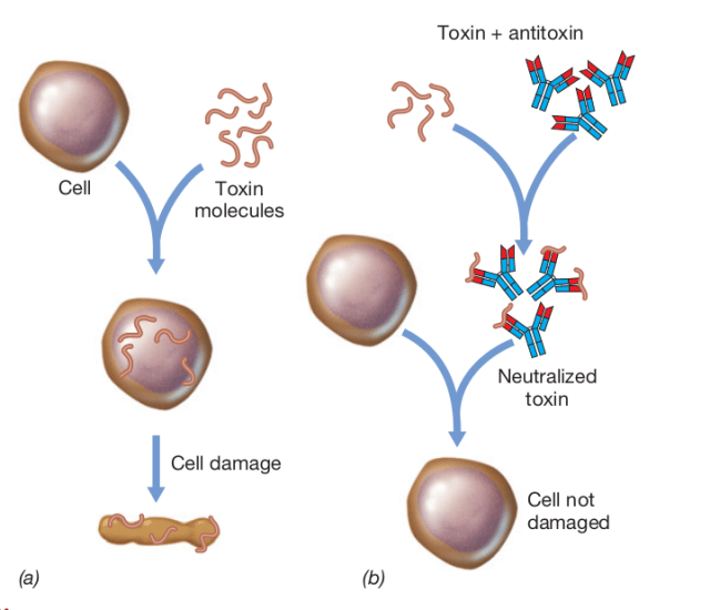 Figure: Toxin neutralization assay. A. Cell death caused by toxin. B. Neutralization of toxin and prevention of cell death. Image source: Brock Biology of Microorganisms
A microbial toxin is neutralized when a specific antibody binds it so that the active portion of the toxin is blocked. Neutralization can block the effects of many bacterial exotoxins.
Examples of toxin-antitoxin neutralization tests:
- Schick test: a diphtheria toxin-antitoxin neutralization test used to assess a person's immune status, historically to find individuals susceptible to Corynebacterium diphtheriae. A standardized dose (0.1 mL) of purified toxin is injected intradermally. If the person has no antitoxin, the toxin causes inflammation at the site 4 to 7 days later (a positive test: susceptible). If no inflammation occurs, antitoxin is present and the person is immune.
- Nagler's reaction: the opalescence produced on egg-yolk agar by the alpha-toxin of Clostridium perfringens is inhibited when anti-alpha-toxin is added to the medium, because the antibody neutralizes the toxin.
Toxin applications. Antitoxin therapy is used to neutralize the toxins of Corynebacterium diphtheriae, Clostridium tetani, and Clostridium botulinum, preventing diphtheria, tetanus, and botulism respectively.
Virus Neutralization Assay
A virus neutralization test determines whether antibody in a patient's serum (or on a mucosal surface) can neutralize the infectivity of a virus. For example, antibodies against the hemagglutinin and neuraminidase proteins of influenza prevent the virus from adsorbing to host cell receptors, protecting the cells from infection. Similarly, mucosal secretory IgA gives intestinal immunity against poliovirus.
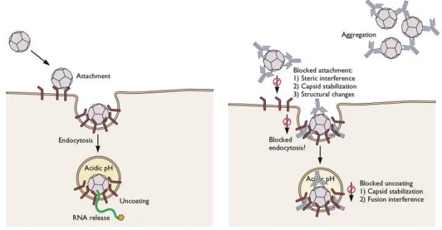 Figure: Prevention of viral infection by neutralizing antibodies
Neutralizing antibodies may block virion binding to receptors, block uptake into the cell, prevent uncoating of the genome in endosomes, or clump virus particles together. A neutralizing antibody is directed against the surface proteins of the virus. Antibodies against internal components (for example, the core antigen of hepatitis B virus) do not neutralize infectivity.
Procedure and how to read the result. The patient's serum is mixed with a standard virus preparation and poured onto a cell line, which is then watched for cytopathic effect (cell death):
- Neutralizing antibody present: the antibody binds the virus's surface antigen and blocks it from infecting the cells. The cell line stays healthy. A positive neutralization test means the patient has functional, neutralizing (protective) antibody, not merely that they were exposed.
- Neutralizing antibody absent: the virus is not neutralized, infects the cells, and the cell line shows cytopathic effect or cell death.
Neutralization tests have been developed for arboviruses, rabies virus, and many others.
Applications of the neutralization test
- Disease diagnosis: for example, herpes simplex virus infection.
- Passive immunization: neutralizing rabies and hepatitis A and B viruses early in the incubation period by injecting antibody against them.
- Active immunization: the live attenuated Sabin poliovirus vaccine elicits a strong mucosal IgA response and gives intestinal immunity against poliovirus.
- Serotyping of viruses: a virus neutralization assay serotypes viruses; for example, poliovirus types 1, 2, and 3 were distinguished using neutralizing antibodies.
How to Remember
Only surface-exposed targets can be neutralized, because neutralization is about physically blocking a step in entry. An antibody against hepatitis B core antigen (HBcAg) can bind its target perfectly well, but HBcAg is packaged inside the virion, inaccessible until after the virus has already entered a cell and begun disassembling. By then, neutralization has already succeeded or failed by other means. Only antibodies against surface proteins, the parts exposed before entry, can block attachment, uptake, or uncoating.
This is the single fact that explains why anti-HBs (surface antibody) indicates protection while anti-HBc (core antibody) does not. Read more hepatitis B serology interpretation article.
Antibody-dependent enhancement (ADE) is the dangerous exception where more antibody does not mean more protection. Picture a non-neutralizing antibody as a key that fits the lock but does not turn it: it binds the virus without blocking its function. Some cells carry Fc receptors that recognize the antibody's tail end regardless of what its other end is doing, so an antibody-coated virus particle can be pulled into cells that would not normally let it in, using the antibody as a delivery mechanism rather than a blocker. This is the mechanism behind why a second dengue infection with a different serotype can be more severe than the first.
Key exam facts
| Concept | Detail |
|---|---|
| Neutralizing antibody target | Surface-exposed viral proteins only |
| Non-neutralizing antibody target | Internal/core viral components (e.g. HBcAg) |
| Mechanisms of neutralization | Block receptor binding, block uptake, prevent uncoating, aggregate virions |
| Antibody-dependent enhancement (ADE) | Non-neutralizing antibody facilitates viral entry via Fc-receptor-bearing cells |
| Classic ADE example | Secondary dengue infection with a different serotype |
| Toxin neutralization examples | Schick test (diphtheria), Nagler's reaction (C. perfringens alpha-toxin) |
| Virus neutralization gold-standard use | Confirming protective immunity (e.g. rabies post-vaccination titers) |
| Positive vs standard antibody test | Neutralization test confirms functional/protective antibody, not just past exposure |
| Other major applications | Diagnosis (e.g. HSV), passive immunization (rabies, hepatitis A/B immunoglobulin), serotyping (poliovirus types 1-3) |
Where Students Get Confused
- "Having antibodies against a virus always means you are protected." Not necessarily. An antibody can bind a virus without blocking its function, and in antibody-dependent enhancement it can make a later infection more severe. Whether antibody helps or harms depends on what it targets and how it works, not just whether it is present. Non-neutralizing antibodies can even enhance infection: Fc receptors on monocytes and macrophages bind the antibody's tail end whether or not the antibody neutralizes the virus, pulling antibody-coated virus into cells that would not normally be susceptible. This is the basis of severe secondary dengue with a heterologous serotype.
- "A positive antibody test and a positive neutralization test are the same thing." They measure different properties. A standard antibody test (like ELISA) confirms that antibody is present (exposure or vaccination happened). A neutralization test confirms that the antibody actually blocks infectivity at a given concentration, which is why it is used to confirm protective immunity (such as rabies titers) rather than simple past exposure.
- "Antitoxin and antibody are different things." They are not. An antitoxin is simply the name for an antibody when it neutralizes a toxin, the same antigen-antibody interaction as virus neutralization, applied to a bacterial exotoxin instead of a virion.
Frequently Asked Questions
If someone has antibodies against a virus, are they automatically protected from it?
Why is a second dengue infection sometimes more dangerous than the first?
What's the difference between an antibody test and a neutralization test?
Is an antitoxin a different kind of molecule from a regular antibody?
References
- Racaniello V. Virus neutralization by antibodies. Virology Blog. 2009. https://virology.ws/2009/07/24/virus-neutralization-by-antibodies/
- Madigan MT, Bender KS, Buckley DH, Sattley WM, Stahl DA. Brock Biology of Microorganisms. 16th ed. Pearson; 2021.
- Willey JM, Sherwood LM, Woolverton CJ. Prescott's Microbiology. 11th ed. McGraw-Hill Education; 2020.

Tankeshwar Acharya, MSc (Medical Microbiology)
Tankeshwar Acharya is an Assistant Professor in the Department of Microbiology at Patan Academy of Health Sciences (PAHS), Nepal, where he has been teaching and practicing clinical microbiology for over 14 years. He is the founder of Microbe Online, one of the leading free microbiology education resources on the web, covering bacteriology, mycology, parasitology, immunology, and clinical laboratory diagnostics written from direct experience in both the classroom and the diagnostic laboratory.
Comments
No comments yet. Be the first to share your thoughts.
Leave a comment
All comments are reviewed before they appear.