

The lecithinase this test detects is not a laboratory curiosity. It is the alpha-toxin of Clostridium perfringens, the enzyme responsible for the catastrophic tissue destruction of gas gangrene (clostridial myonecrosis).
The alpha-toxin is a phospholipase C: it hydrolyzes lecithin, a phospholipid in host cell membranes, lysing red cells, platelets, and muscle cells and spreading through tissue. So when the Nagler reaction shows an opaque halo that is neutralized by antitoxin, it is identifying the same enzyme that dissolves cell membranes in a limb infection.
That is the elegance of the Nagler test: it does not just detect lecithinase activity, it proves the activity is due to a specific, antitoxin-neutralizable toxin, which is what points to C. perfringens rather than the other lecithinase producers. The clinical stakes, gas gangrene is a surgical emergency, are why a rapid presumptive identification matters.
Lecithinase test or Nagler’s reaction is a biochemical test used to identify organisms that liberate phospholipases (lecithinases) e.g. Clostridium perfringens. The alpha (α) toxin of C. perfringens has phospholipase activity and hence, helps in the differentiation of C. perfringens from other Clostridium spp that also produce lecithinase (C. baratti, C. absonum, C. bifermantans, C. sordelli, and C. novyi) by neutralization of its lecithinase C (phospholipase C) activity by an antitoxin
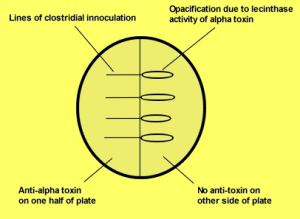 Figure: Lecithinase test or Nagler’s reaction
Bacillus cereus also exhibits strong lecithinase activity but can be differentiated by its strong hemolytic property on sheep blood agar and motility. Among Bacillus species, B. thuringiensis and B. anthracis are lecithinase positive or weakly positive. B. anthracis, however, is a nonmotile organism and produces nonhemolytic colonies.
Principle
Lecithin is a normal component of egg yolks. Bacterial lecithinase breaks down lecithin into phosphorylcholine and an insoluble diglyceride, which forms a precipitate in the medium. This precipitate appears as a white opaque halo surrounding the lecithinase-producing colony grown on the egg yolk agar medium.
Modified egg yolk agar is a differential and enriched medium used to isolate and differentiate different species based on their lecithinase and lipase production and proteolytic activity. The degradation of lecithin present in the egg yolk results in the formation of opaque precipitate around the colonies. The lipase enzyme hydrolyzes the fats within the egg yolk, which results in an iridescent sheen on the colony surface.
Note the two egg-yolk-agar reactions are distinct and read differently: lecithinase produces an opaque white halo in the medium (this test), while lipase produces an iridescent, oil-on-water sheen on the colony surface. They detect different enzymes on different substrates. The lipase reaction is the basis of the separate lipid hydrolysis (lipase) test (usually on tributyrin agar, where a positive is a clear zone, the visual opposite of lecithinase's opaque halo). C. perfringens is the clean split: lecithinase-positive here, lipase-negative there. See the lipid hydrolysis test for that reaction's full method.
Requirements
- Media: Basal egg yolk media is prepared by dissolving the standard amount in distilled water, in which 10% of egg yolk is added after autoclaving and cooling the media before dispensing it into sterile Petri dishes. Various original modifications like modified egg yolk agar media, mannitol egg yolk media, etc., are also available and can be used for specified purposes.
- Others: Inoculating loop, spreader, pipettes
- Equipment: Anaerobic gas pak or candle jar or anaerobic incubator for anaerobic incubation
Quality control
Inspect egg yolk agar for freezing, contamination, cracks, and dehydration before storage and use. Discard the opaque media. Perform QC on each new lot of media before using them.
Following strains can be used for quality control testing in naglers reaction
- Clostridium perfringens ATCC 13124: Lecithinase positive
- Clostridium sporogenes ATCC 11437: Lecithinase negative
- Bacteroides fragilis ATCC 25285: No activity on agar
Procedure
The procedure of Nagler reaction is as follows:
- Label and dry an egg yolk media plate and mark the plate into two halves.
- Inoculate 60 µl of Clostridium perfringens type A antitoxin in half of the plate, spread over the agar surface using a spreader, and allow to absorb and dry.
- Mark the side of the plate in which the antitoxin is inoculated.
- Streak the test organism in a straight line from the toxin-free agar half of the plate to toxin containing side. Repeat the same procedure with control strains on the same plate.
- Incubate anaerobically at 35-37°C for 24-48 hrs.
- Examine the plate for an opalescent halo around the inoculum and inhibition by antitoxin.
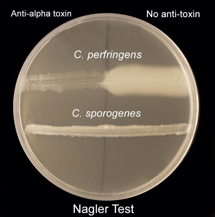 Figure: Naglers test
Result and interpretation
| Observance | Inference |
|---|---|
| A zone of opacity in the antitoxin-free half only but not on another half due to neutralization of the alpha-toxin. | Lecithinase positive |
| A zone of opacity on both sides of the plate or no reaction on the agar. | Lecithinase negative |
- Catalase-positive, spore-forming, Gram-positive rods that are lecithinase positive, with large zones of opacity, belong to the B. cereus group. Lack of motility separates B. anthracis from the other members of the group.
- Lecithinase and lipase are useful as part of the identification of Clostridium to the species level.
C. perfringens is lipase negative and lecithinase positive, which can be neutralized by adding anti-α-toxin prior to inoculation of the agar (the Nagler reaction). C. sporogenesis lipase positive. C. difficile is both lipase and lecithinase negative.
- A Gram-positive rod that is catalase-negative, hemolytic, and lecithinase positive is A. haemolyticum.
- Among the fluorescent group of non-glucose-fermenting, Gram-negative rods, P. putida is lecithinase negative and most P. fluorescens isolates are lecithinase positive. This test can substitute for gelatin hydrolysis.
- Burkholderia spp are often lecithinase positive.
Limitations of the test
- Maintenance of anaerobic conditions is compulsory.
- A negative lecithinase test should be compared to an un-inoculated control plate, as lecithinase can diffuse throughout the entire agar plate and make interpretation difficult.
- C. perfringens type A antitoxin is not specific for C. perfringens; a positive Nagler reaction can also be produced by C. bifermentans , C. sordelli , and C. baratti if heavy inoculum is used.
- Non-glucose fermenting rods may give small zones of opacity.
Egg Yolk Agar Medium Composition
McClung and Toabe agar, modified for lecithinase and lipase tests
| Ingredients | Gram/Liter |
|---|---|
| Proteose no. 2 peptone or Polypeptone (BBL) | 40.0 g |
| Disodium phosphate | 5.0 g |
| Monopotassium phosphate | 1.0 g |
| Sodium chloride | 2.0 g |
| Magnesium sulfate | 0.1 g |
| Glucose | 2 g |
| Hemin solution, 5 mg/ml | 1.0 ml |
| Agar | 20.0 g |
| Water | 1.0 liter |
Preparation
- Suspend ingredients, and adjust the pH to 7.6.
- Mix, and boil to dissolve.
- Dispense 20 ml per tube, and autoclave at 118°C for 15 min.
- Cool to 50°C, and to each tube, add 2 ml of commercial egg yolk emulsion or 1 ml of egg yolk emulsion prepared as follows: Scrub, and then soak, an antimicrobial-agent-free hen egg in 95% ethanol for 1 h. Aseptically aspirate or separate the egg yolk. Add equal volumes of egg yolk to sterile saline, and stir to make a smooth suspension.
- Mix, and pour into plates.
References
- Leber AL, editor. Clinical Microbiology Procedures Handbook. 4th ed. Washington, DC: ASM Press; 2016. doi:10.1128/9781555818814
- Procop GW, Church DL, Hall GS, Janda WM, Koneman EW, Schreckenberger PC, Woods GL. Koneman's Color Atlas and Textbook of Diagnostic Microbiology. 7th ed. Philadelphia: Wolters Kluwer; 2017.
- Jousimies-Somer H, Summanen P, Citron DM, et al. Wadsworth-KTL Anaerobic Bacteriology Manual. 6th ed. Belmont, CA: Star Publishing; 2002.
- Tille PM. Bailey and Scott's Diagnostic Microbiology. 15th ed. St. Louis: Elsevier; 2022.

Comments
No comments yet. Be the first to share your thoughts.
Leave a comment
All comments are reviewed before they appear.